Human Anatomy - H. I. Koliadenko 2009
Structure of the Human Body
The Cell
The structural unit of both animal and plant organisms is the Cell. Cells form Organs, while organs make up Organ Systems and apparatuses.
The cell is a living, self-regulating, and self-renewing system. All cells are interconnected; therefore, an Organism can exist only through its integrity and close interaction with the external environment. The integrity of the organism is maintained by Neurohumoral regulation of Functions. Academician I. P. Pavlov demonstrated that the Central Nervous system plays the leading role in innervating The activity of all organs and systems and in mediating the organism's interactions with the environment.
Cells vary in shape and size (Fig. 1) and perform diverse functions within the organism. All cells consist of essential and accessory components: the essential components include the Cytoplasm, nucleus, and Organelles, while inclusions are considered accessory.
The cytoplasm—the internal environment of the cell—is heterogeneous. It comprises the cytolemma (cell membrane), hyaloplasm, organelles, and cytoplasmic inclusions.
Externally, the cytoplasm is bounded by the cytolemma (cell membrane), which separates it from the external environment (Fig. 2). It consists of Proteins, Lipids, and Polysaccharides, with a thickness of 9–10 nm. The cytolemma is a semipermeable biological membrane that regulates the Transport of substances into the cell and metabolic products out of the cell into the surrounding intercellular fluid. Like other Membrane structures in the organism, the cytolemma is composed of a bilayer of phospholipid molecules oriented perpendicular to the membrane surface. Lipids account for 40%, proteins for 60%, and CARBOHYDRATES for 1% of the membrane components. Some protein and lipid molecules are bound to carbohydrate molecules (making up about 1% of the membrane's carbohydrates). These molecules are invariably located on the outer surface of the membrane, forming the glycocalyx. Cholesterol molecules are present on the inner surface of the membrane.
Class="center">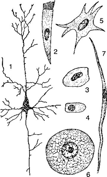
Fig. 1. Various shapes of animal cells:
1 — nerve cell; 2, 3, 4 — epithelial cells; 5 — Connective Tissue CELL; 6 — ovum; 7 — Muscle cell
Proteins in the membrane perform enzymatic, receptor, and other functions. Cell membranes feature cytoplasmic extensions through which cells communicate with one another.
The hyaloplasm encloses The Nucleus, which houses hereditary information in the form of deoxyribonucleic acid (DNA). The nucleus is mostly spherical, with a double-layered membrane known as the karyotheca. Inside the nucleus, there is one or more nucleoli. The thickness of the karyotheca is 7 nm. The nucleus is filled with a colloidal fluid called karyoplasm, which contains Chromatin, achromatin substance, Enzymes, and other BIOLOGICALLY ACTIVE SUBSTANCES.

Fig. 2. Diagram of Cell Structure:
1 — cytolemma; 2 — cytoplasm; 3 — Endoplasmic reticulum; 4 — vacuole; 5 — centrosome (cell center); 6 — nuclear envelope; 7 — ribosome; 8 — nucleolus; 9 — Golgi apparatus; 10 — mitochondrion
During Cell Division, chromatin condenses to form Chromosomes. Human cells contain 23 pairs of chromosomes. Female cells have two X chromosomes (XX), whereas male cells have an X and a Y chromosome (XY). Chromosomes consist of DNA and carry a set of genes, which are the bearers of hereditary traits.
All biochemical processes take place within cell organelles. These include Mitochondria, The endoplasmic reticulum, Lysosomes, the Golgi apparatus, and Microbodies. Organelles are bounded by a membrane 6–7 nm thick.
Non-membranous organelles include the centrosome, microtubules, cilia, and flagella. In addition, cells contain fibrillar structures: myofibrils (in muscle cells), neurofibrils (in Nerve Cells), and tonofibrils.
Mitochondria function as the cell's power plants: they are the site of cellular Respiration—The production of energy utilized by the cell for various functions. Mitochondria are ovoid in shape and are bounded by outer and inner membranes 7 nm thick. The inner membrane forms folds known as cristae, which project into the matrix, a space filled with a colloidal fluid. The surface of the cristae is studded with numerous enzymes that drive the complex biochemical processes resulting in energy production.
Ribosomes synthesize proteins specific to each cell type. Transfer RNA (tRNA) and Messenger RNA (mRNA) participate in this process.
The endoplasmic reticulum comprises rough (granular) and smooth (agranular) networks. The smooth endoplasmic reticulum mainly consists of cisternae and tubules with a diameter of 50–100 nm, which are involved in the synthesis and METABOLISM of lipids and Glycogen. This network is prominent in cells that secrete steroid substances and carbohydrates. The rough endoplasmic reticulum also consists of cisternae, tubules, and lamellae with a diameter of 20 nm or more. The outer surface of these membranes is densely studded with ribosomes, which synthesize and secrete proteins.
Proteins synthesized in the ribosomes of the endoplasmic reticulum are exported outside the cell, whereas ribosomes located free in the hyaloplasm synthesize proteins required for the cell's own vital activities.
Lysosomes are vesicles containing a high concentration of enzymes that facilitate intracellular Digestion.
The Golgi apparatus appears as a system of vesicles, lamellae, tubules, and saccules, which also contain enzymes. It participates in the synthesis of polysaccharides and, by interacting with proteins, helps package and export Metabolic waste products out of the cell. The Golgi apparatus is typically located near the Cell Nucleus.
The centrosome (cell center) is usually located just above the nucleus or near the Golgi complex and contains two dense bodies known as centrioles. Centrioles duplicate prior to cell division.
Microtubules are composed of the protein tubulin, have a diameter of 25 nm, and form the cell's stroma (Cytoskeleton).
Cilia and flagella facilitate cell motility. Both structures are cellular projections anchored by basal bodies at their base. Flagella can reach lengths of 120–150 µm, while cilia are typically 5–10 µm long.
Inclusions are protein granules, fat droplets, or pigment granules found in the cytoplasm. Unlike organelles, they are either present in the cell or absent.
Cell sizes can reach up to 200 μm (such as egg cells), but typically do not exceed 20–30 μm. The Human Body contains cells of A wide variety of shapes: spherical, spindle-shaped, stellate, cylindrical, scaly, and others. All Cells of the organism are capable of division.
Last update: 08/08/2026
Editorial and Educational Adaptation: This material has been compiled based on the primary/original source text. The project team performed an editorial review, corrected technical inaccuracies, structured sections, and adapted the content for an educational format.
What was processed:
- elimination of formatting defects (OCR errors, structural breaks, corrupted characters);
- editorial organization of content;
- standardization of terminology in accordance with academic sources;
- verification of factual statements against the original source text.
All mentions of the author, publication year, and origin of the primary text have been preserved in accordance with the source.