Molecular Biology of the Cell - Volume 3 - Alberts B., Bray D., Lewis J., Raff M., Roberts K., Watson J. 1994
From Cells to Multicellular Organisms
Nervous System
Ligand-Gated Ion Channels and Rapid Synaptic Transmission
The simplest way to transmit a signal from neuron to neuron is via direct electrical coupling Through Gap Junctions. The main advantage of such electrical synapses is that signals are transmitted without delay. On the other hand, these synapses are much less adaptable and suited for regulation than chemical synapses, which mediate the vast majority of neuronal connections. Electrical communication via gap junctions was examined in Ch. 14 (Sec. 14.1.5-14.1.8); here, we will focus exclusively on chemical synapses.
Chemical transmission at synapses operates on the same principles as chemical signaling mediated by Water-soluble Hormones (Ch. 12). In both cases, a Cell releases a messenger molecule that acts on another cell or group of Cells by binding to membrane receptor Proteins. Unlike a hormone, however, the chemical messenger at a synapse—the neurotransmitter—acts over very short distances.
As a result of electrical stimulation, the presynaptic cell releases a neurotransmitter via exocytosis (see Fig. 19-4). Once the neurotransmitter crosses the cleft between the pre- and postsynaptic cells—typically a fraction of a micrometer wide—the chemical signal must be converted back into an electrical one. This conversion is carried out by receptors located in The Plasma Membrane of the postsynaptic cell. There are two MAIN TYPES OF receptors: channel-linked and non-channel-linked (Fig. 19-15). Channel-linked receptors are essentially Ligand-Gated Ion Channels. The conformation of these receptors changes immediately upon neurotransmitter binding, opening a specific ion channel in the membrane and thereby altering membrane permeability. Receptors of this type form The basis of the most common and thoroughly studied mode of signal transmission in chemical synapses, allowing for extremely rapid communication.
Non-channel-linked receptors trigger processes similar to those initiated by water-soluble hormones and local chemical mediators throughout the body (Sec. 12.3). In these receptors, the neurotransmitter-binding sites are functionally coupled to an enzyme that, in the presence of the neurotransmitter, typically catalyzes The production of an intracellular messenger, such as cAMP. In turn, this messenger induces Changes in the postsynaptic cell, including the modification of ion channels in The Cell membrane. Unlike channel-linked receptors, these receptors generally mediate relatively slow but more prolonged neurotransmitter effects. It is believed that the activation of such receptors induces long-lasting changes in Neurons that underlie Learning and Memory (Sec. 19.5.3).
This section explores fast synaptic transmission mediated by ligand-gated ion channels. The Specific features of synaptic transmission involving non-channel-linked receptors and their role in long-term synaptic modifications will be discussed in Sec. 19.5.
19.3.1. The Neuromuscular Junction Is the Most Thoroughly Studied Synapse [14]
Neurons are so densely packed in the Brain that experimenting on individual brain synapses is exceptionally difficult. Consequently, synaptic Functions have been detailed primarily through studies of connections between motor nerves and Skeletal Muscle in frogs and, to a lesser extent, synapses between the giant neurons of squids and other Molluscs.
Vertebrate skeletal muscle fibers, much like Nerve Cells, are electrically excitable; thus, the neuromuscular junction (Fig. 19-16) has served as an excellent model for chemical synapses in general. A motor nerve and its innervated muscle can be isolated from surrounding tissue and maintained in a functional state in a nutrient medium of defined composition. By stimulating the nerve via external electrodes, researchers can record the response of a single muscle cell using an intracellular microelectrode (Fig. 19-17). Figure 19-18 compares the Fine Structure of the neuromuscular junction with that of a typical synapse between two Central Nervous system neurons.
The neuromuscular junction has been the primary focus of numerous prolonged and fruitful investigations beginning in the 1950s. The groundwork for the earliest experiments was laid by the discovery in the early 1920s that acetylcholine, released upon Vagus nerve stimulation, slows The Heart rate. This provided the first unequivocal proof of the Chemical Nature of neuromuscular transmission. Soon after, in the 1930s, it was demonstrated that stimulating a motor nerve innervating skeletal muscle also triggers acetylcholine release, which in turn causes the skeletal muscle to contract. Thus, acetylcholine was identified as the neurotransmitter at the neuromuscular junction. But how is acetylcholine released, and how does it act on the muscle?
Class="center">
Fig. 19-15. The action of a neurotransmitter on a postsynaptic cell can be mediated by two fundamentally Different types of receptor proteins: channel-linked receptors and non-channel-linked receptors. Channel-linked receptors are also referred to as ligand-gated channels.

Fig. 19-16. The frog neuromuscular junction. A. Low-magnification scanning electron micrograph of a single axon terminal on a skeletal muscle cell. B. Schematic representation of the area outlined by the red box in A, showing key details visible under a transmission Electron microscope. The branching pattern of small axon terminals within the synaptic region varies depending on animal species and muscle fiber type. Because of the specialized shape of mammalian axon terminals, the neuromuscular junction is often referred to as a motor endplate. (A-J. Desaki, Y. Uehara, J. Neurocytol, 10, 101-110, 1981, reprinted with permission from Chapman & Hall.)
19.3.2. Voltage-Gated Calcium Channels Are Responsible for Coupling Action Potentials to Mediator Release [15]
The opening and closing of sodium channels propagate a Nerve Impulse along the axon until it reaches the contact site with the muscle cell. Here, this impulse triggers the opening of voltage-gated channels located in the plasma membrane of the axon terminal, allowing Ca2+ ions to enter the axon and thereby prompting the release of acetylcholine (Fig. 19-19).
As three simple observations have demonstrated, an influx of Calcium Ions into the axon terminal is essential for synaptic transmission. First, if these ions are absent from the extracellular fluid surrounding the axon terminal when a nerve impulse arrives, no transmitter is released, and signal transmission fails. Second, if Ca2+ is artificially injected into the Cytoplasm of the nerve terminal via a micropipette, neurotransmitter release occurs immediately, even without electrical stimulation of the axon (this is difficult to perform at the neuromuscular junction due to The small size of the axon terminal, so the experiment was conducted on the synapse between giant squid neurons). Third, artificial depolarization of the axon terminal (also in the giant neuron synapse) in the absence of a nerve impulse and under conditions where sodium and potassium channels are blocked by specific toxins causes Ca2+ to enter the terminal and triggers neurotransmitter release. However, if depolarization shifts the Membrane Potential such that the electrochemical driving force forcing Ca2+ inward drops to zero, neurotransmitter release does not occur.

Fig. 19-17. Experimental setup for studying synaptic transmission at the neuromuscular junction.

Fig. 19-18. A. Electron micrograph of a portion of a neuromuscular junction. B. Electron micrograph of a small region of the rat brain, clearly showing two synapses. One can distinguish the pre- and postsynaptic membranes, the synaptic cleft between them, and synaptic vesicles within the axon terminals, similar to photo A. The two synapses shown in photo B differ in the size and shape of their synaptic vesicles: in type I synapses, the vesicles are round, whereas in type II synapses, they are flattened and are believed to contain a different neurotransmitter. Note the characteristic thickening of both the postsynaptic and (to a lesser extent) presynaptic membranes, visible in both images. Unlike brain synapses, central nervous system synapses lack a basal lamina between the pre- and postsynaptic membranes, although some extracellular material is still discernible there. The absence of a basal lamina is a key structural feature distinguishing central nervous system synapses from neuromuscular junctions. (Kindly provided by John Heuser (A) and J. Campbell, A. R. Lieberman (B).)
The protein forming the channel for Ca2+ entry into the cell—the voltage-gated calcium channel—plays a critically important role. It provides the sole known mechanism for converting electrical signals (brief membrane depolarizations) into chemical changes within the neuron. As illustrated in Panel 19-1, voltage-gated channels for Na+, K+, or Cl- cannot serve this purpose: a single nerve impulse passes such minuscule ion currents through these channels that they alter cytosolic ion concentrations only negligibly. The ion flux through calcium channels is also relatively small on its own, and its contribution to the overall transmembrane electrical current is generally minor. However, this flux is extremely substantial relative to the intracellular concentration of free calcium, which is normally maintained at around 10-7 M (corresponding to fewer than 100 Ca2+ ions per μm3). Driven by the membrane potential and the relatively high extracellular calcium concentration (typically ~1-2 mM), several hundred Ca2+ ions flow through a single open calcium channel in just 1 ms. Thus, when a small number of voltage-gated calcium channels open in the presynaptic terminal in response to a nerve impulse, the intracellular free calcium concentration can easily spike 10- to 100-fold. The incoming free Ca2+ ions act as intracellular messengers, triggering neurotransmitter release at a rate that increases steeply with rising Ca2+ concentration.

Fig. 19-19. Key events occurring at a chemical synapse following the arrival of an impulse at the axon terminal.
The concentration of free calcium ions remains elevated only briefly, as Ca2+-binding proteins, Ca2+-sequestering Organelles, and Mitochondria rapidly take up the Ca2+ ions that have entered the axon terminal. Additionally, plasma membrane calcium pumps—powered by ATP Hydrolysis or the sodium electrochemical gradient—actively extrude calcium ions from the cell (see Sec. 6.4.7 and 12.3.7). This rapid clearance enables the axon terminal to transmit subsequent signals the very moment another nerve impulse arrives.
19.3.3. Neurotransmitters Are Rapidly Released via Exocytosis [16]
The axon terminal at the neuromuscular junction is packed with thousands of identical (~40 nm in diameter) secretory vesicles known as synaptic vesicles, each containing acetylcholine (see Fig. 19-18). Calcium entering the cell triggers a wave of exocytosis, causing the vesicles to fuse with the presynaptic membrane, releasing their contents into the synaptic cleft and acting on the postsynaptic cell. Exocytosis occurs exclusively at specialized sites called active zones, positioned directly opposite the receptors of the postsynaptic cell; this arrangement ensures that the delay in signal transmission caused by neurotransmitter diffusion across the synaptic cleft is rendered negligibly small. Subsequently, the membranes of the "discharged" synaptic vesicles are retrieved from the presynaptic plasma membrane via endocytosis.
Evidence suggests that Ca2+ ions entering the axon terminal not only trigger exocytosis but also activate a Ca2+-calmodulin-dependent protein kinase (Ca-kinase II — see Section 12.4.3). This kinase phosphorylates numerous proteins within the axon terminal, including synapsin I, a protein attached to The surface of synaptic vesicles. Phosphorylation is thought to release synapsin I, allowing the vesicles to migrate to the active zone of the presynaptic membrane and take THE PLACE OF those lost to exocytosis. The entire cycle of events triggered by a single nerve impulse has been vividly demonstrated by rapidly freezing the synaptic region and examining the preparations using Electron Microscopy. Some of these results are presented in Fig. 19-20.
19.3.4. Neurotransmitter is released in random "quanta" [17]
In response to a single nerve impulse, only a few hundred of the many thousands of synaptic vesicles present at the neuromuscular junction are typically released from the axon terminal. Each vesicle, by discharging its contents into the synaptic cleft, contributes to A change in the membrane potential of the postsynaptic muscle cell, which can be recorded using an intracellular electrode (Fig. 19-21). Consequently, the muscle cell membrane is depolarized to threshold, generating an Action Potential. This excitation propagates across the entire cell (Fig. 19-22), triggering its contraction as described in Section 11.1.11.
Even when no impulses arrive at the axon terminal, random, transient shifts in the muscle membrane potential toward depolarization can be observed near the synapse. These so-called miniature synaptic potentials have a remarkably uniform amplitude of only about 1 mV, which is well below the threshold level. Such potentials occur stochastically with a fairly low probability, on average about once per second (Fig. 19-23). Each miniature potential results from the fusion of a single synaptic vesicle with the presynaptic membrane — that is, from the release of the contents of one vesicle. The amplitude recorded in a given muscle cell is more or less constant because the vesicles contain roughly the same number of acetylcholine molecules, approximately 5,000. This represents the minimum portion, or "quantum," of the released neurotransmitter. Signals of greater strength correspond to multiples of this fundamental unit. Calcium ions entering the axon terminal during an action potential boost the vesicle release frequency by a factor of over 10,000 within fractions of a millisecond compared to The rate of spontaneous release in a resting terminal. Nevertheless, the process remains probabilistic; a single nerve stimulus does not always produce the exact same postsynaptic effect. For instance, if an average of 300 quanta of neurotransmitter are released, the actual number in any individual instance may be slightly higher or lower.

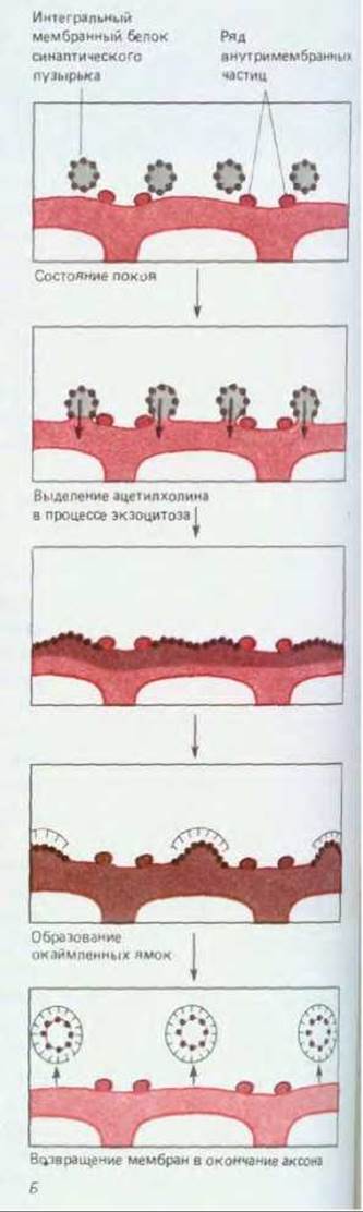
Fig. 19-20. The cycle of events occurring at the axonal terminal membrane in the neuromuscular synapse following stimulation. To trace The sequence of events, tissue samples were subjected to rapid freezing at various time intervals. For simplicity, stimulation was performed under conditions specifically modified to slow the overall process by a factor of 5 to 10 and to increase the number of vesicles undergoing exocytosis. A. Schematic representation of the neuromuscular junction, showing active zones where neurotransmitter release takes place. B. The boxed region from figure A shown at a higher magnification, schematically depicting the events occurring in this area at various time intervals following nerve stimulation. C–H. Appearance of the membrane under the electron microscope (photographs kindly provided by John Heuser). Left: freeze-fracture Cytology/cytology/93.html">ELECTRON MICROGRAPHS OF presynaptic membrane preparations (cytoplasmic face); right: thin-section electron micrographs. C and F. Resting state. D and G. Fusion of synaptic vesicles with the plasma membrane at the active zone (indicated by rows of intramembrane particles). E and H. Retrieval of synaptic vesicle membranes via coated pits and coated vesicles. As can be seen, synaptic vesicles begin to fuse with the plasma membrane 5 ms after stimulation (D, G); each "pore" in the plasma membrane visible in micrograph D results from the fusion of a single synaptic vesicle. Fusion is complete 2 ms later. The first signs of membrane recovery become noticeable after approximately 10 s with The formation of coated pits (see Section 6.5.4), and another 10 s later, these pits begin to pinch off to form coated vesicles (E, H). These vesicles incorporate original synaptic vesicle Membrane Proteins as well as molecules scavenged from the external environment. The cycle concludes with the shedding of the clathrin coat from the vesicles, their refilling with acetylcholine, and the formation of conventional smooth-surfaced synaptic vesicles. This scheme likely accounts for the remarkable uniformity in the size of synaptic vesicles; their volume is determined by the dimensions of the surrounding clathrin coat (see Section 6.5.5).

Fig. 19-20 (continued). Further insights into this regeneration process can be gained by stimulating the nerve in the presence of electron-dense markers such as ferritin. These markers rapidly appear within coated vesicles and subsequently within synaptic vesicles. It should be noted, however, that experts remain skeptical of such experiments, considering some of the observed phenomena to be artifacts.
19.3.5. Ligand-gated channels reconvert the chemical signal into an electrical one [18]
At the synapse, the muscle cell membrane acts as a transducer, converting a chemical signal in the form of a neurotransmitter into an electrical signal. This conversion is carried out by ligand-gated ion channels (i.e., channel-linked receptors), which are proteins embedded in the postsynaptic membrane. The binding of the neurotransmitter to these proteins induces a conformational change, opening the channels to allow ions to flow across the membrane and thereby altering the membrane potential. In turn, if this shift in membrane potential is sufficiently large, it triggers voltage-gated channels to open, resulting in an action potential (Fig. 19-24). Unlike voltage-gated channels, ligand-gated channels are relatively insensitive to Changes in membrane potential and are therefore incapable of self-amplifying, all-or-none excitation. Instead, they generate an electrical signal whose strength depends on the intensity and duration of the external chemical signal — that is, on how much neurotransmitter is released into the synaptic cleft and how long it remains there. As we will see later, this property of ligand-gated channels is crucial for information Processing at synapses.

Fig. 19-21. Postsynaptic response to a single nerve impulse at the neuromuscular junction: a potential change curve in a frog muscle cell, obtained using an intracellular electrode placed near the synapse, as in Fig. 19-17. Normally, the postsynaptic potential (PSP) — the depolarization arising from the direct action of the neurotransmitter on the muscle cell membrane — is large enough to trigger an action potential, which can interfere with the experiment. A "clean" PSP, uncompromised by a nerve impulse, can be obtained by introducing moderate concentrations of curare into the extracellular medium. This poison binds to a fraction of the receptors and blocks their response to the neurotransmitter, reducing the PSP amplitude below the threshold required to elicit an action potential.

Fig. 19-22. Electrical changes in the muscle cell membrane at the neuromuscular junction. The opening of acetylcholine-gated ion channels (A) triggers an action potential (B), which propagates along the muscle fiber membrane (C) and causes it to contract.
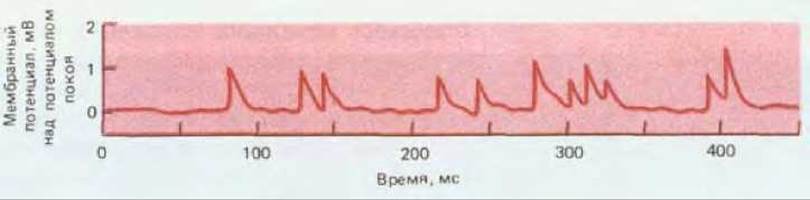
Fig. 19-23. Miniature synaptic potentials (or "miniature end-plate potentials") recorded in frog muscle using an intracellular electrode placed near the neuromuscular junction. Each peak represents a miniature synaptic potential resulting from neurotransmitter release from a single synaptic vesicle. (P. Fatt, B. Katz, J. Physiol., 117, 109–128, 1952.)

Fig. 19-24. Schematic diagram illustrating the function of ligand- and voltage-gated channels. Arrows indicate causal relationships.
Postsynaptic ligand-gated channels possess two other important properties. First, as receptors, they resemble Enzymes in that they interact exclusively with specific ligands and thus respond only to the single neurotransmitter released from the presynaptic terminal; other Transmitters produce virtually no effect. Second, as channels, they exhibit distinct ionic specificities: some selectively permit the passage of K+, others Cl-, and so on, while still others may be relatively non-selective among various cations while excluding anions. As we will see, The Nature of the postsynaptic response depends on the specific ionic permeability of the ligand-gated channels.
19.3.6. The Acetylcholine Receptor is a ligand-gated cation channel [19]
Among all ligand-gated ion channels, the acetylcholine-gated channel (also known as the acetylcholine receptor) in the muscle fiber membrane is the most thoroughly studied. Its molecular properties were discussed in Chapter 6 (Section 6.4.18).
Much like the voltage-gated sodium channel, the acetylcholine receptor can exist in several alternative Conformations (Fig. 19-25). Upon binding acetylcholine, the channel immediately opens and remains open for a brief period with the ligand bound; this duration varies stochastically, averaging about 1 ms or even less depending on Temperature and species. In the open state, the channel is equally permeable to various cations, including Na+, K+, and Ca2+, but is completely impermeable to anions (Fig. 19-26).
Since there is no marked selectivity among cations, THE CONTRIBUTION OF each cation to the current flowing through the channel depends primarily on its concentration and the electrochemical driving force. If the muscle cell membrane potential is at its resting level, the net driving force for K+ is close to zero because the voltage gradient is nearly balanced by the K+ concentration gradient. For Na+, on the other hand, the directions of the voltage and concentration gradients coincide, and their combined effect drives these ions into the cell. (This is also true for calcium ions, although the extracellular concentration of Ca2+ is so much lower than that of Na+ that calcium's share of the total inward ionic current is small.) Consequently, the opening of acetylcholine receptor channels leads primarily to a massive influx of Na+ ions, causing the membrane to depolarize.

Fig. 19-25. Response of acetylcholine receptors to acetylcholine. Upon prolonged exposure to high concentrations of acetylcholine, the receptor transitions into yet another state (not shown here) in which it becomes inactivated and fails to open even in the presence of acetylcholine.

Fig. 19-26. Measurement of current through an open acetylcholine receptor channel at various membrane potentials. Such measurements make it possible to determine the ion selectivity of channels. The current carried through an open channel by a specific type of ion will vary with changes in membrane potential in a manner determined by the ion species and its concentration gradient across the membrane. Knowing the concentration gradients of the major ions present, one can determine channel ion selectivity by simply measuring the current-voltage relationship; more complete information can be obtained by repeated measurements at different ion concentrations. A. Single-channel current recorded using the patch-clamp technique from a channel in a solution with a fixed acetylcholine concentration at three different membrane potential values. In each case, the channel randomly transitions between closed and open states, but at a certain membrane potential—known as the reversal potential—the current is zero even when the channel is open. In this case, the reversal potential is close to 0 mV. B. The same phenomenon can be observed by measuring the total current through A large number of single acetylcholine receptor channels located in the postsynaptic membrane of a neuromuscular junction following a single nerve stimulation. The graphs show changes in this current measured using intracellular electrodes under voltage-clamp conditions. The channels open upon a brief exposure to acetylcholine, but if the membrane potential is held at the reversal potential, the current is zero. Since open channels are permeable to both Na+ and K+, and the electrochemical driving forces for these ions differ, the "zero current" actually corresponds to balanced, opposing fluxes of Na+ and K+. (These channels are also permeable to Ca2+, but the current carried by calcium ions is very small due to their low concentration.) The magnitude of the reversal potential and its sensitivity to extracellular ion concentrations provide insight into the relative permeability of the channel to different ions. For example, some ligand-gated channels are selectively permeable to Cl-, and such channels can be identified by a reversal potential of -60 mV, which is close to the equilibrium potential for Cl-; furthermore, the reversal potential depends on extracellular Cl- concentration, but not on Na+ or K+. (A based on data from V. Sakmann et al., Cold Spring Harbor Symp. Quant. Biol., 48, 247-257, 1983; B based on data from K. L. Magleby, C. F. Stevens, J. Physiol., 223, 173-197, 1972.)
19.3.7. Acetylcholine is removed from the synaptic cleft by diffusion and hydrolysis [20]
For the state of a postsynaptic cell to be successfully regulated by signals arriving from a presynaptic cell, postsynaptic excitation must decay rapidly as soon as the presynaptic cell returns to a resting state. In the neuromuscular junction, this is achieved by the rapid removal of acetylcholine from the synaptic cleft through two mechanisms. First, acetylcholine dissipates via diffusion, which occurs very rapidly due to the short distances involved. Second, acetylcholine is cleaved by acetylcholinesterase into acetate and Choline. This enzyme, secreted by muscle cells, is anchored via a Collagen-like "tail" to the basal lamina that separates the nerve terminal from the muscle cell membrane. A single molecule of acetylcholinesterase can hydrolyze up to 104 molecules of acetylcholine in 1 ms, and therefore all of the neurotransmitter is cleared from the synaptic cleft within a few hundred microseconds of its release from the nerve terminal. Consequently, the period during which acetylcholine can bind to receptors and switch them into an open conformation—thereby altering postsynaptic membrane conductance—is extremely short (Fig. 19-27). As a result, the temporal pattern of presynaptic signals is faithfully mirrored in the pattern of postsynaptic responses.
19.3.8. Rapid synaptic transmission is mediated by a small number of neurotransmitters [13, 21]
The entire STRUCTURE OF THE neuromuscular junction is optimized for the fastest possible signal transmission. Contributing factors include: a large-diameter myelinated motor axon; active zones at the axon terminal where synaptic vesicles stand ready to release acetylcholine precisely opposite postsynaptic receptors; a narrow synaptic cleft; ligand-gated channels in the postsynaptic membrane that open immediately upon neurotransmitter binding; and, finally, acetylcholinesterase in the synaptic cleft, which rapidly halts transmission. The synaptic delay between the peak of the presynaptic action potential and the peak of the postsynaptic impulse is approximately one millisecond or less. Accumulating evidence indicates that rapid chemical synapses in the central nervous system also apparently utilize ligand-gated channels and share the same structural design principles: the presence of active zones, a narrow synaptic cleft, and the localization of receptors opposite sites of exocytosis. Moreover, rapid signaling here also appears to be mediated by only a small group of neurotransmitters. However, this generalization is not yet fully definitive, as it has proven very difficult to determine with certainty which specific neurotransmitter operates at a given synapse.

Fig. 19-27. Changes in postsynaptic membrane conductance in a frog neuromuscular junction induced by a single "quantum" of acetylcholine (the contents of one synaptic vesicle). At the peak of conductance, approximately 1,600 channels are open, each remaining open for an average of 400 µs.

FIG. 19-28. Chemical structures of major neurotransmitters believed to be involved in rapid synaptic transmission via channel-linked receptors.
Rapid synaptic transmission likely evolved very early, as the same neurotransmitters are utilized by evolutionarily distant animal species ranging from Mollusks to mammals. Fast-acting neurotransmitters include acetylcholine, gamma-aminobutyric acid (GABA), Glycine, glutamate, and presumably aspartate and ATP (Fig. 19-28). Typically, each neuron secretes only a single type of neurotransmitter, which is common to all output synapses of that neuron (though on rare occasions there may be two). Direct evidence obtained via the patch-clamp technique confirms that receptors for acetylcholine, GABA, glycine, and glutamate are linked to ion channels. For other receptors, this is also highly probable, though not yet formally proven. DNA Sequencing studies have established structural Homology among the receptors for acetylcholine, GABA, and glycine, suggesting a common evolutionary origin for all ligand-gated ion channels.
19.3.9 Acetylcholine and glutamate mediate fast excitation, whereas GABA and glycine mediate fast inhibition [19, 22]
Neurotransmitters can be classified According to the effects they elicit. As we have seen, acetylcholine, upon binding to its respective receptor in a skeletal muscle fiber membrane, opens a cation channel and depolarizes the membrane, bringing it closer to the threshold for an action potential. Thus, this receptor mediates an excitatory effect. Glutamate appears to act on receptors of a similar type. It has been demonstrated that glutamate serves as an excitatory neurotransmitter at insect neuromuscular junctions, and it is believed to be the major excitatory neurotransmitter in the vertebrate central nervous system, much like acetylcholine plays this role in the Peripheral Nervous System (while also fulfilling vital central functions). Aspartate may act on the same receptors as glutamate, eliciting similar effects. Evidence also suggests that ATP serves as the fast excitatory neurotransmitter at synapses on certain types of smooth muscle.
In contrast to all these transmitters, GABA and glycine mediate rapid inhibition. The receptors that bind these neurotransmitters are coupled to channels that, upon opening, permit the influx of small negatively charged ions—primarily Cl-—while remaining impermeable to positive ions. The extracellular Cl- concentration is much higher than the intracellular concentration, and the Cl- equilibrium potential is close to or even more negative than the normal Resting Potential. Consequently, open chloride channels hold the membrane in a polarized or even hyperpolarized state, thereby hindering cell depolarization and, consequently, excitation (Fig. 19-29). GABA and glycine are thought to be the principal neurotransmitters mediating fast inhibition in the vertebrate central nervous system. GABA is also known to perform this function in the neuromuscular junctions of insects and crustaceans. The Importance of inhibitory neurotransmitters is underscored by the effects of poisons that block their actions; for instance, strychnine binds to glycine receptors and blocks glycine action, causing muscle spasms, convulsions, and death.
19.3.10. A single type of neurotransmitter often has multiple receptor subtypes [23]
The action of a neurotransmitter depends not only on its chemical nature but also on the specific receptor to which it binds. In fact, the same neurotransmitter frequently binds to several different types of receptors. For example, in vertebrates, acetylcholine exerts opposite effects on skeletal and cardiac muscle cells, exciting the former and inhibiting the latter. Different acetylcholine receptors mediate these two responses. It is believed that the inhibitory effect—which develops much more slowly than skeletal muscle excitation—involves non-channel-linked receptors. Channel-linked receptors that mediate the fast excitatory actions of acetylcholine are termed nicotinic receptors because they can be activated by nicotine. Receptors that are not linked to channels and mediate the slow effects of acetylcholine (which can be either inhibitory or excitatory) are called muscarinic receptors, as they are activated by muscarinine (a mushroom toxin). In addition to such substances that specifically activate certain receptors (so-called agonists), there are potent receptor-specific blockers (antagonists) that selectively suppress the function of one or another type of acetylcholine receptor. For example, curare and a-bungarotoxin specifically bind to nicotinic receptors and block their activity, whereas atropine acts similarly on muscarinic receptors. Other Agonists and Antagonists exhibit Specificity toward receptors for different neurotransmitters. Various receptors are very frequently studied, identified, and localized using the agonists and antagonists that bind to them.

Fig. 19-29. Behavior of channel-linked GABA receptors. Upon binding GABA, these receptors form an open channel selectively permeable to Cl-. In this manner, they produce an inhibitory effect: open Cl- channels clamp the membrane potential near the equilibrium potential for Cl-, which in turn is close to the resting potential.
19.3.11. Synapses serve as major targets for pharmacological agents [23, 24]
Neurotransmitter receptors play a crucial role as targets for toxins and drugs. A snake paralyzes its prey using a-bungarotoxin, which blocks nicotinic acetylcholine receptors. By blocking these exact receptors with curare, muscle relaxation can be induced during surgical Procedures while the heart continues to function normally, as curare does not bind to muscarinic receptors. Thus, differences in the ligand-binding properties of these Two Types of acetylcholine receptors allow for highly targeted pharmacological intervention.
Most psychotropic drugs act on synapses, and many of them bind to specific receptors. A case in point is GABA receptors; the best-characterized among them—GABAA receptors—are ligand-gated chloride channels involved in fast inhibition, as discussed above. They are modulated by both benzodiazepine tranquilizers (such as Valium and Librium) and barbiturates, which are used to treat insomnia, anxiety, and Epilepsy. GABA, benzodiazepines, and barbiturates bind cooperatively to three distinct sites on the same receptor protein; these drugs likely exert their psychoactive effects by lowering the threshold concentration of GABA required to open the chloride channels, thereby potentiating GABA-mediated inhibition.
Synaptic transmission can also be disrupted in numerous other ways, such as by inhibiting the degradation of the neurotransmitter or its clearance from the synaptic cleft. Certain drugs inhibit acetylcholinesterase activity at the neuromuscular junction, which prolongs the duration of acetylcholine action on the muscle cell. This helps alleviate muscle weakness in patients suffering from myasthenia gravis, who have a reduced number of functionally active acetylcholine receptors (Section 18.1.9). Other neurotransmitters, such as GABA, are not degraded by enzymes in the synaptic cleft, but are instead reabsorbed by presynaptic terminals or neighboring glial cells. As a rule, specialized transport proteins for the active uptake of neurotransmitters reside in the Plasma Membranes of nerve terminals and glial cells. Certain psychotropic drugs either block or activate this process at specific classes of synapses, yielding beneficial clinical effects.
Nerve signals are transmitted from Cell to Cell via synapses, which can be either electrical (gap junctions) or chemical. In a chemical synapse, depolarization of the presynaptic membrane triggered by a nerve impulse opens voltage-gated calcium channels, leading to an influx of Ca2+ ions, which in turn prompt the release of neurotransmitter from synaptic vesicles via exocytosis. The transmitter diffuses across the synaptic cleft and binds to receptor proteins in the postsynaptic membrane. From the synaptic cleft, the transmitter is rapidly cleared by diffusion, enzymatic degradation, or reuptake into the axon terminal or glial cells. Neurotransmitter receptors can be subdivided into channel-linked and non-channel-linked receptors. Channel-linked receptors (ligand-gated ion channels) mediate rapid postsynaptic effects that manifest within a few milliseconds. Only a small number of neurotransmitters are known to interact with such receptors. Specifically, acetylcholine and glutamate (as well as likely aspartate and ATP) open ligand-gated channels permeable exclusively to cations, leading to fast excitatory postsynaptic potentials, whereas GABA and glycine open homologous channels that predominantly conduct Cl- ions, resulting in fast inhibitory postsynaptic potentials. All of these neurotransmitters, along with many others, can also act on non-channel-linked receptors, eliciting slower and more complex effects.
Last update: 12/08/2026
Editorial and Educational Adaptation: This material has been compiled based on the primary/original source text. The project team performed an editorial review, corrected technical inaccuracies, structured sections, and adapted the content for an educational format.
What was processed:
- elimination of formatting defects (OCR errors, structural breaks, corrupted characters);
- editorial organization of content;
- standardization of terminology in accordance with academic sources;
- verification of factual statements against the original source text.
All mentions of the author, publication year, and origin of the primary text have been preserved in accordance with the source.