Biological Membranes - A. N. Ogurtsov 2012
Electrogenesis of Biomembranes
Electrical Potentials of Biomembranes
Membrane Potential
One of the most vital Functions of a biological membrane is the generation and transmission of biopotentials. This phenomenon underlies cellular excitability, The regulation of intracellular processes, the functioning of The Nervous system, the control of Muscle contraction, and sensory reception.
In medicine, diagnostic Methods such as electrocardiography, Electroencephalography, and electromyography are based on The Study of electrical fields generated by the biopotentials of Organs and Tissues. Furthermore, exposing Cell Suspensions to external electrical impulses serves as the foundation for biotechnological techniques like electroporation and biomembrane electropermeabilization.
During metabolic activity, Two Types of electrical potential differences may arise within Cells and Tissues:
1) oxidation-reduction (redox) potentials, resulting from The transfer of electrons from some molecules to others;
2) membrane potentials, resulting from ion concentration gradients and The transport of ions across the membrane.
The biopotentials recorded in a living Organism are predominantly membrane potentials.
The membrane potential is defined as the potential difference between the inner (cytoplasmic) and outer surfaces of the membrane.
Class="center">
Hereafter, the value of ∆φМ will be denoted simply as φM. Progress in the study of biopotentials has been driven by:
✵ The Development of the microelectrode technique for intracellular potential measurement;
✵ the invention of specialized biopotential amplifiers;
✵ the Selection of suitable research models, particularly large cells such as the giant squid axon.
The diameter of the squid axon reaches 0.5 mm, which is 100 to 1,000 times larger than the diameter of vertebrate axons, including those of humans. Compared to vertebrates, these giant dimensions are of great physiological significance, ensuring the rapid propagation of nerve impulses along the nerve fiber.
When studying the Electrogenesis of Biomembranes, the giant squid axon served as an excellent model organism for investigating biopotentials.
A microelectrode can be inserted through the membrane into the giant squid axon without causing significant damage to The Cell. Figure 88 illustrates the schematic setup for measuring intracellular potential using the microelectrode technique.
A Glass microelectrode is essentially a glass micropipette with an extremely tapered tip (Figure 88 (3)). A metal electrode of such thickness is malleable and cannot puncture The cell membrane; moreover, it polarizes. To prevent polarization, non-polarizable electrodes are employed—for instance, a silver wire coated with AgCl salt and immersed in a KCl or NaCl solution (gelatinized with Agar-agar) that fills the microelectrode.
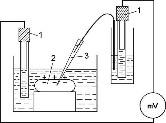
Figure 88 - Schematic diagram of intracellular potential measurement: 1 - calomel reference electrodes; 2 - cell; 3 - glass microelectrode
The second electrode—the reference electrode—is placed in the solution near the outer surface of the cell. The recording device (a millivoltmeter, mV) measures the membrane potential φМ.
The microelectrode technique has made it possible to measure biopotentials not only in the giant squid axon, but also in cells of normal size, including nerve fibers of other animals, Skeletal Muscle cells, myocardial cells, and others.
Last update: 13/08/2026
Editorial and Educational Adaptation: This material has been compiled based on the primary/original source text. The project team performed an editorial review, corrected technical inaccuracies, structured sections, and adapted the content for an educational format.
What was processed:
- elimination of formatting defects (OCR errors, structural breaks, corrupted characters);
- editorial organization of content;
- standardization of terminology in accordance with academic sources;
- verification of factual statements against the original source text.
All mentions of the author, publication year, and origin of the primary text have been preserved in accordance with the source.