Human Anatomy (with the Fundamentals of Dynamic and Sports Morphology) - Ivanitsky M. F. 2008
Introduction to Anatomy
Cells and Tissues
Body Organs are formed by tissues, and the latter by cells. Their study is the subject of two morphological sciences: Histology (The Study of tissues) and Cytology (the Study of the Cell). The Cell is a living, self-regulating, and self-renewing system that serves as the structural, developmental, and functional basis of all animal and plant organisms. In The Human Body, cells vary in shape, size, internal Structure, and functional significance (Fig. 1). Spherical, spindle-shaped, cubic, cylindrical, stellate, and other cell types are distinguished.
Cell size ranges from 7 to 200 microns (µm).
Despite their diverse shapes, cells share a common structural plan (Fig. 2). The main PARTS OF THE cell are the Cytoplasm and The Nucleus.
Cytoplasm. The Cell Cytoplasm is heterogeneous; it comprises the cytolemma (Plasmalemma), hyaloplasm, Organelles, and cytoplasmic inclusions.
The cytolemma (plasmalemma) separates the cell from its external environment, regulates cellular METABOLISM, and ensures the constancy of its internal environment. The thickness of the cytolemma is 9—10 nm*. It is composed of protein and lipid compounds bound with CARBOHYDRATES (Glycoproteins and Glycolipids). According to A. Ham and D. Cormack (1982), the carbohydrate-containing parts of these molecules are located in the surface layer of the cytolemma—the glycocalyx. Conversely, Cholesterol is situated primarily in its deeper layers. On the surface of the cytolemma are so-called receptors—molecules that interact with BIOLOGICALLY ACTIVE SUBSTANCES such as Hormones, mediators, etc., facilitating their "recognition" by the cell. The cytolemma participates in the cellular uptake of large particles (phagocytosis) or macromolecules (pinocytosis), which undergo intracellular Digestion. It ensures The excretion of unwanted metabolic products from the cell (exocytosis). In its composition, the cytolemma is similar to membranous cytoplasmic organelles, which, together with the cytolemma, account for up to 1/2 of the cell mass.
The hyaloplasm is the ground substance of the cell, in which all intracellular formations possessing a definite structure and performing specific Functions are embedded. Organelles are classified into membranous and non-membranous, general and special. General organelles include The Endoplasmic reticulum, Ribosomes, Mitochondria, Lysosomes, the Golgi apparatus (internal reticular apparatus), and centrioles.
Class="center">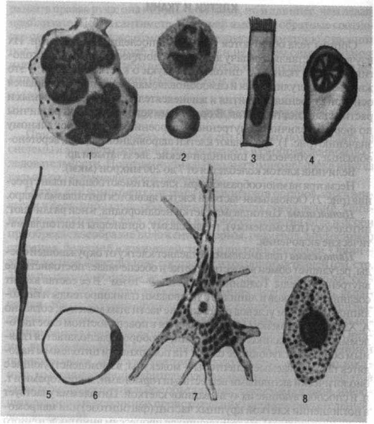
Fig. 1. Polymorphism of Cell Structure:
1 - Bone Marrow megakaryocyte; 2 - neutrophilic leukocyte (top) and erythrocyte (bottom); 3 - ciliated epithelial cell; 4 - plasma cell; 5 - smooth Muscle cell; 6 - fat cell; 7 - neuron; 8 - heparinocyte
The endoplasmic reticulum is a system of tubules that ensures the Transport of substances from the extracellular environment and within the cell. The agranular (smooth) endoplasmic reticulum participates in the synthesis of Introduction/36.html">Carbohydrates and Lipids, while the granular reticulum, studded with ribosomes on its surface, is involved in Protein Synthesis.
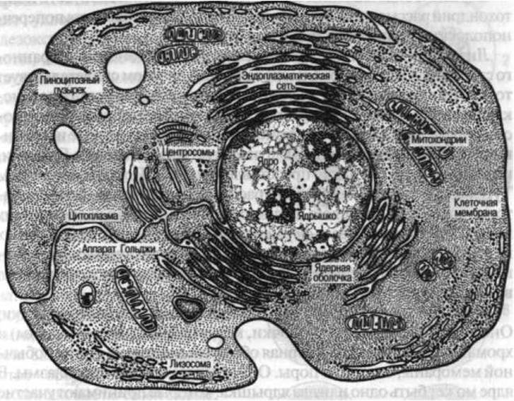
Fig. 2. Cell structure (according to Prokofieva-Belgovskaya)
Ribosomes produce Proteins, which are specific to each type of cell.
Mitochondria contain macroergic compounds and serve as a source of energy.
Lysosomes contain a large amount of Enzymes and carry out intracellular digestion.
The Golgi apparatus (internal reticular apparatus), consisting of cisternae, tubules, and vesicles, is the site of accumulation for substances secreted by the cell.
Centrioles, which form the centrosome, take part in Cell Division.
Special organelles are associated with specific cellular functions. These include myofibrils in muscle cells, neurofibrils in Nerve Cells, and tonofibrils in epithelial cells.
Inclusions are protein granules, fat droplets, or pigment granules that may or may not be present in the cell, unlike organelles, which are its permanent structures.
The structure of the endoplasmic (sarcoplasmic) reticulum and mitochondria is examined in greater detail using the example of Striated Muscle tissue fibers (see p. 35).
Lysosomes differ significantly from other membranous organelles. Their functional importance is evidenced by the fact that they are found in all cell types of mammals, with the sole exception of mature erythrocytes. The structural Variability of lysosomes is determined by the fact that their membranes may originate from various sources: the endoplasmic reticulum, the Golgi apparatus, the cytolemma, etc. According to A. A. Pokrovsky and V. A. Tutelyan, enzyme receptors are located in the lysosomal membranes, exhibiting activity during emergency states of the cell. This provides enzymatic defense for cells against unwanted or foreign metabolic products and pathogenic microorganisms—a phenomenon first highlighted by I. I. Mechnikov long before the discovery of lysosomes.
Nucleus. Like the cytoplasm, the nucleus is a core part of the cell. It consists of the nuclear envelope, karyoplasm (nucleoplasm), and Chromatin structures. The nuclear envelope is constructed on THE PRINCIPLE OF a conventional membrane and contains pores; it separates the nucleus from the cytoplasm. The nucleus may contain one or two nucleoli, which take part in metabolism, including The formation of ribosomes.
The karyoplasm, which is a reticular formation, contains enzymes and other chemically active substances. Protein synthesis takes place within it.
Chromatin structures in a dividing cell form Chromosomes—the physical carriers of hereditary information. There are 23 pairs of them, one of which consists of chromosomes known as sex chromosomes—the X chromosome and the Y chromosome.
Cells of the human Organism multiply through indirect division, or mitosis, and direct division, or amitosis. Germ Cells are characterized by a different type of division known as Meiosis, which results in a reduction of genetic material (unlike somatic cells, germ cells possess a haploid rather than a diploid set of chromosomes). It is believed that mitosis, as opposed to amitosis, is characterized by a higher level of Cell Nucleus activity. The process of mitosis, or karyokinesis (Fig. 3), based on morphologically visible changes, is conventionally divided into four phases: prophase, metaphase, anaphase, and telophase.
Their total duration ranges from one to one and a half hours. The period between cell divisions is called interphase. It is subdivided into three periods lasting approximately 19 hours, during which DNA (deoxyribonucleic acid) molecules are duplicated to provide genes for the two daughter cells. A cell undergoing division continuously, cycle after cycle, cannot perform its specialized functions. To carry them out, it must temporarily or permanently halt mitotic divisions.
1. Prophase. Cellular changes during mitosis begin with the Swelling of the nucleus, whereupon the chromatin substance takes the form of a continuous, tangled thread forming a loose skein. Distinct, morphologically heterogeneous regions—chromosomes—then clearly emerge within it (they are also present in the interphase nucleus in a less distinguishable form), each dividing into two daughter chromosomes. The nuclear envelope dissolves. The nucleolus disappears. The centrosome divides into two centrioles that migrate toward the opposite poles of the cell.
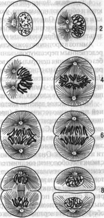
Fig. 3. Indirect cell division (mitosis):
1 — resting cell; 2 — prophase (early stage); 3 — prophase (late stage); 4 — metaphase; 5 — anaphase; 6 — telophase; 7–8 — division of the cytoplasm and nucleus
2. Metaphase. By this time, a spindle formed of achromatic fibers, which take part in the movement of chromosomes, develops between the centrioles occupying the polar positions in the cell. The chromosomes arrange themselves in the central, equatorial plane of the cell, forming a star-like figure. They become shorter and thicker; the subdivision of each into two daughter chromosomes becomes even more pronounced.
3. Anaphase. Daughter chromosomes migrate toward the poles of the cell and congregate near the centrioles, forming a double-star figure.
4. Telophase. The daughter chromosomes gather together, condense, and form new nuclei equipped with a nuclear envelope and nucleoli. Simultaneously, the body of the cell gradually constricts in the equatorial plane and divides in two, resulting in the formation of two new cells.
Amitosis, in contrast to karyokinesis, is characterized by the direct constriction of the nucleus and cytoplasm (occurring without the formation of a central spindle or chromosome coiling), As a result of which two new cells arise from a single parent cell. Most commonly, amitosis manifests as the division of the nucleus rather than The Cell as a whole, leading to multinucleated or binucleated cells. It is observed primarily in pathological conditions or in cells completing their life cycle.
Mitotic cell division ensures the self-renewal of tissues during physiological (inherent to the normal life cycle) or reparative (following tissue damage) regeneration. According to D. S. Sarkisov, a distinction should be made between cellular and intracellular tissue regeneration. The former is characterized by an increase in cell number (due to division) with unchanged cell size. It is typical of the epithelial cover of the Skin and mucous membranes, as well as Connective Tissues. Intracellular regeneration manifests as an increase in the size of cells and their components, along with heightened activity of intracellular structures. This form of regeneration is characteristic of ganglion cells of the Central Nervous system; presumably, it predominates in cardiac and Skeletal Muscle tissue. In many cases, both forms of regeneration manifest simultaneously, such as in smooth muscle, Liver, Kidneys, and other organs.
Along with Cellular forms of living matter Organization, non-cellular structures also exist. These include the intercellular ground substance. Cellular structures may have a simplified (erythrocytes, Blood Platelets) or complex (striated muscle fiber, neuron) structure. According to A. N. Studitsky, the appearance of cells in living nature was preceded by phylogenetically older pre-nuclear structures (prokaryotes), from which cells evolved in the course of living matter evolution.
Tissues
A tissue is a historically evolved system of cells and non-cellular structures sharing common structure and origin, and specialized in performing specific functions.
The structure of any given organ is formed not by a single tissue type, but by various types of tissues. For instance, Bone tissue consists of bone cells with intercellular substance located between them, whereas the formation of a bone involves not only bone tissue, but also muscle tissue (in the walls of Blood Vessels supplying the bone), Nervous Tissue (in structures innervating the bone), and other tissue types. Tissues are classified into epithelial tissues, Tissues of the Internal Environment (or connective tissues), Muscle Tissues, and nervous tissue.
Epithelial Tissues
Epithelial tissues perform protective, secretory, excretory, and absorptive functions.
The protective function of these tissues lies in the fact that, by forming the outer layer of the skin (its epidermis) and lining all Internal Organs, they shield deeper structures from injury and the penetration of microbes and other harmful substances, and in the gastrointestinal tract, from the destruction of its wall by digestive juices. Epithelial tissues facilitate the absorption of digested food within the gastrointestinal tract, as well as the Elimination of Metabolic waste products from the body—both non-essential and toxic ones (for example, urine, the retention of which leads to death).
The secretory function of epithelial tissues is manifested through their Participation in the formation of glands—specifically their terminal secretory portions and duct walls—where they produce secretions such as digestive juices (Glands of the Digestive System), sebum (Sebaceous Glands), and sweat (Sweat Glands).
All epithelial tissues share common structural features.
They contain a negligible amount of intercellular substance. They form sheets of tightly contiguous cells resting on a basement membrane. Epithelial tissues possess high regenerative capacities, continuously retaining cells capable of mitosis.
Epithelial tissues originate from the inner (endodermal), middle (mesodermal), and outer (ectodermal) germ layers (see p. 44).
The Classification of epithelial tissues is based on their Structural and functional characteristics, as well as their participation in the formation of specific organs (Fig. 4).
According to the number of cell layers, epithelia are distinguished as simple (for example, the lining of the intestine) and stratified (for example, the integument of the skin or the anterior corneal epithelium of the eye).
Based on cell shape, epithelia are classified as squamous, cuboidal, columnar (and its variant, ciliated), and transitional. Squamous epithelium lines serous membranes (Pleura, Peritoneum); cuboidal epithelium forms the walls of renal tubules; columnar epithelium lines the mucous membrane of the digestive organs (Stomach, intestines); ciliated epithelium lines the respiratory tract and fallopian tubes; and transitional epithelium lines the urinary tracts (renal pelvis, Ureters, Urinary Bladder).
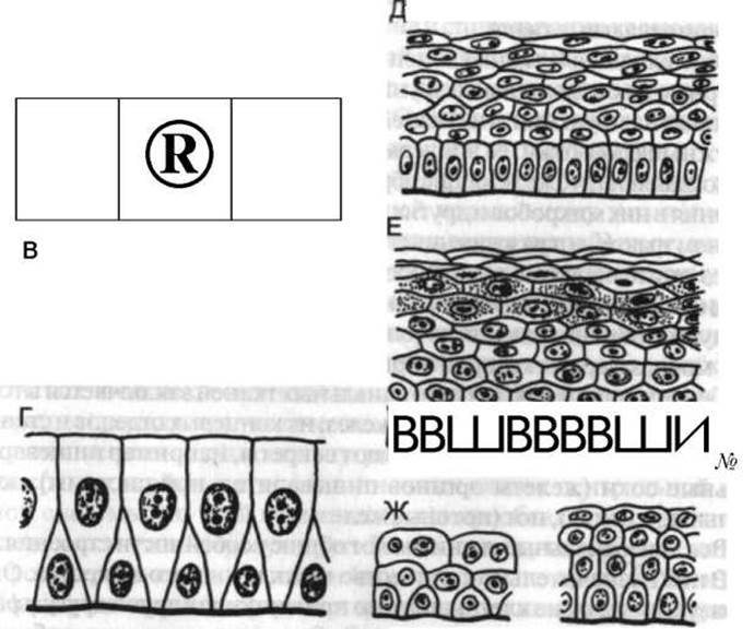
Fig. 4. Schematic diagram of the structure of various epithelia:
A, B, C – single-layered (A – squamous, B – cuboidal, C – columnar); D – pseudostratified; E, F – stratified (E – non-keratinized, F – keratinized); G – transitional: left — when the organ is distended, right — in its collapsed state (after E. A. Shubnikova, 1981)
Stratified Epithelium (typically squamous) has two varieties: keratinized, which lines the skin surface, and non-keratinized, which lines the inner surface of the Oral Cavity, Pharynx, initial segment of the Esophagus, rectum, Vagina, and the Cornea of the eye.
Glandular Epithelium secretes its products onto the epithelial lining surface into hollow organs or directly into the BLOOD AND Lymph, i.e., it forms exocrine and Endocrine glands (organs). Glands can be unicellular (e.g., mucus-secreting goblet cells of the intestine) or multicellular, which are formed by the outward invagination of the epithelial layer. They vary in shape and function. Alveolar, tubular, and mixed (i.e., tubuloalveolar) glands are distinguished. In addition, glands may be simple, branched, or compound.
Simple tubular glands are tube-like invaginations (e.g., intestinal glands).
Branched tubular glands consist of several simple glands opening into a common excretory duct (e.g., glands of the pyloric region of The Stomach, uterine glands).
Compound tubular glands are formed by several branched tubular glands opening into a common excretory duct (e.g., lacrimal glands, liver, Testes).
Simple alveolar glands look like small vesicles or pouches (e.g., sebaceous glands).
Branched alveolar glands consist of several simple alveolar glands opening into a common excretory duct (e.g., tarsal glands of the eyelids).
Compound alveolar glands are formed from several branched alveolar glands that share a common excretory duct (e.g., Pancreas, Mammary Glands).
Simple tubuloalveolar glands are tubes with expansions at their ends (found, for example, in the pyloric region of the stomach).
Branched tubuloalveolar glands consist of several simple tubuloalveolar glands (e.g., urethral glands).
Compound tubuloalveolar glands are constructed from branched tubuloalveolar glands (e.g., Lungs, Prostate Gland). In some cases, the described glands exhibit structural complexities. For instance, simple tubular glands may have coiled ends (e.g., sweat glands).
The constituent parts of a multicellular gland are: the base (fundus), body, neck, orifice, and, in larger glands, a well-defined excretory duct as well.
The expanded, blindly ending region of the gland—its body and base—is lined with secretory epithelium. The process of secretion takes place in this part of the gland. Secretion production occurs differently across various glands: in one group of glands without cellular damage (e.g., goblet cells), in another (e.g., the mammary gland) it involves partial cell death, and in a third (e.g., sebaceous glands) it is accompanied by the destruction of cells. Destroyed cells are replaced by new ones formed through the proliferation of deeper-lying cells that are adjacent to the basement membrane and capable of division (reproduction).
Unlike exocrine glands, endocrine glands lack excretory ducts (see p. 409).
Internal environment tissues, or connective tissues, are characterized by a significant development of Extracellular matrix. They are subdivided into tissues with predominantly trophic, supportive, or protective functions.
The trophic (i.e., nutritive) function is performed mainly by blood, lymph, and partly by loose Connective Tissue, which participate in supplying the body with nutrients and oxygen, as well as in removing Metabolic waste products and carbon dioxide.
These tissues also perform a protective function, which consists in producing substances that destroy microbes entering the body, generating antitoxins and immune bodies, and utilizing the ability of certain cells to engulf and digest microbes and foreign substances (phagocytic capacity).
The supportive function is performed primarily by Dense connective tissue, Cartilage, bone, and partly by loose connective tissue. Individual types of internal environment tissues participate in forming the Hard and Soft skeletons. These include not only Bones and Their Joints, but also ligaments, fasciae, interosseous and intermuscular membranes, the framework of internal organs, etc.
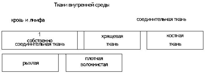
There are A number of tissue classifications, in particular those separating connective tissue, blood, and lymph as independent tissue types. The most common classification is the following (see Scheme 1).
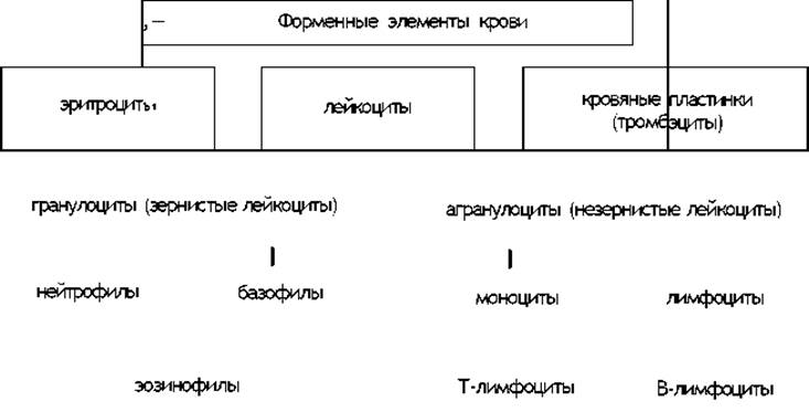
Blood and lymph. These are fluid types of internal environment tissues. They contain about 80% Water and about 20% organic substances, with a specific gravity of approximately 1.055. The total blood volume reaches approximately 4.5 liters in men and 4 liters in women. Blood and lymph consist of plasma and formed elements (Fig. 5). The total mass of formed elements accounts for 1/3–2/5 of the total blood volume, while plasma constitutes 2/3–3/5 of this volume. Among the formed elements of blood, red and white Blood Cells are distinguished (see Scheme 2).
Scheme 1.
Unlike white blood cells, human red blood cells, or erythrocytes, lack nuclei. They are shaped like disks with a central indentation on both sides. On average, 1 mm2 of blood contains about 4.5–5 million erythrocytes. The total surface area of erythrocytes is approximately 3200 m2, and with each breath, roughly 250 m2 of this total surface area participates in Gas Exchange in the lungs. Erythrocytes contain the blood pigment Hemoglobin, which makes up about 1/3 of their total mass. Hemoglobin has the property of binding with oxygen, thereby playing a vital role in gas exchange. This causes the blood to change from a cherry-red to a bright red color. The body of an adult human contains about 660 g of hemoglobin. Erythrocytes are produced in the Red bone marrow, and their lifespan is 80–120 days.
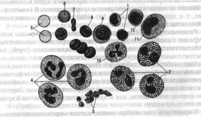
Fig. 5. Human blood. General view of erythrocytes, leukocytes, and thrombocytes in a stained preparation (magnified 1,000x):
1 - small lymphocytes; 2 - eosinophilic leukocytes; 3 - group of blood platelets; 4 - neutrophil leukocytes; 5-8 - erythrocytes in various positions; 9 - large lymphocyte; 10 - basophilic leukocyte; 11 - monocyte; 12 - medium lymphocyte
Diagram 2.
White blood cells, or leukocytes, contain a single segmented or non-segmented nucleus in their cytoplasm. The number of leukocytes is significantly smaller than that of erythrocytes, numbering 6–8 thousand per 1 mm3 (note: manuscript terminology retained as 1 mm5), meaning there is roughly one WHITE BLOOD CELL for every 500–1000 red blood cells.
Leukocytes are divided into granular forms (neutrophils, eosinophils, basophils) and agranular forms (lymphocytes, monocytes). Leukocytes are capable of changing their shape, moving independently, and even exiting capillaries via amoeboid movement. Some leukocytes (primarily neutrophils, monocytes, and lymphocytes) possess phagocytic properties. The primary sites of lymphocyte proliferation are the Thymus gland and Lymph Nodes. Granular leukocytes, much like erythrocytes, develop in the red bone marrow.
The blood also contains specialized tiny cells known as blood platelets, or thrombocytes, measuring 2–5 µm (i.e., 2–4 times smaller than red blood cells and 5–7 times smaller than white blood cells). They participate in blood clotting. There are approximately 200–300 thousand thrombocytes per 1 mm3 of blood.
Lymph, much like blood, consists of lymphoplasma and formed elements. In its composition, lymphoplasma is similar to Blood Plasma but contains fewer proteins. The formed elements in lymph are predominantly lymphocytes and monocytes (see p. 323).
The framework of blood-forming organs—the bone marrow, lymph nodes, and Spleen—is formed by reticular tissue. Additionally, it is found in the mucous membrane of the gastrointestinal tract, respiratory tract, and several other organs. Reticular cells form a unique network and, in some cases, can break away from their connections, becoming mobile and independent. They exhibit phagocytic properties.
Phagocytic properties are also characteristic of the endothelial cells of capillaries, the liver, and other organs, which are closely associated with reticular tissue. This served as the basis for defining the so-called reticuloendothelial system, which encompasses reticular tissue, the endothelium of certain capillaries, cells of loose connective tissue capable of active movement and phagocytosis, as well as blood monocytes and lymphocytes. All of these structures perform a protective function in the body.
Loose connective tissue. This tissue is widespread throughout the body: it accompanies blood Vessels and nerves along their entire length, is located between organs, and is present in the subcutaneous fat layer. Cells of this connective tissue type can accumulate fat in their cytoplasm and transform into fat cells, while the tissue itself can convert into adipose tissue.
The intercellular substance of loose connective tissue contains Collagen and elastic fibers. Collagen fibers are thick, durable, poorly stretchable, and arranged in bundles. Elastic fibers are thin and stretch easily, returning to their original state afterward.
The cellular elements of loose connective tissue include undifferentiated mesenchymal cells and cells that have migrated into it from the blood and lymph, such as granular and agranular leukocytes. The primary cells of this tissue are fibroblasts and histiocytes.
Fibroblasts are characterized by a large, oval-shaped nucleus with one or more nucleoli. The cytoplasm of these cells may be granular or homogeneous, featuring cytoplasmic processes. Fibroblasts are found at various stages of differentiation. The poorly differentiated elements of loose connective tissue include young fibroblasts and reticular cells, as well as certain endothelial cells separating the blood vessel lumen from the surrounding loose connective tissue.
Histiocytes feature a smaller, oval or irregularly shaped nucleus that is often indented on one side compared to that of fibroblasts. They differ from fibroblasts in that their outlines are well-defined and do not merge with the main intercellular substance anywhere.
In addition to fibroblasts and histiocytes, loose connective tissue contains other cell types (pigment cells, mast cells, fat cells, plasma cells, lymphocytes, and leukocytes).
Dense Fibrous connective tissue. It occurs in the form of fibrous connective tissue of the skin, tendons, and as elastic connective tissue. Dense regular fibrous connective tissue of the skin is characterized by significant strength, which is driven by well-developed, interlaced collagen bundles as well as elastic fibers. This tissue contains fully matured fibrocytes and histiocytes.
In dense regular fibrous tissue of tendons, thick bundles of collagen fibers run parallel to one another. In fasciae, they are arranged in various directions. Between the bundles are small clefts housing fibrocytes, known as tendon cells. This tissue forms tendons, fasciae, and ligaments.
Dense regular elastic tissue contains a significant number of elastic fibers that form entire large cords or plates without subdividing into bundles. The ligamenta flava (yellow ligaments) of THE Vertebral Column are constructed from this tissue.
Cartilage tissue. Depending primarily on The Nature of the intercellular substance, cartilage tissue occurs in the form of hyaline, fibrocartilage, and elastic cartilage.
Hyaline, or glassy, cartilage (Fig. 6, A) in its natural state features a structureless, homogeneous intercellular substance consisting of chondrin, chondromucoid, and chondroitin sulfuric acid. Its cells are located within small cavities, filling them completely. The intercellular substance contains fine connective tissue fibers. This type of cartilage tissue is widespread. It covers the articular surfaces of bones and is found in the costal cartilages, laryngeal cartilages (except the epiglottis), Trachea, and Bronchi.
Fibrocartilage is found in the intervertebral and articular disks and represents a fibrous connective tissue containing cartilage cells, impregnated with chondrin much like hyaline cartilage.
Elastic cartilage (Fig. 6, B) contains a high concentration of elastic fibers in its extracellular matrix, imparting a yellowish hue that sharply distinguishes its appearance from that of hyaline cartilage. Elastic cartilage is primarily found in the epiglottis and the auricle of the ear.
Cartilages, particularly hyaline cartilage, are prone to calcification. This is one of their characteristic age-related features.
In this regard, the laryngeal cartilages (primarily the thyroid cartilage) and the costal cartilages rank first.

Fig. 6. Structural diagrams of mature hyaline (A) and elastic (B) cartilages:
1 - perichondrium; 2 - chondroblasts; 3 - chondrocytes; 4 - cartilage extracellular matrix; 5 - elastic fibers (after Steopov, from the book by E.A. Shubnikova)
Bone tissue. Bone tissue is characterized by high mechanical strength and elasticity, enabling it to perform its supportive functions effectively. It consists of cells and an extracellular matrix containing organic (ossein and osseomucoid) and Inorganic Compounds (primarily calcium salts). There are two main Types of bone tissue: coarse-fibered (woven) and lamellar. In humans, coarse-fibered bone tissue is found only at the attachment sites of tendons to bones and in the areas of fusing cranial sutures.
Human bones predominantly exhibit a lamellar structure. The parallel bundles of collagen fibers located within the lamellae run in specific directions corresponding to the mechanical forces acting upon them. They are impregnated with inorganic compounds, which provides this type of bone tissue with a strength second only to tooth enamel. The cells of bone tissue—osteocytes—are housed within lacunae in the extracellular matrix. These cavities interconnect via tiny canaliculi through which the processes of the osteocytes communicate, giving the entire Bone Structure a syncytial character.
In addition to osteocytes, bone tissue contains osteoblasts and osteoclasts. The former are responsible for Bone Formation, while the latter break it down, thereby facilitating continuous bone remodeling.
Microscopic examination demonstrates that bone is composed of osteons and interstitial lamellae (Fig. 7). The osteon serves as the fundamental structural unit of bone. It is a system of concentric bone cylinders resembling nested tubes, formed by osteon lamellae. Several layers of concentrically arranged lamellae encircle the central canal of the osteon. The collagen fibers run in varying directions across different lamellar layers, which enhances the mechanical strength of the osteon. Interstitial lamellae are located in the spaces between osteons. Externally, the layer of osteonal bone tissue is bounded by a continuous layer of outer circumferential lamellae, while internally, facing the marrow cavity, it is bounded by a layer of inner circumferential lamellae.
Blood vessels and nerves pass through the central canal of the osteon. The diameter of these canals ranges from 1/50 to 1/5 mm, depending on the size of the bone and the canal's Location relative to the external surface. Larger bones feature wider bone canals, and superficially located canals are generally wider than those situated deeply.
Osteons, interstitial lamellae, and circumferential lamellae undergo continuous remodeling throughout life in response to mechanical loading conditions, age, nutritional status, and other factors. It is estimated that the lifespan of an osteocyte reaches up to 25 years. In adults, the annual turnover rate is 2.5% for compact bone and 10% for spongy bone. In children, The rate of bone remodeling is significantly higher.
The mechanical properties of connective tissue can be evaluated by its resistance to tension, compression, shear, torsion, and bending. Data of this nature constitute a novel branch of Morphology known as biosopromatics (biomechanical strength of Materials).
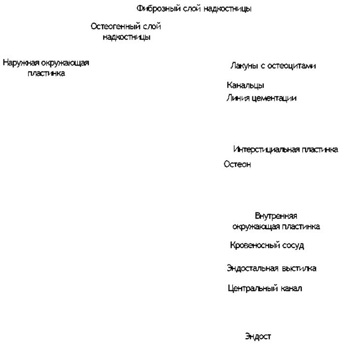
Fig. 7. Microscopic structure of osteonal bone (after Ham)
As is well known, tensile strength is defined as the minimum load per unit of cross-sectional area (1 mm2) required to cause tissue rupture or deformation. Fresh compact bone can withstand a tensile resistance of up to 10–12 kg, costal (hyaline) cartilage withstands 0.5 kg, and tendons—representing dense fibrous tissue—withstand 7 kg.
In terms of compressive resistance, bone is approximately 10 times stronger than cartilage. The compressive strength of bone (12–16 kg) is about 1.5 times greater than its tensile strength. The compressive strength of hyaline cartilage (1.5 kg) is 3 times greater than its tensile strength. Fresh bone is 5 times stronger than reinforced concrete in both compression and tension. Crushing the Femur requires a compressive force of approximately 3,000 kg, whereas crushing the Tibia requires at least 4,000 kg.
Bending resistance at a given bone weight reaches its maximum when The ratio of the inner diameter of a tubular bone to its outer diameter is approximately 8:11. This resistance is higher in younger individuals than in older adults.
A distinction is made between tensile elasticity and compressive elasticity. Practically speaking, tensile elasticity is of paramount importance for tendons, whereas compressive elasticity is vital for articular cartilages. Compared to the elasticity of bone tissue, the tensile elasticity of tendons is 15 times greater, and the elasticity of costal cartilage is 1.5 times greater.
It should be noted that the mechanical properties of connective tissue exhibit substantial individual variation. They may differ not only among various individuals but also within the same person, varying in response to nutritional status, functional demands, and age-related factors.
Muscle tissues
The primary functional property of muscle tissue is contractility, which stems from the ability of the contractile structures within its cells to change length—becoming shorter and thicker (contraction, shortening) or longer and thinner (relaxation, lengthening).
There are three varieties of muscle tissue: smooth, striated, and cardiac.
Smooth (non-striated) muscle tissue. Unlike Other types of muscle tissue, microscopic examination reveals that smooth muscle lacks cross-striations. It is located in the walls of blood vessels, glandular excretory ducts, the gastrointestinal tract, and many hollow organs, as well as within the skin (forming arrector pili Muscles), inside the Eyeball, and elsewhere. The structural unit of this muscle tissue is the myocyte—an elongated cell, occasionally possessing small processes. The cell length is 15–500 µm, and its diameter is 10–20 µm. It contains a centrally located nucleus. As observed under an Electron microscope, myofibrils—the contractile structures characteristic of striated muscle tissue—are absent here. However, there is an Abundance of contractile filaments, or myofilaments (protofibrils), measuring 1–2 µm in length and 5–8 nm (thin myofilaments) or 10–30 nm (thick myofilaments) in thickness. When the myocyte is relaxed, only thin myofilaments are discernible. Contractions of Smooth muscle tissue occur slowly, rhythmically, and involuntarily (e.g., peristaltic Movements of the intestinal tract, Changes in the lumen of blood vessels and glandular ducts, and contractions of the iris Muscles of the eye, etc.).
Striated (skeletal) muscle tissue. It is named for its characteristic cross-striations visible under a microscope. This tissue forms the muscles that drive the Skeletal System and is therefore also referred to as skeletal muscle tissue. Functionally, it is voluntary, as its contraction and relaxation are subject to conscious control. However, the contraction of Respiratory Muscles, which are composed of this tissue, can also occur involuntarily, such as during Sleep. The fibers of striated muscle tissue, serving as its structural units, resemble very long cylinders in shape, with diameters ranging from 1/100 to 1/10 mm.
The fiber length ranges from 1 to 40 mm, but can occasionally reach 10–12 cm. Each muscle fiber consists of cytoplasm (sarcoplasm), a bilayer membrane (sarcolemma), and A large number of elongated nuclei typically located at the periphery. Mitochondria, which are responsible for the Energy supply of the muscle cell, are located in the sarcoplasm (Fig. 8). The size of mitochondria ranges from 0.3–1.7 µm × 0.2–1 µm. They are enclosed by a double membrane: an outer membrane with a thickness of 5–10 nm and an inner membrane with a thickness of 10–20 nm. A narrow space of about 20 nm is formed between them. The inner membrane features folds called cristae, where the enzymes of Oxidative Phosphorylation are localized.
The sarcoplasmic reticulum includes longitudinal and transverse tubular systems. The longitudinal system (L-system) appears as thin-walled tubules running alongside the myofibrils and anastomosing with one another. The transverse system (T-system) consists of transversely oriented tubules (intermediate vesicles). The lumen of the T-system is connected to the extracellular space, although the longitudinal and transverse tubules do not communicate with each other:

Fig. 8. SCHEMATIC STRUCTURE OF a section of a striated muscle fiber based on Electron Microscopy data
Of great interest is the Fine Structure of the contractile elements of muscle fibers, known as myofibrils. Each myofibril passes through numerous thin membranes (telophragms or Z-lines) that lie transversely relative to it and represent an extension of the sarcolemma, with which they are directly connected. Thus, it appears as though the myofibril is divided into distinct small segments—sarcomeres—each of which, in turn, is divided across the middle by an extremely thin partition (thinner than the telophragms) called the mesophragm. Both mesophragms and telophragms serve to anchor the myofibrils passing through them.
The Regions of the myofibrils adjacent to the telophragm consist of a light, isotropic substance, whereas the regions adjacent to the mesophragm are dark and anisotropic.
Consequently, the striated banding pattern is caused by the alternating light and dark regions of the myofibrils, referred to as light and dark discs. Using an electron microscope, it has been established that each myofibril consists of protofibrils of two varieties: thick (16 nm) and thin (5–7 nm) filaments (see Fig. 8). A thick filament contains 180–360 longitudinally oriented protein molecules of Myosin. A thin filament is constructed from molecules of another protein, Actin, which form a double helix. Bridges are formed between the thick (myosin) and thin (actin) filaments (mediated by the "HEAD region" of the myosin molecules).
Muscle fiber contraction occurs due to the sliding of actin filaments between myosin filaments (sliding filament theory). The sarcomere shortens like a collapsible telescope. Its volume remains constant, while its transverse diameter increases. The sliding filament theory was proposed by A. Huxley in 1957. Other explanations for the Mechanism of muscle contraction also exist.
The morphological and functional heterogeneity of skeletal muscle fibers has been established. Type I fibers (red) and type II fibers (white) are distinguished. Red fibers have a small diameter. They are characterized by a high activity of oxidative enzymes due to the predominance of aerobic oxidative processes, as well as a high content of the protein Myoglobin. These fibers are surrounded by 2–3 blood capillaries, meaning they have a high level of blood supply. Red muscle fibers are classified as slow, tonic fibers.
White fibers are thicker. They contain large amounts of phosphorylase and ATP, which support anaerobic processes. The primary energy source is Glycogen. Their blood supply is lower than that of red fibers: there is an average of one blood capillary per muscle fiber. White fibers are considered fast, tetanic fibers.
An intermediate type of muscle fiber also exists.
Under electron microscopy and histochemical analysis, these fiber types differ in their content of mitochondria, lipids, and the ratio of oxidative enzymes to glycolytic enzymes, as well as in the width of the Z-lines between sarcomeres. Red fibers (slow) are rich in mitochondria and lipids (which serve as their main energy source); oxidative enzymes predominate over glycolytic enzymes, and the Z-line is wide. White fibers (fast) contain few mitochondria, lipids, and oxidative enzymes, but are rich in glycolytic enzymes, with a narrow Z-line. No significant differences in glycogen content or myofibrillar structure have been established between these fiber types.
A comparison of several limb muscles revealed a mosaic pattern in the distribution of type I and type II fibers.
The fiber sheath—the sarcolemma—helps strengthen the interconnection of muscle fibers. Its inner layer, the plasmalemma, is analogous to the membranes of other cells. The outer layer, the basement membrane, consists of fine fibrils and is closely associated with the surrounding connective tissue, the endomysium. For the structure of a muscle as an organ, see page 127.
Striated muscle fibers, together with the neuron that innervates them, form a motor unit. It includes either red or white muscle fibers. Their number is quite substantial (for example, 1,634 in the medial head of the gastrocnemius muscle and 667 in the tibialis anterior muscle).
At the site where the axon of a motor nerve cell approaches a muscle fiber, a neuromuscular synapse is formed in the shape of a motor end plate. The axon terminals lie in depressions on The surface of the muscle fiber lined by the sarcolemma. The nerve cell axon does not penetrate into the interior of the muscle fiber. Synaptic clefts with a width of 20–60 nm remain between them. Here, the impulses prompting the muscle to contract are transmitted from the nerve cell axon to the muscle fiber (see Fig. 9). Concurrently, Calcium Ions are released from the sarcoplasmic reticulum of the fiber into the myofibrils. As a result, actin acquires The ability to interact with myosin, leading to fiber contraction (see page 36).
Cardiac muscle tissue. This tissue is striated, but possesses structural features that set it apart into a distinct group. One such feature is that cardiac muscle cells, or cardiomyocytes, form numerous junctions with one another. Cardiac muscle contractions occur involuntarily.
The nuclei of cardiomyocytes occupy a central position, while the myofibrils are located at the periphery of the cell. Unlike skeletal muscle tissue, cardiac muscle cells contain significantly more mitochondria with a high density of cristae. This indicates a robust energy supply for The Heart muscle. Conversely, the sarcoplasmic reticulum is less developed here than in skeletal muscle tissue.
Contacts between cardiac muscle cells occur in the region of intercalated discs and lateral cytoplasmic projections. Intercalated discs not only connect cells to one another but also participate in the transmission of excitation from one cell to another. The lateral projections and intercalated discs ensure that the myocardium contracts as a single functional unit. Among the cells responsible for contraction are those that conduct impulses within the heart. These are large cells rich in sarcoplasm, with few myofibrils and mitochondria, and a large nucleus that is not always located in the center of the cell.
Nervous tissue
Nervous tissue consists of nerve cells with their processes and the endings of these processes. It also includes structures that provide supportive and trophic functions for the nervous tissue proper, collectively known as neuroglia (macro- and microglia).
Each nerve cell contains cytoplasm and a rounded or slightly oval nucleus. Mitochondria and the Golgi apparatus are prominent in the cytoplasm. Tigroid deposits, formed as modifications of the endoplasmic reticulum, are also present. Under the electron microscope, neurofilaments—filaments about 10 nm thick—can be identified.
A nerve cell with its processes is called a neuron (Fig. 9). It represents the structural unit of nervous tissue. Nerve cells whose processes extend to organs (such as muscles) and transmit impulses that stimulate them to activity are called motor, efferent, or centrifugal. Nerve cells whose processes conduct impulses from the periphery to the center are sensory, afferent, or centripetal. In addition to motor and sensory Neurons, There is a vast number of interneurons (association neurons) that connect sensory and motor neurons to one another. The size of nerve cell bodies varies, ranging from 25 to 150 µm in diameter. Some nerve cells have bodies so large (for example, the cells of the anterior horns of the Spinal Cord) that they are on the verge of visibility to the naked eye. The shape of nerve cell bodies is also diverse: polygonal, fusiform, elongated, or round. This shape is related to the number of cell processes, as the neuron body forms a protrusion corresponding to each process.

Fig. 9. Structure of a motor neuron:
1 — perikaryon; 2 — axon and nerve fiber; 3 — nerve endings in muscle; 4 — dendrites; 5 — myelin sheath; 6 — nodes of Ranvier. The diagram compares light and electron microscopy (after G.F. Ivanov and Kovalsky, modified)
The pattern of process emission allows neurons to be classified into unipolar (having a single process that T-bypasses into two branches), bipolar (with two processes), and multipolar (with many processes).
Some nerve cell processes are short, protoplasmic, and tree-like branching—these are dendrites; others are long neurites, or axons. The length of nerve cell processes can be substantial (exceeding 1 m in some locations). Impulses travel away from the cell body along neurites, whereas they travel toward the cell body along dendrites.
These processes continue within nerve fibers as axis cylinders, typically enveloped by glial sheaths of varying structural complexity. Only in the Gray matter of the Brain do nerve cell processes lack such sheaths. Conversely, the processes that constitute the White matter OF the central nervous system are enclosed by a sheath.
Nerve cell processes form nerve endings: receptors (on dendrites) and effectors (on neurites). Nerve cells connect with each other and with the innervated organ (muscle, skin, or gland) via specialized structures known as synapses, which exhibit an extreme diversity of forms.
Cell processes serve not only for the conduction of nerve impulses but also for The transport of proteins and other substances to or from the neuron body. There are Two Types of intracellular flow: a slow rate of 1—2 mm per day and a fast rate of 5—10 mm per hour.
It is well established that certain neurons possess neurosecretory capabilities and are thus termed secretory neurons. The Formation of the secretion is associated with tigroid substance and the Golgi complex. Neurosecretory granules migrate along the axon away from the cell body; however, unlike other transported substances, they are released into the blood or CEREBROSPINAL FLUID (much like hormones) rather than into synaptic regions.
Nerve fibers vary in diameter. One group of axis cylinders is enclosed within a sheath of myelin (a lipid-like substance), forming myelinated (or medullated) fibers, whereas another group lacks this covering and constitutes unmyelinated (or non-myelinated) fibers. The myelin sheath increases the conduction velocity of nerve impulses; consequently, its presence is functionally advantageous. Unmyelinated fibers form a polyaxonal sheath system in which the axons are surrounded by satellite (Schwann) cells. During the formation of myelinated fibers, the axon initially lies on the periphery of a satellite cell and then becomes 'invaginated' into it, leading to the formation of a 'mesaxon' fold (Fig. 10). The mesaxon spirals outward around the axon, and myelin is formed at the contact sites of the proliferating mesaxon folds. Along the course of a myelinated fiber, the myelin covering becomes intermittently thinned, forming the nodes of Ranvier. These are biologically active regions of the nerve characterized by an accumulation of mitochondria, numerous ions, and metabolic products.
Nerve fibers coated with a layer of connective tissue, known as the endoneurium, form bundles surrounded by thicker layers of the perineurium, collectively constituting a nerve. Depending on the number of fibers and bundles comprising a nerve, its thickness varies significantly—ranging from microscopic branches invisible to the naked eye to very thick trunks (for instance, the thickness of the sciatic nerve can exceed 1 cm).
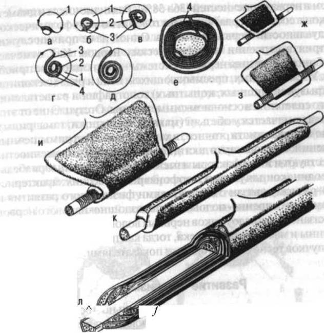
Fig. 10. Diagram of nerve fiber myelination: a—e — successive stages of mesaxon rotation around the axon in cross-section; f—l — process Overview; 1 — Schwann cell; 2 — axon; 3 — mesaxon; 4 — myelin layers (after Geren)
There are two extreme variants of peripheral nerve structure: oligofascicular (the nerve is thin, consisting of a small number of large bundles with a compact arrangement of fibers within each bundle) and multifascicular (the nerve is thick, formed by numerous small-diameter bundles with a loose arrangement of fibers). The number of fibers within individual nerves is highly variable: the median nerve at the mid-arm level contains 19—32 thousand fibers, the ulnar nerve at the same level contains 13—18 thousand, and the musculocutaneous nerve contains 3—12 thousand. Nerve trunks differ in the diameter of their constituent fibers: small and medium myelinated fibers account for 11–45% in the median nerve, 9–37% in the ulnar nerve, and 10–27% in the radial nerve. Nerves supplying the skin contain a higher proportion of these fibers (60–80%) compared to muscle-innervating nerves (18–40%); intercostal nerves likewise contain more (70–80%) than limb nerves (36–38%). Variations in fiber count and diameter within a nerve give rise to what is termed the 'morphological individuality' of nerve trunks. One contributing factor is the structural Asymmetry of peripheral nerves. As V.V. Bobin notes, the asymmetry of The Nervous System is an evolutionary acquisition. In amphibians, reptiles, birds, and several mammals (rodents, carnivores, ungulates), the topography and branching of the Brachial Plexus are largely symmetrical. Departures from this rule begin to appear in primates (macaques, guenons, baboons), particularly within the hand nerves, specifically regarding their intramuscular branching patterns. In humans, this asymmetry reaches its maximum development.
Other factors of variability also exist. The degree of variability differs across morphoneurological characteristics and correlates differently with physical development indicators and age. For instance, in the ilioinguinal and genitofemoral nerves, the number of nerve fiber bundles has been shown to be independent of age, body length, and body weight, whereas the cross-sectional area of the bundles correlates closely with these parameters.
Last update: 08/08/2026
Editorial and Educational Adaptation: This material has been compiled based on the primary/original source text. The project team performed an editorial review, corrected technical inaccuracies, structured sections, and adapted the content for an educational format.
What was processed:
- elimination of formatting defects (OCR errors, structural breaks, corrupted characters);
- editorial organization of content;
- standardization of terminology in accordance with academic sources;
- verification of factual statements against the original source text.
All mentions of the author, publication year, and origin of the primary text have been preserved in accordance with the source.