Molecular Biology of the Cell - Volume 3 - Alberts B., Bray D., Lewis J., Raff M., Roberts K., Watson J. 1994
From Cells to Multicellular Organisms
Germ Cells and Fertilization
Meiosis
The realization that sex Cells are haploid and must therefore be formed through a specialized mechanism of Cell Division emerged from observations that also hinted, perhaps for the first time, that Chromosomes contain Genetic information. In 1883, it was discovered that the egg and sperm nuclei of a certain species of worm contain only two chromosomes each, whereas the fertilized egg contains four. The chromosome theory of heredity could thus explain the long-standing paradox that the paternal and maternal contributions to offspring traits often appear equal, despite the vast difference in size between the egg and the sperm.
Another profound implication of this discovery was that sex Cells must be formed by a unique type of nuclear division in which the entire chromosome Complement is divided precisely in half. This type of division is called meiosis (a Greek-derived word meaning "reduction." The term for the other type of cell division, mitosis, comes from the Greek word *mitos*, meaning "thread"; this name reflects the thread-like appearance of chromosomes as they condense during nuclear division—a process that occurs in both standard and meiotic division). The behavior of chromosomes during meiosis, when their number is reduced, proved more complex than previously assumed. Consequently, the crucial features of meiotic division were only established by the early 1930s through a vast body of meticulous research that bridged Cytology and genetics.
15.2.1. Meiosis involves two nuclear divisions rather than one
Diploid nuclei contain two copies of each chromosome (with the exception of sex chromosomes), one inherited from the male parent and the other from the female. These two copies are called homologs, and in most cells, they behave as entirely independent chromosomes. When each chromosome is duplicated via METABOLISM/36.html">DNA Replication, its two copies initially remain joined together (referred to as sister chromatids). In standard cell division (described in Chapter 13), sister chromatids align along the equatorial plane of the spindle in such a way that their kinetochore fibers point toward opposite poles. As a result, the sister chromatids separate from one another in anaphase (now called chromosomes), and each daughter cell inherits one copy of each homolog (see Section 13.5). Meanwhile, haploid Gametes produced by the division of a diploid cell via meiosis contain only a single chromosome from each homologous pair (of either paternal or maternal origin)—that is, exactly half the original chromosome number. Consequently, The Cell division machinery faces an additional requirement here: homologs must be able to "recognize" one another and pair up before aligning at the spindle equator. Such pairing, or synapsis, of maternal and paternal homologous chromosomes occurs exclusively in meiosis (Fig. 15-8); the details of this process will be discussed later.
Given a mechanism for the synapsis of paternal and maternal homologous chromosomes and their subsequent segregation, meiosis could, in principle, be carried out by modifying a single mitotic cycle—if the chromosome duplication phase (S) were omitted and homologs paired prior to the M phase. In that case, two haploid cells could be generated directly through the next cell division. In reality, however, the meiotic process is more complex. Prior to synapsis, each homolog undergoes duplication, forming a pair of closely linked sister chromatids, much like what happens in standard cell division. The distinctive features of meiosis manifest only after DNA replication is complete. Rather than separating from each other, the sister chromatids behave as a single entity (as if chromosome duplication had not occurred): each duplicated homolog pairs with its partner to form a four-chromatid Structure called a bivalent. The bivalent positions itself at the spindle equator, and in anaphase, the duplicated homologs (each consisting of two sister chromatids) separate from one another and migrate to opposite poles, with the two sister chromatids remaining joined in each. Thus, During the first meiotic division, each daughter cell inherits two copies of one of the two homologs and therefore contains a diploid amount of DNA. However, it differs from ordinary diploid cells in two respects: 1) both DNA copies of each chromosome originate from only one of the two homologous chromosomes present in the original cell (although, as we shall see, genetic recombination causes some mixing of maternal and paternal DNAs), and 2) the cell receives these two copies as tightly linked sister chromatids that form a single chromosome (Fig. 15-8).
The formation of haploid gamete nuclei can now proceed very simply through the second meiotic division, in which chromosomes align on the equator of a new spindle and, without further DNA replication, sister chromatids separate from each other just as in standard mitosis, producing cells with a haploid complement of DNA. Thus, meiosis consists of two cell divisions following a single round of chromosome duplication, so that each cell entering meiosis ultimately yields four haploid cells (Fig. 15-8). Sometimes meiosis proceeds abnormally, and homologs fail to separate—a phenomenon known as nondisjunction. Some of the resulting haploid cells receive an insufficient number of chromosomes, while others acquire extra copies. Such gametes give rise to defective embryos, the majority of which perish.
Class="center">
Fig. 15-8. Comparison of meiosis with standard mitosis (schematic). For simplicity, only a single pair of homologous chromosomes is shown. Pairing of homologous chromosomes occurs only in meiosis; because each chromosome duplicates and consists of two sister chromatids prior to pairing, two cell divisions are required to produce haploid gametes. Consequently, any diploid cell entering meiosis gives rise to four haploid cells. During meiotic synapsis of homologous chromosomes, Crossing Over takes place between them; The Nature of this phenomenon is explained below.
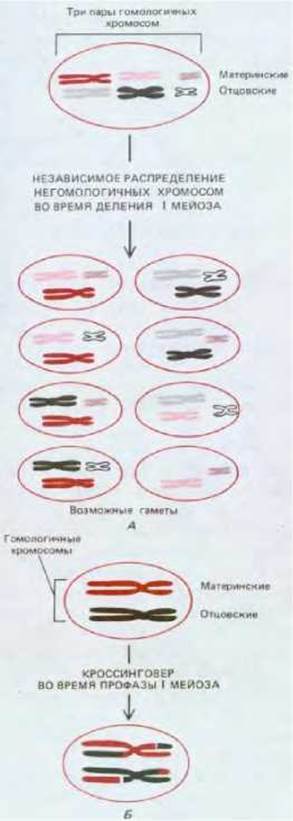
Fig. 15-9. Schematic illustrating the two primary mechanisms for redistributing genetic material during meiosis. A. In an Organism with n chromosomes, independent assortment of homologous chromosomes during the first meiotic division can produce 2n different haploid gametes. In this case, n = 3, yielding 8 different gamete types. B. During prophase I of meiosis, crossing over occurs—homologous chromosomes exchange segments, leading to Gene reassortment. Because the DNA sequences of two homologs always exhibit numerous minor differences, both mechanisms enhance hereditary Variability in sexually reproducing organisms.
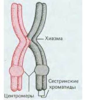
Fig. 15-10. Schematic representation of paired homologous chromosomes transitioning into metaphase I of meiosis. A single crossover event has occurred in the preceding prophase, resulting in the formation of one chiasma. Note that the four chromatids are grouped into two pairs of sister strands, with the strands in each pair closely apposed not only at the centromere but along their entire length. Therefore, this entire group of chromatids is frequently referred to as a bivalent.
15.2.2 Gene shuffling is enhanced by crossing over between homologous non-sister chromatids
As we have seen, genes can be reshuffled through the fusion of gametes from two different individuals. However, this is not the only route for genetic variation. No two offspring from the same parents (unless they are identical twins) are ever completely identical. This is because, long before gamete fusion, two distinct types of gene shuffling take place during meiosis.
One type of shuffling results from the random assortment of various maternal and paternal homologs among daughter cells during the first meiotic division; each gamete receives its own unique assortment of maternal and paternal chromosomes (Fig. 15-9A). From this fact alone, it follows that the cells of any individual can, in principle, generate 2n genetically distinct gametes, where n is the haploid chromosome number. For humans, for instance, each individual is capable of producing at least 223 = 8.4 × 10⁶ genetically distinct gametes. In reality, however, the number of possible gametes is immeasurably greater due to crossing over—a process that occurs during the prolonged prophase of the first meiotic division, when homologous chromosomes exchange segments. In humans, crossing over takes place on average two to three times per pair of homologous chromosomes. As shown in Fig. 15-9B, this process "shuffles" The genes of any chromosome within the gametes.
Crossing over involves a break in the DNA double helix within one maternal and one paternal chromatid, after which the resulting segments are rejoined crosswise (a process of genetic recombination). The molecular details of this mechanism are outlined in Chapter 5. Recombination occurs during prophase I of meiosis, when the two sister chromatids are so tightly apposed that they cannot be resolved individually (see below). Much later in this extended prophase, the two separate chromatids of each chromosome become clearly distinguishable. At this stage, they are seen to be linked by their centromeres and closely aligned along their entire length. The two homologs remain connected at the points where crossing over occurred between the paternal and maternal chromatids. At each such point, termed a chiasma, two of the four chromatids are seen to cross over one another (Fig. 15-10). Thus, chiasmata are the morphological manifestation of prior crossing over, which cannot be observed directly in action.
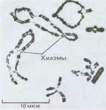
Fig. 15-11. Light micrograph of bivalents with multiple chiasmata at the diplotene stage. The large chromosomes of the orthopteran insect shown here are an especially favorable subject for cytological studies. (Courtesy of Bernard John.)
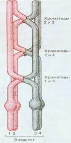
Fig. 15-12. Three chiasmata are shown—the result of three separate crossover events. Each of the two chromatids of one chromosome can cross over with any chromatid of the other chromosome in the bivalent. For example, in this case, chromatid 3 has exchanged segments simultaneously with two chromatids, 1 and 2.
At this stage of meiosis, the homologs in each pair (or bivalent) remain connected by at least one chiasma. Many bivalents exhibit a greater number of chiasmata due to multiple crossovers between homologs (Figs. 15-11 and 15-12).
15.2.3. Chromosome synapsis involves the synaptonemal complex
During prophase I of meiosis, chromosomes undergo highly complex morphological changes associated with synapsis and Separation. In accordance with these changes, prophase is divided into five sequential stages: leptotene, zygotene, pachytene, diplotene, and diakinesis (Fig. 15-13). The most striking phenomenon is the initiation of tight chromosome alignment during zygotene, when a specialized structure called the synaptonemal complex begins to form between pairs of sister chromatids within each bivalent. The point of complete chromosome pairing marks the onset of pachytene, which typically lasts for several days; following chromosome separation, the diplotene stage begins, during which chiasmata first become visible.
Genetic recombination requires close apposition of the recombining chromosomes. The synaptonemal complex, which forms immediately before pachytene and disassembles right after it, holds homologous chromosomes in close proximity by binding them along their entire length; it is thought to be essential for crossing over to occur. The synaptonemal complex is a long, proteinaceous structure resembling a rope ladder, against the opposite sides of which the two homologs are tightly apposed (bivalent, Fig. 15-14). The sister chromatids of each homolog remain closely associated, with their DNA forming numerous loops on the same side of the protein "ladder." Thus, although homologous chromosomes are brought close together along their entire length within the synaptonemal complex, the maternal and paternal chromatids that will subsequently exchange segments remain on opposite sides of the "ladder," separated by a distance exceeding 100 nm.
Cytological studies indicate that chromosome synapsis is preceded by the formation of a proteinaceous axial element along each homolog. As synapsis proceeds, these axes apparently converge to form the lateral elements of the synaptonemal complex, constituting the Two Sides of the protein "ladder." Both the initial axial elements and these lateral elements contain a protein that stains very intensely with silver, allowing these structures to be visualized using both light Microscopy and electron micrographs (Fig. 15-15). It remains unknown what prompts homologous chromosomal regions to align precisely opposite one another during the zygotene stage. Base-pair complementarity along the entire length of the interacting chromosomes is unlikely to be required, since the Chromatin of one homolog in the synaptonemal complex lies quite far from that of the other, and in some cases, the synaptonemal complex can link regions of two non-homologous chromosomes. One possible explanation is that chromosome synapsis initiates through the interaction of complementary DNA Base Pairs in small, specific regions of each chromosome. The synaptonemal complex then zips up the remaining aligned Regions of the chromosomes. Some mechanism of local matching of this sort is necessary to account for the fact that an inverted segment in one of two homologous chromosomes typically (though not always) leads to a local disruption of normal synapsis during zygotene, allowing homologous genes to pair even within the inversion region (Figs. 15-16 and 15-17). Individual stages of meiosis are illustrated in Fig. 15-18, which also provides a detailed Description of the corresponding processes.

Fig. 15-13. Sequence of events during synapsis and chromosome separation in prophase I of meiosis. A fully formed synaptonemal complex persists throughout the pachytene stage.
15.2.4. Exchanges between chromatids are believed to be mediated by recombination nodules
Although the synaptonemal complex provides the structural framework necessary for recombination events, it is probably not directly involved in them. Instead, a crucial role in these events is thought to be played by recombination nodules, which are very large Protein Complexes measuring about 90 nm in diameter (by comparison, a large globular protein molecule with a mass of 400,000 daltons has a diameter of roughly 10 nm). Recombination nodules are spaced at intervals along the "ladder" of the synaptonemal complex, positioned between the two homologous chromatids (see Fig. 15-14). They are hypothesized to represent the sites of large multi-enzyme "recombination machinery" that bring local DNA segments of the maternal and paternal chromatids into close proximity across the 100-nm-wide region of the synaptonemal complex.
Several indirect lines of evidence support this proposed function of recombination nodules:
1. The total number of nodules roughly corresponds to the total number of chiasmata observed later in prophase.
2. The distribution of nodules along the synaptonemal complex mirrors that of crossovers; for instance, much like crossovers, nodules are absent in regions where the synaptonemal complex joins segments of heterochromatin. Furthermore, genetic and cytological studies show that the occurrence of one crossover inhibits the formation of another in the adjacent chromosomal region. Similarly, nodules are generally not found in close proximity to one another.
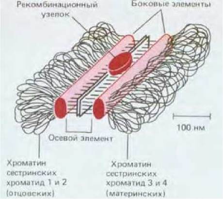
Fig. 15-14. Schematic diagram of a typical synaptonemal complex. Only a small segment of this long, ladder-like structure is shown, displaying the lateral and axial (central) elements of the complex, as well as a recombination nodule. Similar synaptonemal complexes are found in A wide variety of organisms, ranging from Yeast to humans. However, almost nothing is known about the protein molecules that make up these complexes.
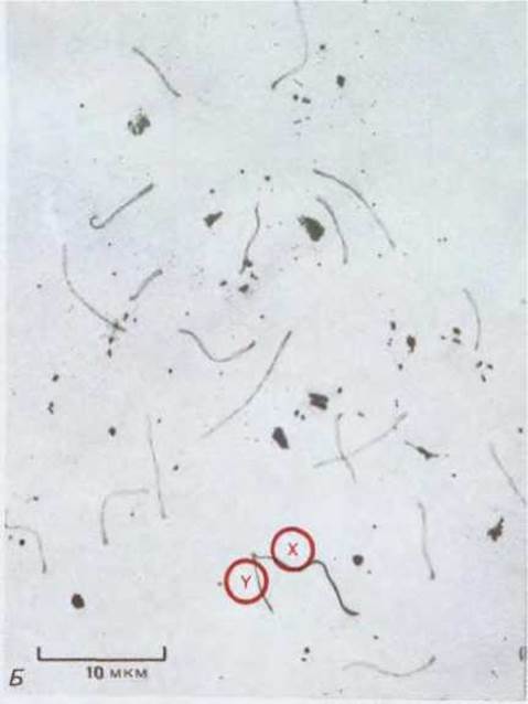

Fig. 15-15. ELECTRON MICROGRAPHS OF complete chromosome sets in silver-stained squashed preparations of mouse spermatocytes at (A) early (zygotene), (B) middle (pachytene), and (C) late (diplotene) stages of prophase of the first meiotic division. (Photographs kindly provided by Montrose J. Moses.)
A. Pairing (zygotene). Prior to chromosome conjugation, the protein filaments are separated from each other; they then approach one another, and once the proper distance is established at one or more initiation sites for synapsis between the chromosomes, the synaptonemal complex begins to form (often starting from the chromosome ends). As seen with the sex chromosomes (X and Y), pairing frequently requires traversing vast distances within The Nucleus, though The Mechanism of this movement remains unknown. Dark bodies represent nucleoli.
B. Completion of pairing (pachytene). Synapsis is complete when synaptonemal complexes link all homologous autosomes in pairs. The X and Y chromosomes conjugate only partially. Crossing-over takes place between chromatids, which are indistinguishable in such micrographs.
C. Chromosome separation (diplotene). Prior to The breakdown of the protein filaments, they pull apart from one another, signaling the end of synapsis. In the preparation, they remain connected in certain places by persisting segments of the synaptonemal complex, which are believed to mark the sites where crossing-over occurred. Later, as the chromatin condenses and individual chromatids become distinguishable, chiasmata indicate the positions of previous crossovers.
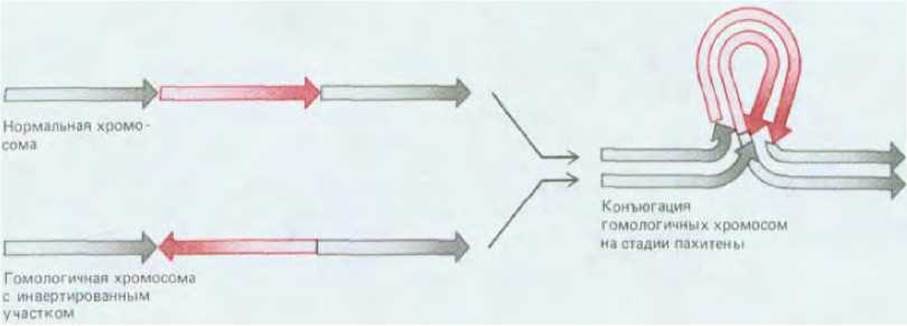
Fig. 15-16. Diagram of synaptonemal complex formation between a normal chromosome and a homolog containing an inverted segment.
Structures of this type indicate that homologous chromosomes conjugate through local Sequence Homology between specific regions.
Compare this figure with Fig. 15-17.
3. Certain Mutations in Drosophila lead to an abnormal distribution of crossovers along the chromosome length and a drastically reduced recombination frequency; correspondingly, mutant flies have fewer recombination nodules, and their spatial arrangement along the chromosome is altered in the same manner as the crossover distribution. This correlation provides strong evidence that each crossover event is determined by the localization of a single nodule.
4. It is believed that During genetic recombination, a certain amount of DNA Synthesis takes place in the region of each crossover (see Section 5.4.5). Autoradiography combined with Electron microscopy demonstrates that radioactive precursors are incorporated into pachytene DNA primarily at or near the recombination nodules.
Since the number of recombination nodules roughly matches the number of crossovers, it is reasonable to suggest that these nodules act with high efficiency to drive recombination between the chromatids of two homologous chromosomes. Unfortunately, The structure of recombination nodules and their MECHANISM OF ACTION remain entirely unknown.
15.2.5. Chiasmata play a vital role in chromosome segregation during meiosis
Crossing-over not only promotes genetic shuffling but also plays a crucial role in ensuring the proper segregation of two homologs into daughter nuclei. Specifically, chiasmata are what hold maternal and paternal homologs together until anaphase I, performing a function analogous to that of centromeres in ordinary mitosis. In mutant organisms with a reduced frequency of meiotic crossing-over, individual pairs of chromosomes fail to form chiasmata, and such chromosomes are unable to segregate properly. As a result, a significant proportion of the resulting gametes end up with too many or too few chromosomes—an instance of chromosome nondisjunction.
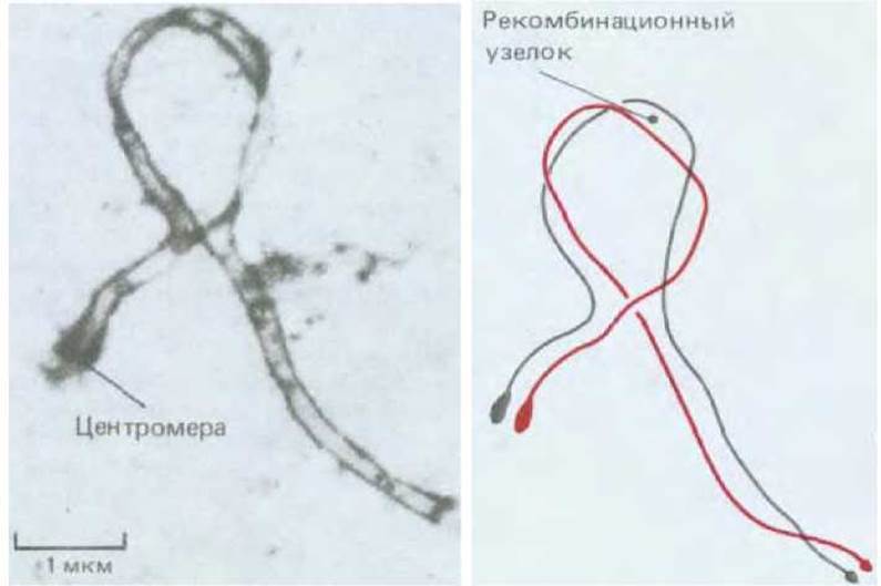
Fig. 15-17. Tight conjugation of two homologous mouse chromosomes, one of which contains an inversion, at the pachytene stage. A recombination nodule is visible within the loop (see Section 15.2.4). Left: electron micrograph; right: explanatory diagram. (P. A. Poorman et al., Chromosoma, 83, 419, 1981.)
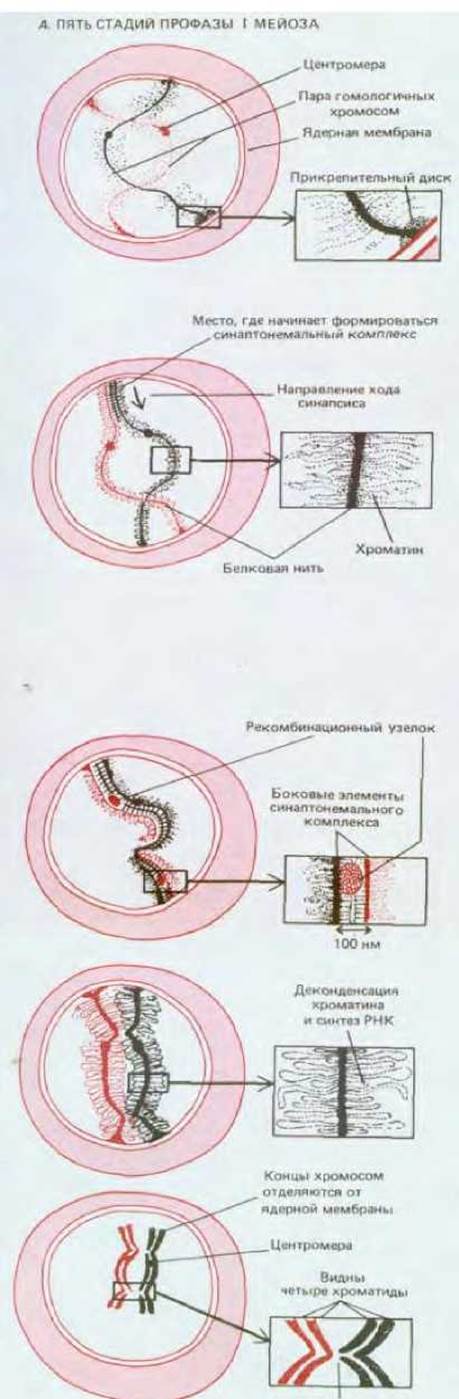
Fig. 15-18. Diagrams showing visible changes in two homologous chromosomes throughout meiosis. The course of events is illustrated for mammalian cells, although a very similar pattern can be observed in cells of many other organisms. A - five Stages of the first meiotic prophase (A); B - subsequent stages of meiosis.
LEPTOTENE. Prophase I begins with the leptotene stage, when each chromosome is seen to alter its interphase conformation and enter a condensed state, forming a long, thin thread with a protein axial core. Each chromosome is attached at both ends to the nuclear membrane by a specialized structure called the attachment plate. Although each chromosome has already replicated and consists of two sister chromatids, these chromatids lie very closely together, making each chromosome appear single (individual chromatids remain indistinguishable until late prophase—the diplotene or diakinesis stage).
ZYGOTENE. The transition from leptotene to zygotene is marked by the initiation of synapsis—the close pairing of two homologs. Conjugation frequently begins when the homologous ends of two chromosomes come together on the nuclear membrane, after which the pairing process extends inward along the chromosomes from both ends. In other cases, synapsis may initiate at internal sites and proceed toward the ends with the same ultimate result. It is believed that every gene comes into contact with its homologous counterpart on the other chromosome. As the homologs conjugate, their protein cores draw together to form the two lateral elements of an elaborate ladder-like structure known as the synaptonemal complex. Each pair of chromosomes formed during meiotic prophase I is generally termed a bivalent; however, because each homologous chromosome of the pair consists of two closely apposed sister chromatids, the term tetrad is even more appropriate for each pair.
PACHYTENE. Once synapsis is complete along the entire length of the chromosomes, cells enter the pachytene stage, where they may remain for several days. During this stage, prominent recombination nodules appear within the longitudinal groove of the synaptonemal complex and are thought to play a vital role in the exchange of chromosomal segments. Such exchanges lead to crossovers between two non-sister chromatids, involving one chromatid from each of the two paired chromosomes. Crossovers are not yet visible in pachytene, but all of them subsequently manifest as chiasmas.
DIPLOTENE. The diplotene stage of meiotic prophase I begins with the separation of the conjugated chromosomes. The synaptonemal complex disassembles, allowing the two homologous chromosomes of the bivalent to pull somewhat apart from each other. Nevertheless, they remain held together by one or more chiasmas—that is, the sites where crossing-over has occurred. In oocytes (developing egg cells), the diplotene stage can be protracted for months or even years, as it is during this phase that chromosomes condense and synthesize RNA to supply the egg cell with nutritive reserves. In special cases, diplotene chromosomes become exceptionally active in RNA Synthesis; such lampbrush chromosomes are found in amphibians and certain other organisms. DIAKINESIS. Diplotene merges imperceptibly into diakinesis—the pre-metaphase stage when RNA synthesis ceases and chromosomes condense, thicken, and detach from the nuclear membrane. It is now clearly visible that each bivalent contains four distinct chromatids, with each pair of sister chromatids joined by a centromere, while the non-sister chromatids that underwent crossing-over are linked by chiasmas. Following the Conclusion of this prolonged prophase I, two nuclear divisions without an intervening period of DNA synthesis bring meiosis to completion. These stages typically occupy no more than 10% of the total time required for meiosis and share the same names as the corresponding stages of mitosis. The remainder of the first meiotic division comprises metaphase I, anaphase I, and telophase I. By the end of the First Division, the chromosome complement is reduced from tetraploid to diploid, much as in mitosis, and one cell yields two. The crucial difference is that in the first meiotic division, each cell receives two sister chromatids joined at the centromere, whereas in mitosis it receives two separated chromatids. Next, following a brief interphase II in which chromosomes do not duplicate, the Second Division ensues rapidly—prophase II, anaphase II, and telophase II. As a result, four haploid nuclei are produced from each diploid cell that entered meiosis.
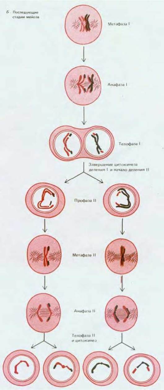
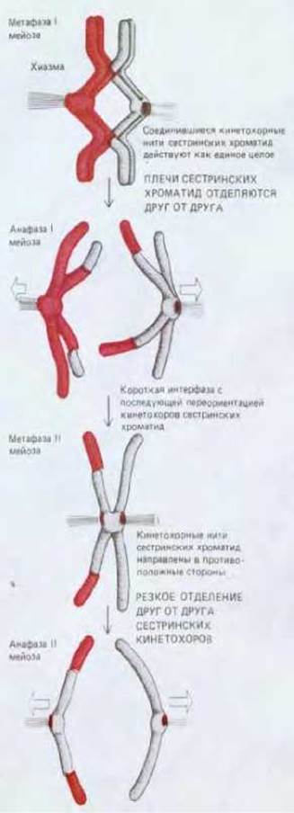
Fig. 15-19. Comparison of the mechanisms governing the ordered arrangement of chromosomes in metaphase and their segregation in anaphase during the First and Second meiotic divisions. The second division employs the same mechanisms as standard mitosis (see Chapter 13).
There are at least two significant differences between the mechanisms of chromosome segregation in conventional mitosis and in the first meiotic division:
1) whereas in mitosis spindle fibers attached to the kinetochores of two sister chromatids pull in opposite directions, in the first meiotic metaphase these fibers pull in the same direction for both sister chromatids (Fig. 15-19);
2) in mitosis, the poleward migration of chromatids is triggered by the separation of sister kinetochores from one another (initiating anaphase, Section 13.5.7), whereas in the first meiotic anaphase, this movement appears to be initiated by the breakdown of poorly understood forces that hold the arms of sister chromatids in close apposition, which in turn leads to the dissolution of the chiasmas connecting the maternal and paternal chromosomes (see Fig. 15-9). This not only accounts for the fact that chiasmas are required in many organisms for the proper alignment of chromosomes in metaphase I, but also Answers why the chromosomes formed in anaphase I typically do not have their sister chromatid arms stuck together, giving them an unusual "unfolded" appearance that makes them resemble mitotic chromosomes (Fig. 15-19).
15.2.6. Segregation of sex chromosomes also relies on their conjugation
We have explained how the conjugation of homologous chromosomes drives their segregation into two daughter cells. But what about the sex chromosomes, which are non-homologous in male mammals? Females possess two X chromosomes, which conjugate and segregate just like other homologs. Males, however, harbor one X and one Y chromosome, and these chromosomes must conjugate during the first metaphase to ensure that spermatozoa contain either an X or a Y chromosome, preventing them from carrying both sex chromosomes or neither. The required conjugation is made possible by a small homologous region located at the end of each of these chromosomes; this homology enables the X and Y sex chromosomes to pair during the first meiotic prophase (Fig. 15-15, B). This guarantees the conjugation of the X and Y chromosomes and The production of only Two Types of sperm: those carrying a Y chromosome, which give rise to a male embryo, and those carrying an X chromosome, which give rise to a female embryo.
15.2.7. The second meiotic division resembles conventional mitosis
Meiosis consists of two successive cell divisions, the first of which lasts almost as long as the entire process of meiosis and is far more complex than the second (Fig. 15-20). The first division is distinguished by A number of unique features. For example, DNA replication during the preparatory S phase generally takes significantly longer than in mitosis. In addition, cells may remain in meiotic prophase I for days, months, or even years, depending on the organism's species and the type of gametes produced. (This protracted phase of the first meiotic division is traditionally called prophase, although it bears a strong resemblance to the G2 phase of ordinary mitosis in that the nuclear envelope remains intact throughout and only dissolves as spindle fiber formation begins—that is, as prophase I transitions into metaphase I.)
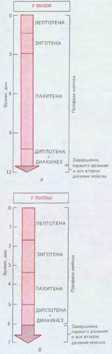
Fig. 15-20. Comparison of the durations of various meiotic stages shown in Fig. 15-18. Approximate time intervals are given for a male mammal (mouse) and a plant (lily). These intervals vary between female and male gametes (eggs and sperm) of the same species and between identical gametes of different species. For instance, meiosis in human males lasts 24 days, whereas in male mice it lasts 12 days. In all cases, however, prophase of the first meiotic division is vastly longer than all other stages combined.
Upon completion of the first meiotic division, nuclear envelopes re-form around the two daughter nuclei, and a brief interphase ensues. During this time, the chromosomes decondense somewhat; shortly thereafter, however, they condense again, and prophase II begins. Because no DNA synthesis occurs during this period, it appears that in some organisms chromosomes transition directly from one division into the next. Prophase II is brief across all organisms: the nuclear envelope breaks down as a new spindle forms, followed by the rapidly succeeding stages of metaphase II, anaphase II, and telophase II. Just as in mitosis, sister chromatids develop kinetochore fibers extending from the centromere in opposite directions. The two sister chromatids are held together at the metaphase plate until anaphase, when they separate due to the sudden poleward pulling of their kinetochores (Fig. 15-19). Thus, the second meiotic division is akin to standard mitosis (which cannot be said of the first meiotic division). The only essential difference is that here there is only one copy of each chromosome rather than two, as in mitosis.
Meiosis concludes with the formation of nuclear envelopes around the four haploid nuclei generated in telophase II (see Fig. 15-18, B). As we will see, in vertebrates the egg cell is already fully formed by the end of meiosis (and in some cases already fertilized), whereas the sperm is only just beginning its development.
Summary
During meiosis, four haploid cells are produced from a single diploid cell through two successive cell divisions following a single round of DNA replication. In animals, the initial phases of egg and sperm formation are similar. In both cases, meiosis is dominated by prophase I, which may consume 90% of the total meiotic duration. Throughout this period, each chromosome consists of two closely apposed sister chromatids. Crossing-over between chromosomes takes place at the pachytene stage of prophase I, when the pairing of each homologous chromosome pair is stabilized by the synaptonemal complex. Each crossover is thought to be mediated by a prominent recombination nodule and results in the formation of a chiasma that persists until anaphase I. As a result of the first meiotic division, each daughter cell receives one chromosome from each homologous pair, which at this stage consist of joined sister chromatids. The second division then proceeds rapidly without DNA replication, distributing each sister chromatid into a separate haploid cell.
Last update: 12/08/2026
Editorial and Educational Adaptation: This material has been compiled based on the primary/original source text. The project team performed an editorial review, corrected technical inaccuracies, structured sections, and adapted the content for an educational format.
What was processed:
- elimination of formatting defects (OCR errors, structural breaks, corrupted characters);
- editorial organization of content;
- standardization of terminology in accordance with academic sources;
- verification of factual statements against the original source text.
All mentions of the author, publication year, and origin of the primary text have been preserved in accordance with the source.