Evolution of Viruses - Zhdanov V. M. 1990
Special Section
Conclusion
Academician of the USSR Academy of Medical Sciences V. M. Zhdanov dedicated his entire creative life to Structure/149.html">The problem of organic evolution. In approaching these issues, he adhered to a broad biological perspective on The Nature of the phenomena under study. V. M. Zhdanov believed that The Development of matter leads to The Emergence of biological life, and the culmination of its refinement can be the appearance of intelligence. This is a natural stage of biological evolution. A species endowed with intelligence gains immense selective advantages and opportunities for survival and prosperity. Nature made three attempts to create intelligence. Two of them—the brains of social insects and the brains of octopuses—ended in failure, while the last one brought success and led to the ascending branch of progressive evolution in the form of a distinct species: Homo sapiens. V. M. Zhdanov was deeply interested in cosmological problems. He supported the idea of multiple centers of Life in the Universe. One such center—namely, our planet Earth and the biosphere existing upon it—became the subject of his scientific work. Exploring the evolutionary METABOLISM/13.html">History of the biosphere, V. M. Zhdanov, as a virologist, studied the Evolution of the realm Vira. Bridging the particular and the universal, he approached the evolution of Viruses from general biological standpoints and revealed The Significance of virological data for evolutionary theory. V. M. Zhdanov's creative legacy on evolution includes 5 monographs published between 1953 and 1984. Work on his final book, Evolution of Viruses, was cut short in 1987. These books have greatly influenced The formation of scientific views on The process of viral evolution and organic evolution as a whole. In this regard, it is of interest to examine the current state and Prospects for the further Development of the doctrine of viral evolution, as well as the latest achievements in this field. Our conclusion is dedicated to addressing this task. The period of 1987–1988 is reviewed. This exposition does not claim to be exhaustive either in the range of problems addressed or in the number of bibliographic sources used. The aim of this work was to highlight several key issues deemed most significant. A certain place is also given to the Discussion of controversial evolutionary problems and the definition of B. M. Zhdanov's position within this debate.
Evolutionary Foundations of viral systematics. Viral Taxonomy has made a positive contribution to general systematics. Virology has established a so-called universal Classification in which supraspecific taxa are distinguished not only by phenotypic traits, but also on The basis of fundamental properties. These include the type of nucleic acid, Genome Organization, the mode of Genetic information expression, virion structure, and other molecular-biological, biochemical, and functional indicators. Such an approach is rarely used in the systematics of higher eukaryotes. The practical classification of these organisms is still based on morphological criteria. Having outstripped the systematics of higher eukaryotes in this respect, viral systematics has lagged behind it in terms of the biological principles of construction. In viral systematics, the delineation of high-ranking taxa does not take into account the evolutionary relationships among them. This is due to the fact that until very recently, these relationships remained undisclosed.
Advances in science—and first and foremost the accumulation of data on the Introduction/19.html">Primary Structure of viral genes and their protein products—have created the prerequisites for establishing the evolutionary foundations of viral systematics. V. M. Zhdanov took an active part in this work. The system proposed by him for RNA-genomic viruses is based on the strategy of the viral genome, with the viruses themselves grouped in order of increasing structural complexity.
Currently, data have appeared in the literature that allow for a further analysis of evolutionary relationships among A number of RNA-genomic viruses, between viral and cellular genomes, and also for examining the evolutionary interrelations among Viroids, virioids, and The Cell [Kingsbury D. W., 1988; Goldbach R., 1988]. Based on the results of viral RNA sequencing, comparisons of genome strategies, and protein structures and Functions, R. Goldbaсh (1987), R. Goldbach, and J. Wellink (1988) concluded that positive-sense plant RNA viruses share similarities with animal viruses and are divided into two supergroups: picorna-like and Sindbis-like viruses. The supergroup of plant picorna-like viruses includes como-, nepo-, and poty-like viruses. The commonalities between these viruses and animal-infecting Picornaviruses are as follows: 1. In all cases, a low molecular weight VPg protein is attached to the 5'-end of the genomic RNA, and a poly(A) tract is present at the 3'-end of The Genome. 2. The primary Translation product of the viral genome is a giant precursor polypeptide that is cleaved into functional Proteins via proteolytic Processing. 3. Nonstructural proteins are encoded by an identical set of genes, which can be viewed as a constant Gene module. 4. The degree of Homology among nonstructural proteins exceeds 20%. In all cases, these proteins are involved in RNA Replication.
Class="center">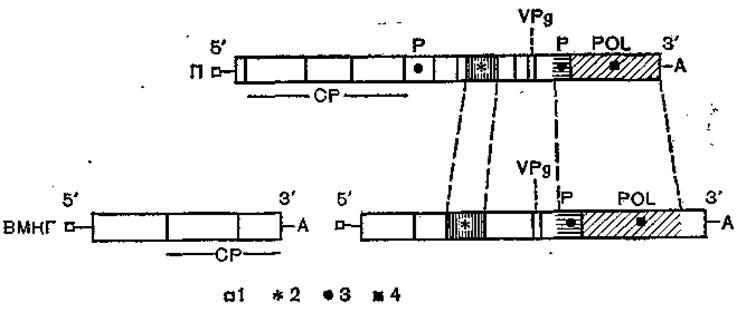
Fig. 63. Comparison of genomes of picornaviruses (poliovirus — II) and comoviruses (cowpea mosaic virus — CPMV) [after Goldbach R., Wellink W., 1988].
Rectangles indicate open reading frames; hatched areas represent regions of high (over 20%) homology; 1 — VPg protein at the 5'-end of the genome; 2 — nucleotide-binding region; 3 — Cysteine protease region; 4 — polymerase region; A — poly(A) sequence at the 3'-end of the genome; VPg — region of VPg capsid proteins; P — protease region; POL — region of RNA-dependent RNA polymerase.
The features of genome organization similarity between poliovirus (a monopartite virus) and cowpea mosaic comovirus (a multipartite bipartite genome) are presented in Fig. 63. Similarities are found in the arrangement and function of the capsid protein genes, the VPg protein, the polymerase complex, and proteases. Homologous sequences are present in the polymerase and nucleotide-binding regions.
The supergroup of plant Sindbis-like viruses includes ilarviruses, bromoviruses, cucumoviruses, tobamoviruses, tobraviruses, furoviruses, hordeiviruses, carmoviruses, and tombusviruses. A common feature of these viruses is the conservation of homologous genome regions, the presence of a cap structure at the 5'-end of the RNA, and the generation of subgenomic RNAs during the viral replicative cycle. The nonstructural proteins of Sindbis-like viruses share homologous regions with the nonstructural proteins of Sindbis virus and are encoded by similarly organized genes. These proteins possess a nucleotide-binding region, exhibit RNA polymerase activity, and are involved in RNA replication. Thus, members of this group show similarities to Sindbis virus in the arrangement and function of their polymerase genes—in other words, all these viruses share a common replicative module. This points to common evolutionary roots. A similar conclusion can be drawn regarding plant picorna-like viruses and animal picornaviruses.
PLANT AND ANIMAL kingdoms diverged in their evolution approximately 1.2×10⁹ million years ago. Given the high evolutionary rate of RNA genomes, the emergence of the ancestors of modern RNA-genomic viruses of plants and vertebrates should be attributed to a later epoch. Subsequently, these proviruses diverged in their evolution, gave rise to numerous branches, occupied various ecological niches, and became established in plant and vertebrate hosts.
Currently known plant picorna-like viruses differ significantly from one another in virion Morphology and a number of other traits. The virions of como- and nepoviruses are spherical, whereas those of poty-like viruses are rod-shaped. The genome of como- and nepoviruses is divided into two particles, whereas poty-like viruses are monopartite. Sindbis-like viruses are characterized by even greater diversity. Most of them are multipartite and exhibit differences in their genomic translational strategies. All these traits are secondary, acquired in the course of evolution. The emergence of multipartite genomes in RNA-containing Viruses is attributed, for example, to the combined action of ecological factors and the Influence of the Nature of the genetic material [Nee S., 1987]. The leading role in this evolutionary direction is played by the high multiplicity of plant virus transfer, the absence of a fully developed host immune system capable of restricting viral replication, and the significant error rate of RNA replication, which confers selective advantages to fragmented genomes.
An important ecological factor in the evolutionary development of positive-sense RNA-genomic viruses was The Use of arthropod vectors, which ensured contact between plants and vertebrate hosts. It is interesting to note that recombination served as one of the mechanisms for generating Variability. The Role of the latter in the evolution of positive-sense RNA genomes has remained insufficiently clear. The outcomes of past recombination events can be observed when comparing the genomes of certain plant picorna- and Sindbis-like viruses [Goldbach R., Wellink J., 1988]. Overall, the outcome of evolution is the formation of two branches of positive-sense RNA-genomic viruses, united by a common origin, retaining elements of Structural and functional unity, and utilizing Representatives of the animal and plant kingdoms as hosts.
V. M. Zhdanov shared the view regarding the evolutionary links between viruses and cellular genetic elements. This idea is gaining increasingly widespread development. Based on structural and functional similarity and genome homology, two phylogenetic lines have been constructed: 1) group I introns — viroids — virusoids — delta agent, and 2) Transposons — Retroviruses — Hepadnaviruses [Kingsbury D. W., 1988]. The commonality of the first phylogenetic line is determined by the fact that the genome of its constituent agents is in all cases represented by a small single-stranded RNA rich in complementary regions. Regions of high homology are present. The ancestral form appears to be mobile cellular genetic elements resembling group I introns, which gave rise to viroids.
Fourteen viroids are currently known. All of them use plants as hosts. They represent "naked" RNA devoid of a protein coat. The genome length ranges from 246 to 371 NUCLEOTIDES. They do not require a helper virus for replication. Viroid replication is directed by the host cell's DNA-dependent RNA polymerase using cRNA oligomers as a template. The lethal effect of viroids on plant Cells is associated with the suppression of pre-mRNA Processing [Smarda S., 1987; Albanese J., La Rosa R., 1988].
Virusoids are considered to be viroids that have lost the capacity for autonomous replication. Virusoid replication is carried out with the assistance of a helper virus. They use plants as hosts. The genome has a length of 350 to 400 bases and contains protein-coding sequences that direct the synthesis of proteins involved in the Formation of the virusoid coat.
A special place in the phylogenetic line under consideration is occupied by the delta agent, which is the only currently known human virusoid-like pathogen. Its replication occurs with the help of hepatitis B virus. The genome of the delta agent consists of 1,683 nucleotides. There are 5 open reading frames, two of which are in genomic orientation and three in antigenomic orientation [Makino Sh. et al., 1987]. The viral genome-encoded protein with a Molecular Weight of 24,000 is phosphorylated at Serine. This phosphoprotein possesses RNA-binding activity and participates in the formation of delta agent particles [Chang Ming-Fu et al., 1988]. The delta agent has no analogues among animal viruses. It remains unclear whether the delta agent originates directly from group I introns or whether its evolution proceeded step-by-step through viroids and virusoids. The delta agent shares significant homology with the latter [Makino Sh. et al., 1987].
The possibility of constructing the phylogenetic line of transposons — retroviruses — hepadnaviruses is due to the fact that the retro- and hepadnaviruses included in it utilize the reverse Transcription mechanism for their replication. The region of the genome encoding Reverse Transcriptase contains areas of high homology. The genomic DNA of hepadnaviruses and the DNA copy of the retroviral genome have The ability to integrate into the cellular genome, thereby forming transposon-like structures. The structural and functional similarity of transposons with integrated retro- and hepadnavirus genomes indicates their evolutionary commonality. However, The complexity of their organization makes it impossible to state which element in this system is primary. The possibility of evolutionary movement in two directions—i.e., from the cell to transmissible genetic elements and from them to the cell—cannot be excluded either. Upon integration into the cellular genome, retro- and hepadnaviruses are capable of causing insertional Mutations. Such processes are likely an important mechanism for shaping the eukaryotic genome during evolution [Jolais F., Sabo A., 1988].
The rate of molecular evolution of DNA-genomic viruses. RNA- and DNA-genomic viruses differ significantly in their level of mutability. RNA replication occurs with low fidelity. Significant errors are introduced into the transmitted information. The mutation frequency per generation reaches 10-3–10-4. This is 100,000 times higher than the mutation frequency of DNA. Over 33 generations, RNA-genomic viruses can accumulate a reserve of variability that would take DNA-genomic viruses 10 million generations to produce. The low mutability of DNA is determined by the nature of this genetic material, the replication mode, and the capacity for repair. These data previously led to the belief that DNA-genomic viruses are characterized by profound hereditary conservatism. Currently, this traditional view requires clarification, if not revision.
The question of the mutability of DNA-genomic viruses is part of the broader issue regarding the rate of molecular evolution of DNA at various levels of the evolutionary ladder. Discussions on this matter in the scientific literature have been ongoing for more than 15 years. Based on accumulated data, it can be considered established that the rate of molecular evolution is not uniform across different taxonomic groups, and a universal molecular clock of evolution does not exist. Based on the results of unique DNA sequence Hybridization and sequencing data of homologous genes and pseudogenes in various species, R. J. Britten (1986) concluded that the rate of molecular evolution can vary by up to 5-fold, ranging from 1.3×10⁻⁹ to 6.6×10⁻⁹ substitutions per site per year. The lowest rates of DNA evolution were found in higher primates and birds, whereas the highest were found in rodents, sea urchins, and Drosophila. Li Wen-Hsiung et al. (1987) believe that the rate of nucleotide substitutions in rodents is 4 to 8 times higher than in higher primates, and 2 to 4 times higher than in ungulates. The tempo of molecular evolution in hominoids is lower than in higher primates. Humans exhibit a lower frequency of nucleotide substitutions compared to chimpanzees and orangutans. Generation length and the number of DNA replications in germline cells are cited as reasons for differences in the rate of molecular evolution. Mention is also made of the Specific features of DNA replication Mechanisms and repair efficiency.
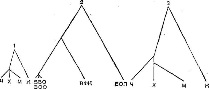
Fig. 64. Phylogenetic trees of vertebrates and Poxviruses [after Gentry G. A. et al., 1988].
1 — vertebrates; 2 — poxviruses; 3 — vertebrates. The evolutionary rate has been equalized with the evolutionary rate of poxviruses (coefficient 2.8). Branch lengths in cases 1 and 2 are proportional to the evolutionary distance expressed as the number of mutations per 100 amino acid residues in the vertebrate or viral thymidine kinase molecule. H — human; Hmr — hamster; M — mouse; C — chicken; VZV — varicella-zoster virus; MPV — monkeypox virus; VV — vaccinia virus; RFV — rabbit fibroma virus; APV — avian poxvirus.
DNA-genomic viruses are no exception to the general rule. As agents with a shorter life cycle, their rate of molecular evolution exceeds that of their vertebrate hosts. A review of data on this issue was provided by G. A. Gentry et al. (1988).
An idea of the differences in the rates of molecular evolution between poxviruses and their hosts is provided by the following clear example. Figure 64 shows the phylogenetic trees of poxviruses and vertebrates, drawn on the basis of comparing the Amino acid sequences of deoxythymidine kinase. In cases 1 and 2, the phylogenetic trees are constructed according to a uniform principle but at different scales. Branch lengths are proportional to the evolutionary distance expressed as the number of mutations per 100 amino acid residues. The mutability of poxviruses exceeds the corresponding figure for vertebrates by 2.8 times. A comparison of tree 1 and tree 2 shows that they differ in height. This indicates that the evolutionary rate of vertebrates is lower than that of poxviruses. Vertebrate tree 3 is constructed to the scale of the poxvirus Phylogenetic Tree. An additional coefficient of 2.8 was applied. The differences in the length of the vertebrate tree and the poxvirus tree, caused by the unequal rates of amino acid residue substitution in deoxythymidine kinase, are thereby leveled out.
Varicella-zoster virus and Herpesviruses types 1 and 2 diverged in their evolution approximately 48.6 million years ago. Over this period, the rate of Amino Acid Substitutions in the small subunit of Ribonucleotide reductase exceeded the corresponding figure for the host enzyme by 38 times. The data examined show that DNA-genomic viruses lag significantly behind RNA-genomic viruses in their level of mutability, yet slightly outpace their vertebrate hosts. This makes it possible to speak of a relatively low hereditary conservatism in DNA-genomic viruses and to distinguish them among organisms utilizing DNA as their genetic material.
The mutability of the viral genome creates Prerequisites for the microevolution of DNA genomic viruses, which, judging by the state of natural viral populations, proceeds quite intensively. This is evidenced by the high mutation load in viral populations and the presence of intrapopulational differences. Particularly interesting data have been obtained in The Study of Adenoviruses. These studies employed a unified methodology for the restriction analysis of virion DNA, making the results of different authors comparable. Li Quan-Gen and G. Wadell (1988) used Bell, Bglll, Hpal, Sail, and Smal endonucleases to analyze 61 strains of adenovirus type 3 isolated across 6 continents. They identified 17 genomic types, which were divided into 3 genomic groups. Strains assigned to group 1 were isolated in Africa, Europe, and North and South America; group 2 strains in Africa; and group 3 comprised strains with a global distribution, with the exception of South American countries. For adenovirus type 16, 16 genomic variants have been described, with variability sites evenly distributed throughout the genome. Among 76 strains of adenovirus type 37 isolated over the past 10 years from patients on three continents, 20 strains were found to differ in their genomic DNA physical maps from the prototype strain. In some cases, Changes in the physical maps were accompanied by serological differences detected by HI and NT assays. However, these differences were minor and did not correspond to the divergence expected from restriction analysis data [Adrian Fh. et al., 1988].
The role of populations in evolution. At first glance, V. M. Zhdanov's evolutionary concept appears unclear due to his denial of the real existence of populations in viruses. In the present book, one can read the following: "Equally inapplicable to viruses is THE CONCEPT OF a population, since the intracellular stage of reproduction, and even more so integration processes, entirely deprive the interpretation of a reproducing virus as a population of any meaning, etc."
Difficulties in introducing the category of "population" into virology are easily overcome if we consider that a population is a historical category. The structure and organizational principles of populations have changed in the course of evolution. Viral populations possess unique characteristics and differ significantly in their structure from the populations of higher eukaryotes, which serve as the population standard for most biologists [Tsilinsky Ya. Ya., 1988]. V. M. Zhdanov overlooked this obvious truth. This is directly related to the state of modern evolutionary biology and Zhdanov's views on the driving forces of evolution.
The Synthetic Theory of evolution is undergoing a deep crisis. Its central dogma—according to which the accumulation of random mutations in populations and their subsequent evaluation by natural Selection are necessary and sufficient conditions for micro- and macroevolution—can no longer claim a monopoly in the Study of the evolutionary process. Modern evolutionary theory must account for genome instability, the neutrality of molecular evolution, and many other phenomena [Antonovics J., 1987; Ho Mae-Wan, 1987; Pollard J. W., 1987; David J. R., 1988]. Zhdanov's denial of the obvious fact that viral populations exist serves as a peculiar protest against the inability of the classical synthetic theory of evolution (which is largely limited to considering population processes) to explain the development of the organic world.
Simultaneously with the revision and modernization of the synthetic theory of evolution, a new philosophy of biology is being born in modern science. V. M. Zhdanov's works on evolution contributed to this process. Zhdanov identified a force that can accelerate evolutionary development: viral Transduction. The idea of the role of viruses in the horizontal transfer of genetic information has been expressed generally on multiple occasions. Zhdanov's merit lies in the fact that, as a professional virologist, he comprehensively substantiated this idea.
The development of evolutionary theory does not mean forgetting the achievements of the past [Futuyma D. J., 1988]. While denying the real existence of viral populations, V. M. Zhdanov essentially did not break away from the synthetic theory of evolution and widely used population and ecological approaches to evolutionary phenomena in his work. Let us examine the development of this trend.
The classical and continuously operating sources of hereditary variability in viruses are mutations, true recombinations, and the reassortment of genome fragments. Accumulated data indicate that these sources create a huge reserve of genetic variability in populations and ensure microevolution. This function can be performed not only by extensive mutations, but also by single point mutations. To the existing Evidence of the impact of point mutations on the phenotype, we can add the work of Jooh Ji-Woon et al. (1988), who established that the diabetogenic variant of encephalomyocarditis virus differs from the non-diabetogenic one by the presence of a single oligonucleotide that is absent from the T1-oligonucleotide maps of the non-diabetogenic variant. The change in the T1-oligonucleotide map of the non-diabetogenic variant was caused by a single point substitution A→C located in the region of the genome corresponding to the missing oligonucleotide. No other substitutions were found in this or adjacent regions. These results suggest that a significant change in the pathogenic potential of encephalomyocarditis virus—expressed as the loss of its ability to cause diabetes—may be caused by a single substitution-type point mutation. Data regarding retroviruses are also of interest. A single mutation in the envelope protein gene leading to a Lysine-to-Arginine substitution disrupts their processing and deprives the endogenous oncotropic murine leukemia virus of the C3H/He strain of its replication ability and several other properties. A reverse mutation at the processing site (replacing arginine with lysine) leads to a reversion to the wild type [Sithanandam G., Rapp U. R., 1988].
Data on the role of genome fragment reassortment in the variability of orbiviruses continue to accumulate. J. L. Stott et al. (1987) studied the formation of reassortants in calves infected with two serotypes of bluetongue virus. The majority (89%) of viral clones isolated from the Blood of infected animals were reassortants, involving 6 out of the 10 segments present in the viral genome in their formation, with 16 types of reassortants identified. P. D. Oberst et al. (1987) demonstrated that genetic interaction between bluetongue virus strains in natural hosts occurs not only during acute infection, but also during persistent infection. The authors isolated reassortants from a bull over a period of 41 days during which viremia persisted. These results indicate that the reassortment of genomic segments makes a significant contribution to the formation of the pheno- and genotypic differences characteristic of naturally circulating bluetongue virus strains.
Due to the high mutability of their genetic material, RNA genomic viruses evolve within the infected host during the infectious process. A virus that has invaded a host Organism is genetically different from the virus that subsequently accumulates in the patient. F. Gebauer et al. (1988) tracked the variability of FOOT-and-Mouth disease virus serotype C3 in cattle over 539 days of persistent infection. The rate of molecular evolution of the viral genome was 0.9×10⁻² to 7.4×10⁻² substitutions per site per year; 59% of the RNA mutations led to amino acid substitutions. During the infection, virus isolates differing antigenically from the parental strain were obtained from the bull. These data indicate that large ruminants not only serve as reservoirs for foot-and-mouth disease virus, but also act as a source of altered variants.
The accumulation of mutations and variability supplied by recombination leads to the formation of intra- and interpopulational differences. In this regard, data on poliovirus reviewed by Yu. Z. Gendon (1988) are extremely indicative. The use of T1-oligonucleotide mapping, nucleotide and amino acid sequencing, and antigenic analysis using a panel of Monoclonal Antibodies established that poliovirus strains circulating in various geographical zones possess phenotypic and genotypic individuality. Ten groups have been identified (Central Asian, North American, South African, Indian, etc.). R. Rico-Hesse et al. (1987) conducted a comparative study of genome sequences encoding Regions of the VP1 capsid protein and the A2 non-capsid protein in 62 strains of poliovirus type 1. The strains were isolated over the past 31 years across 5 continents from paralytic poliomyelitis patients. They were grouped, and foci of Circulation of similar strains were identified. The recombinant origin (wild-wild, vaccine-wild) of certain isolates was established. From a practical standpoint, It is important to note that antigenically atypical strains capable of overcoming the immunological barrier created by vaccination can emerge in natural poliovirus populations. Variability in this direction is associated with incomplete population immunization coverage and low herd Immunity levels [Tsilinsky Ya. Ya., 1988]. Public health faces The Challenge of adopting measures to protect against antigenically altered variants and developing appropriate Vaccines. New opportunities for solving this problem are opened up by successes in producing viable intertypic poliovirus hybrids using Genetic Engineering Methods [Hogle J. M., 1988].
In the course of evolution, viral populations adapt to their environment, occupy specific ecological niches, and diverge genetically. Important data have been obtained from the study of viruses of the genus *Hantavirus* within the family *Bunyaviridae* [Le Due J. W., 1987]. The genus includes the CAUSATIVE AGENT OF Korean hemorrhagic fever—Hantaan virus, isolated in Korea in 1978—and Hantaan-like viruses, which are divided into 3 serological groups. All these viruses are similar in ecology, transmission routes, and mechanisms of natural survival, causing a chronic asymptomatic infection in rodents. Despite a high Immune Response, the virus can persist in the host for life. The virus is excreted and spreads into the environment via rodent saliva, urine, and feces; this mechanism drives horizontal transmission within rodent populations. Hantaan and Hantaan-like viruses cause various hemorrhagic fevers in humans. Human infection occurs via airborne, contact, and rodent-bite pathways.
Hantaviruses have a global distribution and vary serologically, in their degree of virulence for humans, and in other traits depending on their geographical zone. Hantaan virus and closely related strains circulate in China, Korea, the Far East, southern Siberia, and the European part of the USSR. In humans, it causes severe hemorrhagic fever with renal syndrome, accompanied by high mortality. In contrast, Hantaan-like viruses circulating in Scandinavia, Western and Central Europe, as well as certain regions of the European part of the USSR (prototype strain Puumala), cause a relatively mild and benign disease—nephropathia epidemica. Hantaan-like viruses prevalent in the USA and Canada (prototype strain Prospect Hill) are not virulent to humans. In addition to geographical isolation, these differences are driven by ecological factors, specifically the use of different hosts. The host for Hantaan virus is the striped field mouse (*Apodemus agrarius*), for the Puumala virus it is the bank vole (*Clethrionomys glareolus*), and Prospect Hill virus is ecologically associated with the California vole (*Microtus californicus*). The Importance of ecological factors in the differentiation of hantaviruses is underscored by the fact that the ranges of *Apodemus agrarius* and *Clethrionomys glareolus* overlap in Europe. Consequently, virus strains that differ both serologically and in their pathogenic potential for humans circulate within the same territory.
Hantaviruses are not the only viral group whose members have diverged As a result of both geographical and ecological isolation. Important data have been obtained regarding viruses of the genus *Bunyavirus* of the family *Bunyaviridae* [Calisher Ch. H., 1988]. Viruses of the C antigenic group belonging to this genus are clearly differentiated serologically by HI, CF, and NT assays and differ in the primary structure of their surface glycoprotein G. Their range includes Brazil and several other South American countries. Group C viruses are capable of genome fragment reassortment and potentially form a single gene pool. Nevertheless, the genetic individuality of group members is preserved because each virus occupies its own ecological niche, preventing their circulation pathways in vertebrate hosts and vectors from intersecting. Ecological isolation is also characteristic of many Bunyaviruses found in the same territory belonging to the Guama, Patois, Simbu, and California antigenic groups.
The heterogeneity of viral populations and their rapid microevolution are general phenomena characteristic of viruses across various taxonomic groups. At the same time, even among closely related viruses, this process has its own specific features. M. Yamashita et al. (1988) studied the Features of the molecular evolution of Influenza B virus and the patterns of strain turnover over the past 40 years.
To this end, the primary STRUCTURE OF THE hemagglutinin (HA) gene and the non-structural protein (NS) gene was determined in viral isolates obtained between 1940 and 1987. DNA copies of the corresponding genome segments were used for sequencing. The evolutionary distance between isolates was determined based on the number of nucleotide substitutions in these genes. The results were compared with similar literature data regarding influenza A and C viruses.
As shown in Fig. 65, which presents data obtained over 7 consecutive epidemic seasons, the patterns of genetic material movement in influenza A, B, and C viral populations differed. For influenza A virus, only a single parental strain serves as the ancestor of the strains prevailing in a given epidemic season, whereas for influenza B and C viruses, there are several. This phenomenon is particularly pronounced in the case of influenza C virus. As a result, varying degrees of trait scatter are created in viral populations. In each epidemic season, the influenza A virus population is represented by a single Lineage, while influenza B and C populations are represented by several. In the latter case, two or more lineages survive. Comparing the lengths of the branches connecting the strains reveals that the evolution of influenza B virus proceeds more slowly than that of influenza A, but faster than that of influenza C.
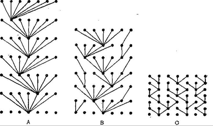
Fig. 65. Trait scatter and strain turnover in influenza A, B, and C viral populations [after Yamashita M. et al., 1988]. Points lying on the horizontal plane correspond to strains isolated in the same epidemic season. The length of the lines connecting these points corresponds to the evolutionary distance between the strains.
The pattern of strain turnover in influenza A virus is apparently driven by the higher mutability of the viral genome, as well as a shorter life cycle and faster generation turnover per unit of time on a population scale. This facilitates the accumulation of mutants—including single mutants capable of overcoming herd immunity—which ensure the continuity and persistence of the virus over time. The rest of the parental population is lost. Influenza B and C viruses experience lower immune pressure. Despite a narrower spectrum of antigenic variability caused by relatively low mutability, this ensures the prolonged circulation of strains over several epidemic seasons. Influenza B virus evolves 2–6 times more slowly than influenza A virus. Indicators of the low mutability of influenza C virus include the absence of mutations in the NS gene among viral isolates separated by a 19-year interval, and changes in only two nucleotides in the HA gene in isolates obtained with a 31-year interval. The molecular evolution of influenza B virus proceeds at a rate of 1.1×10⁻³ nucleotide substitutions per site per year.
The ecological niche occupied by viruses includes not only the host organism and population, but also the susceptible cells in which viral reproduction takes place. For a long time, it was believed that viral host Specificity and the selection of specific target cells within the host organism were ensured by the specificity of virus-cell receptor interactions. Later, evidence was obtained that host range restriction is also determined by the conditions of viral reproduction within sensitive cells. Among new works devoted to this problem, the report by J. Benavente and A. Shatkin (1988) stands out; they studied the reason for the inability of avian reovirus to reproduce in mouse cells. The virus specifically interacted with a culture of mouse L cells and penetrated the Cytoplasm, where viral transcripts were formed. However, viral mRNAs failed to bind to Polysomes, and viral Protein Synthesis did not occur, aborting the infection cycle. Virus-specific RNAs synthesized in L cells were functionally active and translated into proteins *in vitro* in rabbit reticulocytes. Thus, the reproduction of avian reovirus in mouse cells was blocked at the stage of protein synthesis initiation.
One of the pathways of viral evolution is divergence within the host organism. This path was taken by human herpesviruses, which utilize various target cells, exhibit distinct tissue Tropisms, and differ in the Pathogenesis of the diseases they cause. The tissue tropism of these viruses is largely ensured by biochemical mechanisms: modification of transcription regulation, post-translational alteration of structural proteins, and The activity of regulatory genes. These properties were acquired in the course of evolution and contribute to the specificity of virus-cell interactions [Mach M. et al., 1988].
The presented data show that elements of gradualistic morphogensis can be traced in the microevolution of viral populations, with ecological factors playing an important role. Overall, this fits into the Concept of the synthetic theory of evolution regarding the slow accumulation of minor changes and the gradual divergence of populations occupying their ecological niches. The features of gradualistic viral evolution, driven by the evolutionary potential of the genome and the Structure of viral populations, were discussed earlier [Tsilinsky Ya. Ya., 1988]. Gradualistic morphogenesis is not the only path followed by viruses. Viral evolution also occurs as a result of explosions, leaps, and saltations. One opportunity for this arises due to the neutrality of molecular evolution.
The Significance of the neutral theory of molecular evolution for virology. V. M. Zhdanov was quick to grasp new ideas in evolutionary doctrine. He did not overlook M. Kimura's neutral theory of molecular evolution (1985), the postulates of which are outlined in this book.
The vast factual material accumulated in virology confirms Kimura's concept and indicates that the molecular evolution of viruses follows a neutralist path. This applies not only to DNA genomic viruses, but also to RNA genomic viruses. The latter is particularly important for validating the neutralist theory, which was developed based on empirical data obtained from organisms using DNA as genetic material. The nature of the molecular evolution of RNA genomic viruses demonstrates that neutralist principles are universal and extend across the entire biosphere.
Using RNA genomic viruses as research objects has opened up new possibilities for understanding the patterns of molecular evolution. The general course of the development of life on Earth has been traced over 3.5 billion years. Mutational changes accumulate in the genome and proteins of all organisms from generation to generation. Due to the low mutability of DNA, the rate of molecular evolution in organisms utilizing this genetic material is relatively low. The following rates have been determined for mammalian proteins: Insulin—0.44, Myoglobin—0.89, Hemoglobin α-chain—1.2, Lysozyme—2.0, pancreatic RNase—2.1, fibrinopeptides—8.3×10⁻⁹ per site per year, etc. The rate of molecular evolution of papovavirus genes encoding small t-antigen, large T-antigen, and VP1 and VP2 proteins is 4.1×10⁻⁹ to 6.5×10⁻⁹ per site per year. The brevity of human existence precludes direct observation of these processes. To reconstruct the course of molecular evolution, indirect methods are employed—specifically, comparing organisms that share a common ROOT and have evolutionarily diverged. The scale of differences is used to judge the time of origin of organisms and the evolutionary path they have traversed. High RNA mutability, exceeding DNA mutability by 100,000 times, and the rapid accumulation of molecular changes make it possible to reconstruct the course of molecular evolution of RNA genomic viruses using a more advanced method: direct comparison of modern viruses with their ancestors. The research material consists of virus strains isolated at intervals of 5, 10, 20 years or more, separated by an evolutionary distance of 5,000, 10,000, and 20,000 generations, etc. (assuming an average viral Life Cycle of 8 hours). Comparing these strains makes it possible to trace the course of molecular evolution and determine its character. Thus, the molecular evolution of RNA genomic viruses is judged not from the finished results of past history, but directly from the changes observed in archaic forms.
A central tenet of the neutralist theory is its third postulate, which states that during evolution, mutational substitutions leading to minor disruptions in a molecule's Structure and function (conservative substitutions) occur more frequently than those causing more substantial impairments. The theory refers to the preferential accumulation of neutral or nearly neutral mutations—that is, mutations that do not significantly affect the phenotype or produce only a weak negative effect.
Let us consider an example illustrating the fixation of neutral mutations in a viral genome. V. J. Johnson et al. (1986) sequenced DNA copies of the 26S RNA from a virulent strain of Venezuelan equine encephalitis virus (Trinidad) and its attenuated derivative, strain TC-83. Attenuation resulted from prolonged passage and selection of the virus in cell cultures. The 26S RNA molecule contains the genes for three structural virion proteins: the surface Glycoproteins E2 and El, and the internal protein C. It was shown that neither gene order, molecular length, nor THE POSITION OF the Open Reading Frame changed during attenuation. The attenuated strain TC-83 differed from its virulent Trinidad precursor by 13 nucleotide substitutions, of which only 5 led to amino acid changes. All 5 mutations affecting the Amino Acid Sequence were localized in the gene encoding the E2 protein. This gene, as well as the non-coding region at the 5'-end of the 26S RNA molecule, are considered potential genetic determinants of virulence. Functionally, the E2 glycoprotein induces the Formation of protective antibodies and apparently determines the specificity of virus-cell interactions. In Venezuelan equine encephalitis virus complex strains, the E2 protein exhibits significant antigenic variability. All 7 mutations identified in the El and C protein GENES OF THE attenuated TC-83 strain were neutral and did not cause amino acid substitutions.
The possibility of the emergence and fixation of neutral mutations in the genome is determined by The properties of The Genetic Code. The nucleotides making up codons carry unequal informational loads. THE ORIGIN OF these differences is linked to the evolution of the genetic code. Of the three nucleotides comprising a codon, the First and Second bear the greatest informational weight, whereas the third nucleotide is characterized by low specificity. Consequently, the affinity of codons for specific Amino Acids is often determined by the first two nucleotides. The third position may be occupied by any of several nucleotides without altering the meaning of the triplet or its capacity to encode a specific amino acid. Thus, nucleotide substitutions at the third codon position are most frequently neutral; in other words, neutral mutations typically become fixed precisely at this position.
The preferential accumulation of mutations at the third codon position is characteristic of the viral genome. N. Saitou (1987) summarized data on The Nature and localization of nucleotide substitutions found in the genes for hemagglutinins H1 and H2, neuraminidases N1 and N2, as well as the non-structural protein NS and matrix protein M genes of human influenza A virus. Overall, 268 mutations were observed at the third codon position, compared to only 189 in the first and second positions.
Mutations at the third codon position are most often synonymous, meaning they do not lead to amino acid substitutions in the protein molecule. J. R. Wiener and W. K. Joklik (1988) compared The nucleotide sequences of the M2 genomic segment from human reoviruses types 1, 2, and 3, which have diverged evolutionarily and differ serologically as well as in certain other biological properties. The M2 segment encodes the Synthesis of the acidic μ1 protein, which has a low cystine, Histidine, and Methionine content and is rich in Proline. Approximately 27% of The amino acid residues adopt an α-helical configuration. Proteolytic Cleavage of the μ1 protein—occurring between amino acid residues at positions 42 and 43—yields the μ1C protein, which serves as a major structural component of the virion. The M2 segments of serotypes 1 and 2, 1 and 3, and 2 and 3 shared 15%, 23%, and 23% sequence identity, respectively. In contrast, the M2 gene product, the μ1 protein , exhibited a high degree of homology (97%). The conservation of the μ1 protein was achieved because the majority of RNA mutations were fixed at the third codon position and did not result in amino acid replacements. When comparing the M2 genomic segment between serotypes 1 and 3, 87.6% of all identified mutations were located at the third codon position; for serotypes 1 and 2, and 2 and 3, this figure was 86.4% and 86.6%, respectively. The relative frequencies of mutations at the first codon position were 9.6%, 11.8%, and 11.2%, while at the second position they were 2.8%, 1.8%, and 2.2% for serotypes 1 and 3, 1 and 2, and 2 and 3, respectively. Only 18 out of 324 mutations identified in the comparison of serotypes 1 and 3 led to amino acid substitutions. For serotypes 1 and 2, and 2 and 3, these values were 20 out of 501 and 23 out of 507, respectively.
In addition to the Properties of the genetic code, the neutral nature of mutations is determined by the Structural Features of the protein molecule. A protein molecule consists of a small functionally active center and a considerably larger "passive" region. The fixation of mutations within this passive region neither alters protein function nor affects the phenotype. Furthermore, the correlation between the primary structure of a protein and its higher-order structures—which govern its functional activity—is degenerate. Consequently, alterations in the amino acid sequence resulting from mutations may leave higher-order structures and protein function unaffected, rendering the mutations neutral.
A classic example of the preservation of viral protein functional activity amidst the accumulation of neutral mutations is provided by the work of J. Blok and G. M. Air (1982), who compared the primary structures of neuraminidase segments from 8 subtypes of influenza A virus. The neuraminidase molecule possesses a three-dimensional structure comprising a stalk, a HEAD, and a transmembrane segment with a hydrophobic region at the NH2 terminus. The head is associated with the antigenic and enzymatic activity of the protein, while the hydrophobic region anchors the molecule to The Lipid Bilayer of the virion. The stalk connects these two domains, with its stabilization maintained by CARBOHYDRATES and Disulfide Bonds. A comparison of the primary structures of the transmembrane segment and stalk showed that these regions differ in amino acid sequence. Despite low overall homology, the general architecture of The polypeptide chains and their properties were conserved. The transmembrane segment in all neuraminidase subtypes contained a high proportion of hydrophobic amino acids, whereas the stalk structure revealed cysteine residues capable of forming disulfide bonds as well as glycosylation sites. These disulfide bonds and carbohydrate attachments ensure the stabilization of the stalk's three-dimensional structure. It was demonstrated that pronounced mutational remodeling of the primary structure did not affect the Tertiary Structure of the examined neuraminidase regions nor alter their function, confirming that the accumulated mutations were neutral in character.
Thus, neutral mutations fall into two categories. Mutations of the first type are fixed in the genome without altering the amino acid sequence of the protein molecule, whereas those of the second type alter both The nucleotide sequence and the corresponding amino acid residues without disrupting protein function. As the preceding Examples show, both types of neutral mutations occur in viruses. The molecular evolution of viruses proceeds via the preferential accumulation of these mutations, ultimately priming the viral genome for sudden leaps, bursts, or dramatic transformations. This sets the stage for the emergence of new forms or, alternatively, leads to viral extinction.
The Biological Significance of the neutralist theory lies in the premise that evolutionary changes at THE MOLECULAR LEVEL—namely, the reorganization of the genetic material itself—are driven not by external environmental influences, but by internal, independent genetic processes. Changes in DNA and RNA occur primarily through the fixation of neutral and nearly neutral mutations, the accumulation of which is not controlled by selection and proceeds without its intervention. The rate of fixation of these mutations is determined largely by the mutation rate itself. The synthetic theory of evolution holds the contrasting view that fixed mutational changes are adaptive and driven by positive Darwinian selection, whereby mutations with phenotypic expression and positive effects are preferentially retained. The accumulation of neutral and nearly neutral mutations is supported by extensive empirical data from comparative studies of genome and protein primary structures across various organisms, and data on viruses also strongly support the neutralist theory.
Neutral and nearly neutral mutations accumulate in genomes and proteins without initially having a significant impact on the survival and Reproduction of a species. However, when environmental conditions change, this situation can shift: previously neutral mutations may become selectively valuable and serve as the raw material for adaptive evolution. This pre-existing reservoir within the genome increases the evolutionary potential of organisms and is presumably actively utilized during periods of heightened evolutionary activity, which, According to the punctuated equilibrium concept, give rise to speciation.
The Evolutionary Significance of neutral and nearly neutral mutations also lies in their capacity to facilitate abrupt evolutionary shifts. Even a single "fortunate" mutation occurring within a genome saturated with silent, unexpressed mutations can generate a novel combination of genetic factors, giving rise to a gene with novel properties and dramatically altering the phenotype. This process resembles a phase transition, occurring abruptly and saltationally. Such shifts may yield either positive or negative effects—the latter of which will be discussed below in the context of mass extinctions. Both the accumulation of neutral and nearly neutral mutations and the transition of this material into a new qualitative state occur without the participation of selection; consequently, newly emerged traits may lack adaptive value. Examples of such acquisitions include the majority of protein polymorphisms widely prevalent among higher eukaryotes, where the predominant portion of polymorphic alleles in a given species is maintained by the mutation process and random elimination.
A distinctive feature of this evolutionary pathway is that selection does not create new forms; rather, it merely tests and culls them, after which the successful forms are further "fine-tuned" by selection. Conversely, the synthetic theory of evolution assigns selection a leading role in form-building processes. It should be noted that these evolutionary pathways are not mutually exclusive, but rather Complement one another to ensure the ongoing evolution of the biosphere.
Analysis of variability in natural viral populations and disease incidence trends indicates that one primary mode of viral evolution is sudden, saltational form generation. Over the past 20 years, several viruses responsible for novel Human and Animal diseases have been discovered. These include the etiological agents of AIDS (HIV-1 and HIV-2); simian, feline, and bovine immunodeficiency viruses causing immunodeficiency states in their hosts; enterovirus 70, the causative agent of acute hemorrhagic Conjunctivitis in humans; a parvovirus causing myocarditis and diarrhea in puppies; human T-cell leukemia virus types associated with adult T-cell leukemia and tropical spastic paraparesis (such as human T-cell leukemia virus type 1); and human enteric viruses (rotaviruses, Caliciviruses, astroviruses, Norwalk-like agents, etc.). In most cases, this reflects not the appearance of entirely novel viruses, but the identification of previously unrecognized agents and the diseases they cause. Alongside these, however, there are genuine examples of the emergence of new forms, the most reliable being the origins of HIV-1, canine parvoviral myocarditis and diarrhea, and enterovirus 70. These viruses appeared abruptly and spread rapidly among immunologically naive hosts, sparking epidemics of previously unknown diseases. Evaluating the scale of these evolutionary shifts, it is notable that each instance was confined to the expansion of already established genera and families (such as the genera *Parvovirus* and *Enterovirus*, and the family *Retroviridae*) with new representatives—effectively the formation of new species.
The sudden, saltational appearance of altered strains of already known viruses occurs quite frequently and is a hallmark of the intraspecific microevolution of RNA genome-bearing viruses. Such strains play a major role in the Epidemiology and epizootiology of influenza, foot-and-mouth disease, and many other infections. For instance, a novel strain of avian influenza A virus crossed the interspecies barrier to trigger a devastating epizootic of influenza among seals along the Atlantic coast of the USA in 1979–1980. This is the first and thus far only known instance of a highly specialized avian influenza A virus making a direct transition to mammals. It is likely that these evolutionary events are sometimes driven by mutations that "fall on fertile ground"—that is, they arise within a genome already saturated with neutral mutations and primed for reorganization.
In addition to their evolutionary role, neutral mutations fulfill another highly distinctive function in RNA viruses: defense against accelerated evolution. Due to the high mutability of their genetic material, the molecular evolution of RNA viruses proceeds extremely rapidly. Summarizing data on influenza A, B, and C viruses, foot-and-mouth disease virus, reoviruses, HIV, and others, the rate of molecular evolution in the viral genome is estimated at 2×10⁻² to 5×10⁻⁵ substitutions per site per year, though these rates vary among different viruses and genes. Functionally critical proteins and their corresponding genomic regions evolve more slowly than less vital ones. For example, a comparison of the matrix protein of human influenza A (H3N2) isolates from 1972 and 1979 revealed only two amino acid substitutions. The matrix protein carries significant functional responsibilities: it forms a domain on the inner surface of the cytoplasmic membrane where genomic segments aggregate during virion assembly, specifically recognizes viral glycoproteins, and ensures the formation and Stability of the envelope beneath the viral lipid bilayer. Apparently, only those amino acid substitutions that preserved protein function were fixed in the matrix protein sequence. In contrast, the light chain of the influenza A (H3N2) hemagglutinin accumulated substitutions at a rate of 0.24–0.57% per year, and the heavy chain at 0.2–0.91% per year. It remains unclear whether the molecular evolution of RNA viruses proceeds at a uniform pace over time, as available data are contradictory. Evidence for a constant rate of evolution comes from the fact that earlier studies claiming otherwise were based on limited data and were not subsequently corroborated. In particular, our understanding of the molecular evolution of influenza B virus has evolved; through the study of a representative group of strains, M. Yamashita et al. (1988) demonstrated that its evolution proceeds at a steady temporal rate. Overall, it can be concluded that RNA viruses possess a molecular clock of evolution, and this clock ticks several orders of magnitude faster than that of DNA viruses.
The most widely recognized cause of organismal extinction is the inability of a species' gene pool to adapt to changing abiotic and biotic environmental conditions. Given the rapid molecular evolution of RNA viruses, we can point to an additional, intrinsic cause of extinction: the disintegration of the established genetic structure driven by an excess of mutations. The neutral nature of molecular evolution slows this process. Both the genetic material and its protein products exhibit substantial buffering capacity, absorbing the continuous influx of mutations. Neutral mutations accumulate in RNA and Protein molecules without exerting negative effects, allowing high rates of mutational substitution in genes while vital functions are preserved, thereby establishing a defense against runaway evolution. This raises the question of how long this established barrier can withstand the pressure of mutations, leading us to consider mass extinctions as a general biological phenomenon.
A recurrent phenomenon in The history of life on Earth is the mass extinction of species and higher taxonomic groups. During the Mesozoic, these events occurred with a periodicity of approximately 26 million years, and during the Cenozoic, every 35–40 million years. Mass extinctions are not indiscriminate; rather, specific taxa become their selective victims. The causes of mass extinctions remain insufficiently understood. Numerous hypotheses linking them to external factors (global catastrophes triggered by asteroid impacts, supernova explosions, the passage of Nemesis, etc.) can account for specific instances but fail to explain the phenomenon as a whole. According to M. V. Volkenstein and T. S. Rass (1987), mass extinctions depend not only on shifting external biotic and abiotic factors, but also on internal drivers. Species may perish as a result of accumulating neutral mutations that become deleterious when combined. The extinction mechanism is also tied to pseudoneutral mutations, which exert only negligible negative effects. The accumulation of neutral mutations can disrupt the compensatory balance of these pseudoneutral mutations, ultimately degrading organismal fitness and causing the extinction of the species. Extinction is fundamentally an autocatalytic process; "video-disappearance" occurs abruptly and resembles a phase transition. Conversely, these events can sometimes take a different course, where genome reorganization yields positive effects, laying the groundwork for further evolutionary progress. Throughout the evolutionary history of the biosphere, some species perished without leaving descendants, while others branched into subsequent evolutionary lineages.
This hypothesis applies to RNA viruses given their high rates of molecular evolution. It can be postulated that the internal factors reducing viral viability and driving extinction stem from the progressive accumulation of neutral and pseudoneutral mutations. Initially, these mutations inflict only minor harm; however, as changes multiply over time, the capacity for mutation compensation diminishes. This process is as inexorable as Aging. Driven by the degeneracy of the genetic code and the loose correlation between primary and spatial protein structures, alterations accumulate covertly until a phase transition occurs, underpinned by a drastic genomic overhaul and a sudden shift in the spatial conformation of protein molecules. Functional impairments ensue, viral viability drops, and the stage is set for extinction.
Senescence, aging, and death are The Fate of all living things, affecting not only individual organisms but communities as well. Among higher sexually reproducing eukaryotes, the unit of extinction is the species. A species community shares a unified gene pool that is fully engaged in the process of molecular evolution, which in turn generates the preconditions for extinction; all individuals within a species share a common destiny and eventual loss of vital potential. In contrast, populations and species of RNA viruses possess a different genetic architecture, consisting of assemblages of distinct clones that form isolated clonal lineages across successive generations, with little to no genetic exchange between them. Due to stochastic processes, the molecular evolution of individual lineages within a population proceeds independently: in some cases, evolution is favorable, and genomic reorganization does not preclude lineage survival, whereas in other cases, it compromises viral viability, rendering those particular lineages victims of extinction.
The manifestation of these processes during the natural circulation of viruses is the sudden cessation of epidemics and the disappearance of circulating strains. A sharp decline in epidemic outbreaks and a rapid drop in incidence—occurring despite the presence of susceptible populations—have been documented for influenza during the dominance era of any given shifted variant of influenza A. It appears as though the infection gradually Burns out as it transmits from host to host (Kendal, A. P., 1987). Periods of dominance for each shifted influenza A variant also ended abruptly; on a global scale, the virus would vanish from circulation in less than a year. The sudden halt in the circulation of previously dominant strains cannot be explained solely by herd immunity pressure. Apparently, shifted influenza A strains and their drift variants are capable of undergoing only a limited number of generations before failing to sustain transmission from host to host, ultimately reaching an evolutionary dead end. The potential for reduced viability and viral extinction is rooted in the fact that strains driving epidemics and pandemics originate from selectively valuable mutants or reassortants and genealogically represent clonal lines. The unfavorable molecular evolution of many such lines imposes strict limits on their longevity.
It remains unclear whether the extinction process affecting individual viral strains also extends to populations and species as a whole. We cannot rule out the possibility that the lifespan of currently known RNA viruses in their modern forms is finite, leaving them with two potential paths: extinction or transformation. While certain rapidly evolving RNA viruses may succumb to extinction, one must also account for the fact that high rates of RNA evolution coexist with the preservation of vital functions necessary for genetic Homeostasis. Alongside their capacity for rapid evolution, RNA viruses exhibit profound evolutionary stasis, manifested as remarkable hereditary conservatism and the long-term retention of fundamental properties and core biological characteristics (Tsilinsky, Ya. Ya., 1988). Historical records indicate that yellow fever has afflicted humans for at least 190 years, Crimean-Congo hemorrhagic fever for at least 600 years, and rabies for 2,000 years. The etiological agents of these infections have carried their defining species traits—the ability to cause specific characteristic diseases—across centuries. Roughly speaking, this evolutionary distance corresponds to approximately 130,000 generations for the yellow fever virus, and 350,000 and 145,000 generations for Crimean-Congo hemorrhagic fever and rabies viruses, respectively. In terms of generational count, this distance far exceeds the Separation between modern humans and their ape-like ancestors. RNA viruses display prolonged conservation and retention of specific ancient gene modules. The presence of such modules—representing positive-sense single-stranded RNA replication systems—unites evolutionarily distant animal and plant viruses into coherent groups of picorna- and Sindbis-like viruses. These viruses have traversed long evolutionary paths from common ancestors while preserving functionally critical genomic regions. All of this indicates that mass extinctions of RNA viruses, if they occur at all, are selective—a pattern analogous to organismal extinction among higher life forms.
The origin of pandemic variants of influenza A virus. The central and still unresolved question in influenza research concerns the sources and origins of pandemic influenza A variants circulating within human populations—in other words, The Mechanism of antigenic shift. V. M. Zhdanov championed the ecological concept and contributed immensely to its development, positing that the emergence of pandemic variants stems from the incorporation of animal influenza A virus genes into the human influenza A genome.
Proposed roughly 20 years ago, this hypothesis rapidly gained widespread popularity among virologists, molecular biologists, general biologists, physicians, and veterinarians studying influenza. It seemed that the origin of pandemic influenza A variants had been fundamentally resolved: pandemic strains arose through the reassortment of genomic segments between human and animal Influenza Viruses, which were thought to belong to a single viral species sharing a common gene pool. However, as time passed, decisive proof supporting the ecological concept failed to materialize. Moreover, the concept began to encounter difficulties as accumulating evidence highlighted the strict host specificity of influenza A viruses, the challenges of crossing the interspecies barrier, The production of disharmonious gene combinations during reassortment, and the low viability of human-animal influenza reassortants. Laboratory reassortment of genomic fragments between human and animal influenza viruses successfully yielded strains bearing antigenic formulas matching those of known pandemic variants, but this failed to validate the ecological concept. These reassortants served merely as antigenic, rather than biological, analogs of pandemic variants; gene segment reassortment disrupted virus-specific virulence and stripped the viruses of their capacity for transmission and epidemic spread within the population. Furthermore, the ecological concept could not explain the most recent antigenic shift of 1977, which involved the sudden re-emergence of the influenza A (H1N1) virus. The origin of this virus was unrelated to genetic material exchange among whale, swine, and avian influenza viruses; instead, the 1977 strains proved identical to influenza A (H1N1) strains that circulated in human populations from 1950 to 1952. These facts point to a direct lineage continuity and long-term persistence of the virus, which enabled its eventual return to circulation.
All of this heightened interest in the alternative view that human influenza A is an anthroponotic infection caused by strains exclusively associated with the human population. The acquisition of genes from "animal reservoirs," even if possible, has no bearing on antigenic shift; rather, the latter arises from the return to circulation of strains that persist long-term within the human population. As a broad-minded scientist and polymath, V. M. Zhdanov did not confine himself to favored dogmas, but sought to encompass the entire problem holistically—an approach that applied equally to influenza research. A proponent of intellectual pluralism, Zhdanov believed that such an ideology fosters scientific progress. In this light, it is worthwhile to examine the tenets of the anthroponotic hypothesis regarding the origin of pandemic human influenza A variants and its subsequent development.
Modern Perspectives framing influenza as an anthroponosis were substantiated in the work of R. E. Hope-Simpson and D. B. Golubev (1987), who advanced a novel concept of the epidemic process in influenza A. The authors noted that the number of shifted variants causing human influenza A is not three (H1N1 from 1946–1957 and post-1977; H2N2 from 1957–1968; and H3N2 post-1968), but four. The fourth variant is the influenza A (H0N1) virus, which emerged in 1929 and circulated until 1946. Serological data also point to a fifth shifted influenza A variant (Hsω1N1) that circulated from 1918 to 1929 and subsequently spilled over into the swine population, where it has persisted to the present day. Grouping the influenza A viruses (Hsω1N1), (H0N1), and (H1N1) into a single subtype is fundamentally incorrect, as these viruses induce only incomplete cross-protection.
The emergence of each of them was in the nature of an antigenic shift. In the case of the replacement of influenza A virus (H0N1) by (H1N1), this shift did not occur as a result of the reassortment of genome fragments. It has been documented that the emergence of influenza A virus (H1N1) was caused by a significant mutation in the hemagglutinin gene of influenza A virus (H0N1). The limited number of pandemic variants of human influenza A cannot be explained from the standpoint of the ecological concept. According to this concept, free reassortment of genome fragments among human, animal, and avian influenza viruses could lead to the generation of dozens and hundreds of reassortants that would experience no immune pressure from the human "host" and spread freely through the population.
The ecological concept of the origin of pandemic influenza A variants fails to explain not only the limited number of such variants, but also the Cyclical Nature of their appearance in the human population. The sequence of variant replacement has been traced over a span of 100 years. Serological studies of blood sera from elderly individuals have shown that the H2N2 variant apparently circulated during the last quarter of the 19th century. It was succeeded by the H3N2 variant, which predominantly circulated from roughly 1900 to 1918. Around 1907, the H1N1 variant joined the H3N2 variant (much like it does in our time). The subsequent order of strain succession was discussed above.
R. Е. Hope-Simpson and D. В. Golubev (1987) focus on the cyclicity of shift variants of human influenza A and the limitation in their number, not to emphasize the weakness of the ecological hypothesis, but to substantiate the concept they proposed. The authors examine epidemiological data indicating that the development of outbreaks and the progression of influenza morbidity cannot be explained solely by the continuous and sequential transmission of the virus from a sick person to a healthy one, as occurs in measles. Numerous attempts to construct Mathematical models of influenza epidemics based on this principle have yielded no results. In a number of cases, the course of influenza morbidity resembles a model in which the infection arises in multiple primary foci and then subsides upon transmission from host to host. This is supported by epidemiological observations of the simultaneous occurrence of influenza cases across large territories and in disparate regions. The nature of such epidemics once created the illusion that the influenza virus was introduced from outer space.
The authors suggest that the influenza A virus is incapable of indefinitely sustained human-to-human transmission and that, during the interepidemic period, it persists not in animals or birds, but within The Human Body in the form of a latent or persistent infection. Months or even years later, under the Influence of Environmental factors and changes in the host organism's state, the virus reactivates in hidden carriers and spreads throughout the human population. External factors act upon the entire host population; consequently, the virus is shed by a multitude of carriers, and the epidemic begins from multiple primary foci. Through this mechanism, new batches of virus are fed into circulation as the epidemic progresses. The reactivated virus encounters the host's immune defense, resulting in antigenic drift.
It is interesting to note that the epizootic process of influenza in domestic animals can follow a similar pattern. In the 1950s in the USA, the simultaneous emergence of influenza A outbreaks was observed in pigs across geographically isolated farms. It was hypothesized that the virus persisted in the animals and was activated by meteorological factors.
The periodic activation of the virus in hidden carriers and its subsequent spread within the human population lead both to epidemics—which follow one another at intervals of 1–2 years during the predominance of a particular shift variant—and to antigenic shifts resulting in pandemics. All 5 shift variants of human influenza A persist in the population and undergo periodic reactivation. The H3N2 influenza A virus pandemic had a particularly long latent period; it had persisted in the human population since the beginning of the 20th century without manifesting epidemically until 1988. Isolated virological and serological findings point to the possibility of the long-term conservation of archaic variants.
An important role in substantiating the concept proposed by R. Е. Hope-Simpson and D. В. Golubev (1987) is played by data obtained during the interepidemic period. Between epidemics, against the Background of the cessation of pathogen circulation, isolated virologically confirmed cases of influenza are typically recorded. Their occurrence can be interpreted as the result of virus activation in carriers followed by its limited spread.
A weak point of the anthroponous concept of human influenza A in the past was the lack of evidence for long-term virus carriage. The situation has now changed. As R. Е. Hope-Simpson and D. В. Golubev (1987) justly note, the possibility of latent and persistent human infection with influenza A virus is beyond doubt, although the forms of carriage require further study. Apparently, the action of defective interfering viruses plays an important role in establishing this carrier state. Significant positive contributions to this issue are made by data concerning persistent influenza A virus infection in cell cultures. While this fact does not constitute direct proof of virus persistence in the organism and its preservation in the host population via the specified mechanism, data obtained at the cell culture level nonetheless point to such a possibility.
When discussing the causes and periodicity of influenza A virus reactivation in the human body, one should note the cyclical nature of natural phenomena, which represents a general biological regularity. The repetitiveness and cyclicity of life processes are expressed in multi-year fluctuations in animal populations, reversible restructuring of biocenotic species structures, pulsation of zoonotic infection foci, changes in pathogen properties, increases in virulence, etc. Natural cycles are driven by environmental factors that also affect humans, among which solar energy occupies a prominent place. The significance of the solar energy flux was highlighted by the eminent scientist A. L. Chizhevsky (1976).
In his book *The Terrestrial Echo of Solar Storms*, published in 1937, he noted that solar activity acts as a global-scale regulator of epidemiological processes. Analyzing data on the occurrence and course of influenza epidemics spanning nearly 500 years, A. L. Chizhevsky (1976) concluded that the recurrence period of influenza epidemics (meaning pandemics) averages 11.3 years. The deviation of the initial years of an epidemic from the peak of solar activity in either direction averages 2.3 years. In other words, influenza epidemics tend to begin either 2.3 years before the solar maximum or 2.3 years after it.
The intensity of an epidemic appears to be somewhat dependent on the intensity of solar activity. Solar activity affects the vital activity of organisms and reduces the body's resistance to pathogens. While the biological action of solar energy on various organisms and natural processes is quite evident, the mechanism of this impact remains unclear. At the same time, extensive data on solar radiation factors have been accumulated across various fields of knowledge. In general, periods of heightened solar activity are marked by a sharp surge in short-wave hard radiation. This triggers electrical, magnetic, and electromagnetic storms that affect the macroorganism as well as the virus, both directly and indirectly through the host organism. It is conceivable that during latent influenza infection, solar energy is capable of activating the persistent virus.
An examination of this lesser-known anthroponous concept demonstrates that the study of influenza evolves not only on the basis of ecological views regarding the nature of this infection, but also through other perspectives. V. M. Zhdanov strongly supported such a broad approach to solving scientific problems.
Overall, the question of the origin of pandemic human influenza A strains has evolved into a major evolutionary problem. One of the pathways of viral evolution is slow co-evolution alongside their hosts. As hosts diverge evolutionarily, the viruses diverge as well. Co-evolution leads to specialization and adaptation of the virus to specific species, establishing interspecies barriers. Specialization hinders the virus's ability to switch to a new host [Tsilinsky Ya. Ya., 1988]. Human and animal influenza A viruses are on this evolutionary path. Their common roots are evidenced by a unified genome strategy, a high degree of genetic homology, the capacity for genome fragment reassortment, and serological relationships, among other factors. Indicators of evolutionary divergence include incomplete homology of the genome and proteins, the formation of discordant combinations during genome fragment reassortcation leading to low viability of reassortants, and host-specific specialization that reduces the ability of both the virus and its reassortants to overcome the interspecies barrier. When assessing the strength of this barrier, one must consider that influenza virus host specificity is determined not merely by the receptor apparatus during virus-cell surface interactions. An important role in defining the host range is played by the interaction between the nucleocapsid and cellular components, which is likewise specific in nature and takes place at the intracellular stage of the viral infection cycle.
It follows from the above that the possibility of genes entering the human influenza A virus population from "animal" reservoirs is determined by how far the divergent evolution of influenza A viruses has progressed and how deeply they have adapted to their hosts. We are referring specifically to the acquisition of individual genes rather than an entire virus. Existing data rule out the possibility that an influenza A virus originating from animals could enter circulation in the human population and establish itself there. In 1976, sporadic cases of influenza caused by a swine influenza A virus closely related to the A (HsωlNl) virus were observed among people in direct contact with animals on pig farms in the USA. Despite a complete lack of immunity to this virus in the human collective, the spillover from the animal population failed to trigger an outbreak. The virus was not transmitted further from the initial cases along an epidemiological chain. This outcome occurred even though swine influenza viruses are genetically closer to human influenza viruses than are the influenza viruses of other animals and birds.
V. M. Zhdanov's views on gene exchange between human and animal influenza A viruses evolved over time. Initially, he regarded these viruses as a single species within which intensive gene redistribution takes place. His subsequent statements on this matter were more cautious. The human influenza A virus is described as an emerging species that has not yet completely severed its ties with animal influenza A viruses. Through these retained connections, occasional replenishment of the human influenza A gene pool with genes from animal influenza A viruses remains possible.
Clearly, the possibility of exchange is determined by the degree of evolutionary divergence between the Viruses and Their adaptation to their hosts. In this regard, a fundamentally important line of research initiated by V. M. Zhdanov is the study of the molecular-biological nature of influenza A virus host specificity, as well as the obstacles encountered by the virus and its reassortants in overcoming the interspecies barrier. These studies will help answer whether pandemic variants of influenza A arise as a result of gene introduction from the outside or through the succession of variants already fixed within the human population.
Last update: 13/08/2026
Editorial and Educational Adaptation: This material has been compiled based on the primary/original source text. The project team performed an editorial review, corrected technical inaccuracies, structured sections, and adapted the content for an educational format.
What was processed:
- elimination of formatting defects (OCR errors, structural breaks, corrupted characters);
- editorial organization of content;
- standardization of terminology in accordance with academic sources;
- verification of factual statements against the original source text.
All mentions of the author, publication year, and origin of the primary text have been preserved in accordance with the source.