Chordate Zoology - textbook - Y. V. Tsaryk - 2013
Chapter 5. SUPERCLASS TETRAPODS (LAND VERTEBRATES) TETRAPODA, seu QUADRIPEDA.
ANAMNIOTES (ANAMNIA) AND AMNIOTES (AMNIOTA)
5.2. CLASS REPTILES, REPTILIA
5.2.1. Structural Features of Reptiles
Appearance. The body shape of reptiles is more diverse than that of amphibians. This is related to their modes of locomotion. Serpentine reptiles (snakes, legless lizards) have lost their limbs, yet they successfully move through leaf litter, grassy thickets, and tree crowns, and are also good swimmers.
On a solid substrate, locomotion occurs by means of limbs and lifting the body off the ground. This enabled lizards (agamas, toadhead agamas) to transition to rapid quadrupedal movement, and eventually to bipedal running. In arboreal animals, the hind limbs elongated to assist in leaping between branches, and some developed lateral Skin folds that aid in gliding (flying dragons).
Within the Class Reptilia, various types of locomotion on land, in Water, and in the air have evolved. Adaptation to movement in diverse environments was the primary driver of progressive reptilian evolution. It was based on exploiting the full potential of the mechanical design formed by the central internal supporting Skeleton, paired limbs, and their associated Muscles.
Integument. The upper layers of the stratified epidermis in reptiles undergo keratinization: granules of the protein keratin accumulate in the Cells, displacing the Cytoplasm and Nucleus. Beneath this dead layer lies the deep Malpighian layer, consisting of living epidermal cells capable of division (Fig. 5.17). The proliferation of the stratum corneum gives rise to scutes, scales, horny granules, tubercles, spines, and claws. Beneath the horny scales of some reptile species, bony plates lie within the mesodermal layer of the skin, the corium. In turtles, these fuse to form a bony shell that is fused to THE Vertebral Column.

Fig. 5.17. Cytology/practical/54.html">Longitudinal section of lizard skin (after O. Bütschli)
Pigment cells are located in the Malpighian layer and the upper PARTS OF THE corium. The skin provides reliable protection against water evaporation, mechanical damage, and the penetration of pathogens. At the same time, it has lost the capacity for gas exchange, water evaporation, and excretion of metabolic products. The skin fits tightly to the body. The horny covering is renewed through shedding: the complete or partial sloughing of the old stratum corneum and The formation of a new one. Many species shed several times a year.
Locomotion in reptiles is highly diverse. Living in dense grassy thickets, tree crowns, and forest litter led to a transition to crawling, which resulted in the loss of limbs. Such individuals move by lateral undulation of the body (serpentine movement). This mode of locomotion is also highly effective in aquatic environments. The appearance of lateral skin folds provides the basis for gliding. Thus, reptiles move in a variety of ways (Fig. 5.18).


Fig. 5.18. Body shape and modes of locomotion in some modern reptiles (not to scale):
Slow walking: А - steppe tortoise. Running: Б - sand lizard; В - common basilisk; Г - frilled-neck lizard. Gliding leaps:
Д - flying dragon. Rock climbing: Е - Caspian gecko. Tree climbing: Ж - steppe agama; З - chameleon; І - boomslang. Ground crawling: К - sheltopusik; Л - amphisbaenian; М - sand racer. Swimming: Н - American alligator; О - marine iguana; П - yellow-bellied sea snake; Р - hawksbill sea turtle
Skeleton. The vertebral column of reptiles is divided into five regions: cervical, thoracic, lumbar, sacral, and caudal.
For foraging and orientation, The Development of a flexible neck and increased HEAD mobility were crucial for reptiles. Head mobility is enabled by the presence of the first two cervical vertebrae: the atlas (atlas) and the axis (epistropheus). The atlas is a bony ring divided by a dense ligament into upper and lower halves; the Brain connects to the Spinal Cord through the upper opening, the anterior surface of the lower half articulates with the occipital condyle of the skull, and the odontoid process of the second cervical vertebra, the axis, projects into the lower opening from behind.
Neck movement is determined by the number and Structure OF THE cervical vertebrae, which vary among different groups. Tuataras have amphicoelous vertebrae (fish-like) with remnants of the notochord between them. In crocodilians and most squamates, the vertebrae are procoelous (concave anteriorly). Some of the cervical vertebrae bear short Ribs. The complex neck movements of turtles are facilitated by a variety of vertebral structures: the posterior vertebrae are procoelous, the anterior are opisthocoelous (concave posteriorly), and the middle ones are amphicoelous.
Long ribs articulate with the thoracic vertebrae, and their ventral ends are attached to the Sternum via cartilaginous portions, forming a closed rib cage (snakes lack a rib cage). The Pectoral Girdle articulates with the sternum. Lumbar vertebrae also bear ribs that do not reach the sternum. The Pelvic Girdle is attached to the sacral region, which consists of two vertebrae. The caudal region helps maintain balance during locomotion and sometimes serves as the primary propeller (in aquatic snakes, crocodilians, and some aquatic lizards). In lizards capable of autotomy, the caudal vertebrae can break in the middle, where thin cartilaginous layers divide the vertebral body into two parts.
The total number of vertebrae in different reptile species can range from 50-80 (7-10 cervical, 16-25 thoracolumbar, 2 sacral, 15-40 caudal), 140 (in thick and short snakes), to 435 (in long-bodied snakes).
Paired limbs and their girdles. The pectoral girdle of reptiles is similar to that of amphibians; however, well-developed ossifications of the coracoids (coracoideum) at the junction with the scapula (scapula) form the glenoid fossa—the attachment site for the head of the humerus (Fig. 5.19). A thickened suprascapular Cartilage (cartilago suprascapularis) is fused to the top of the scapula, and a cartilaginous procoracoid (cartilago procoracoidea) is attached to the front of the coracoid. The coracoid and procoracoid of each side are fused with the unpaired bony sternum (sternum); through the rib cage, the pectoral girdle is attached to the Axial Skeleton. A T-shaped dermal bone, the interclavicle (episternum), is fused to the underside of the sternum. Paired dermal clavicles (clavicula) connect the anterior end of the interclavicle to the dorsal portion of each scapula.
The pelvic girdle consists of two innominate bones; each is formed by the fusion of three pelvic bones: the ilium (ilium), ischium (ischium), and pubis (pubis). Together, they form the acetabulum, which articulates with the head of the Femur to form the joint (Fig. 5.20). The iliac bones are connected to the transverse processes of the sacral vertebrae. In all modern reptiles, the pelvis is closed: the right and left pubic and ischial bones are joined to each other along the midline by a symphysis—a cartilaginous bridge.
Paired limbs in different species and groups of reptiles vary depending on their modes of locomotion. However, they share the general structural pattern of paired limbs characteristic of terrestrial vertebrates (Fig. 5.19).
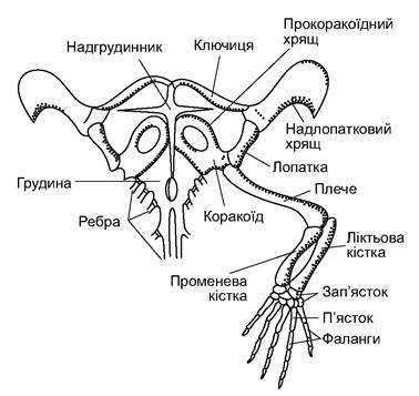
Fig. 5.19. Pectoral girdle and forelimb of the lizard Lacerta
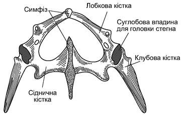
Fig. 5.20. Pelvic girdle of the lizard
The shape of the reptile skull depends mainly on feeding habits and prey capture Methods. The skull features elongated jaws that form a long snout. This jaw shape allows them to tear pieces of flesh from large prey.
At the same time, the mass of the masticatory muscles is increased. The skull consists almost entirely of bone (Fig. 5.21).
The occipital region is formed of four bones (occipital) of chondral origin: the supraoccipital, the basioccipital, and two exoccipitals.

Fig. 5.21. Skull of the lizard Lacerta, after W. Parker.
A - dorsal view; B - ventral view; C - lateral view:
1 - foramen magnum; 2 - exoccipital bone; 3 - supraoccipital bone; 4 - basioccipital bone; 5 - occipital condyle; 6 - basisphenoid bone; 7 - Vomer; 8 - choana; 9 - parasphenoid; 10 - Parietal bone; 11 - interparietal bone with a foramen for the parietal organ; 12 - Frontal bone; 13 - Nasal bone; 14 - premaxillary bone; 15 - maxillary bone; 16 - prefrontal bone; 17 - Lacrimal bone; 18 - supraorbital bones; 19 - postfrontal, or postorbital, bone; 20 - squamosal bone; 21 - supratemporal bone;
22 - Temporal bone; 23 - nostril; 24 - quadrate bone; 25 - Palatine bone; 26 - pterygoid bone; 27 - epipterygoid, or columellar bone; 28 - transverse bone; 29 - articular bone; 30 - dentary bone; 31 - surangular bone; 32 - coronoid bone
They border the foramen magnum, below which is a single occipital condyle formed by the basioccipital and two exoccipital bones. The membrane basisphenoid bone (basisphenoideum) is located anterior to the basioccipital and forms the floor of the skull. Anteriorly, a small parasphenoid (parasphenoideum) is fused to it, and paired vomers (vomer) are located, lateral to which are the choanae. In the region of the otic capsule, there are three otic bones (otici): the prootic (which is independent), the opisthotic (fused with the exoccipital), and the epiotic (fused with the supraoccipital). There are no bones in the olfactory region; it remains cartilaginous.
The skull roof is formed by paired membrane bones: nasal (nasalia), prefrontal (praefrontalia), frontal (frontalia), and postfrontal (postfrontalia); further back are the parietal (parietalia) and the unpaired interparietal (interparietale) bones; the interparietal bone has an opening for the parietal organ. The sides of the skull are formed by membrane bones: paired premaxillary (intermaxillare) (fused in some species), paired maxillary (maxillare), supraorbital (supraorbitale), jugal (jugale), quadratojugal (quadratojugale), and squamosal (squamosum) bones. From the posterior part of the palatoquadrate cartilage arise paired chondral ossifications—the quadrate bones (quadratum). The anterior part of the palatoquadrate cartilage is replaced by membrane bones that form the floor of the skull: paired palatine (palatini) and pterygoid (pterygoidei) bones. Transverse bones (transversi) connect the pterygoids with the maxillaries, and in lizards and tuataras, additionally, the epipterygoid, or columellar (epipterygoidei) bones connect the pterygoid bones with the parietals.
In turtles and crocodilians, the growth of the palatal processes of the premaxillary, maxillary, and palatine bones leads to the formation of a secondary bony palate, which divides the Oral Cavity into upper (nasopharyngeal) and lower (oral cavity proper) compartments.
The lower jaw was formed by the ossification of Meckel's cartilage, replaced by the articular bone (articulare) which articulates with the quadrate, and a series of dermal bones: the dentary (dentale), angular (angulare), surangular (supraangulare), coronoid (coronare), and sometimes a few other small bones.
The upper part of the hyoid arch (hyomandibula) has transformed into the Middle ear ossicle—the stapes. The hyoid apparatus consists of a cartilaginous plate (homologous to the copula) and three pairs of horns, homologous to the hyoids and remnants of the branchial arches.
The Evolution of the masticatory muscles was accompanied by the remodeling of the roof and, partially, the lateral walls of the originally solid stegal skull characteristic of reptile ancestors (Fig. 5.22).
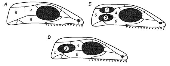
Fig. 5.22. Diagram of the evolutionary transformations of the stegal skull in reptiles (after E. Goodrich):
A - stegal skull; with modifications characteristic of anapsids; B - diapsid skull with two temporal fenestrae; C - synapsid skull with a single lateral temporal fenestra;
1 - upper temporal fenestra; 2 - lower temporal fenestra; 3 - single lateral temporal fenestra; 4 - postorbital fossa; 5 - squamosal bone; 6 - jugal bone; 7 - quadratojugal bone
The structure of the Bone tissue has also changed. While the general type of tubular limb bones was preserved, the coarse-fibered bone tissue characteristic of amphibians was replaced by fine-fibered bone, which is stronger. The limb bones acquired an osteonal structure and a zonar (layered) Organization. The bone element—the osteon—is a bony tube whose walls are formed by thin but strong bone lamellae, with the cavity of the bone tissue occupied by a Blood vessel and a nerve.
The Muscular System. The muscular system of reptiles is significantly modified compared to that of amphibians. The original metameric arrangement of muscles is preserved only in the muscles connecting adjacent vertebrae and in the musculature of the abdominal wall. On the skull, a powerful masticatory Muscle system is developed within the temporal fenestrae, which moves the jaws. The appearance of a mobile cervical region was accompanied by the formation of cervical muscles, which are particularly well-developed in species with elongated and flexible necks. Strap-like muscles participate in body bending and limb movement. In reptiles, the rudiments of subcutaneous muscles appeared, which alter THE POSITION OF the horny scales. This muscular system is well-developed in snakes and legless lizards, participating in locomotion by ensuring coordinated Movements of the ventral scutes.
The transition of reptiles to pulmonary respiration significantly improved lung ventilation. A true rib cage developed, driven by specialized intercostal muscles, alongside strengthened Abdominal muscles.
Digestive System. Modern reptiles are primarily carnivorous. Most feed on small terrestrial and aquatic animals, the populations of which in nature are large and relatively stable; they also consume small vertebrates such as fish, amphibians, mouse-like rodents, and others. Aquatic and semi-aquatic turtles and snakes hunt for aquatic invertebrates and fish. Marine snakes, such as Pelamis platurus, feed mainly on cephalopods. Crocodiles and large snakes attack sizeable prey. Terrestrial and aquatic turtles, as well as tropical agamas and iguanas, are herbivorous reptiles.
Reptiles capture their food using jaws armed with numerous sharp Teeth. These teeth fuse to the jaws and palatal bones (pleurodont and acrodont teeth); only in crocodiles and fossil mammal-like reptiles are the bases of the teeth embedded in special sockets known as alveoli (thecodont teeth). In modern reptiles, the teeth are homodont (uniform); specialized venomous fangs have evolved only in certain snakes.
Crocodiles and turtles are capable of tearing off separate chunks of flesh from large prey. Most species swallow their food whole. Turtles, which lack teeth, shear plant material with the sharp keratinous edges of their jaws. The structure of the snake jaw apparatus allows them to open their mouths extremely wide (Fig. 5.23) and swallow prey that exceeds the snake's own diameter. Consequently, the temporal arches have disappeared from the snake skull, and the jaw apparatus has transformed into a hinge-lever system.

Fig. 5.23. Jaw apparatus of a rattlesnake:
A - closed Mouth; B - open mouth; C - diagram of the main levers; D - feeding process in an egg-eating snake
Salivary Glands are located in the oral cavity of reptiles; their mucous secretion moistens food and facilitates swallowing. In venomous snakes and certain lizard species, some salivary glands have evolved into venom glands, the secretion of which flows through a duct or a surface groove on the fangs. Some glands have acquired The ability to produce digestive Enzymes, although their concentration in reptile saliva is negligible.
A muscular Tongue, capable of being projected far forward, is situated on the floor of the oral cavity. Lizards and snakes have a bifid (forked) tongue used as an Olfactory Organ and, in conjunction with Jacobson's organ, as a chemoreceptor. In chameleons, the tongue can be rapidly shot out; it is expanded at the tip and serves to capture small, moving prey such as insects (Fig. 5.24).

Fig. 5.24. Structure of the chameleon tongue, after J. Smith:
A - position of the tongue during food capture; B - mechanism of tongue projection
The Esophagus is well-defined. In snakes, it possesses strong muscles that push large prey down into The Stomach. The stomach, clearly demarcated from the esophagus, has muscular walls. The intestine is longer than that of amphibians. At the junction between the small and large intestines, a rudimentary cecum emerges; it is better developed in herbivores (such as the steppe tortoise). The intestine opens into the cloaca (Fig. 5.25). The Pancreas is located within the first loop of the intestine. The large Liver contains a Gallbladder, whose duct opens into the intestine adjacent to the pancreas.
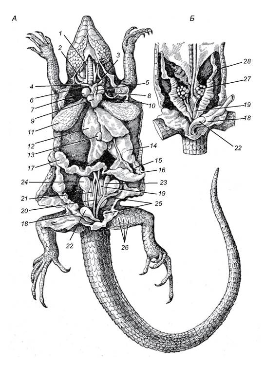
Fig. 5.25. Internal Organs of a lizard: A - male, B - female;
1, 2 - external (1) and internal (2) jugular Veins; 3, 4 - left (3) and right (4) carotid Arteries; 5, 6 - left (5) and right (6) aortic arches; 7, 8 - right (7) and left (8) atria; 9 - ventricle of The Heart; 10 - left Subclavian Artery; 11 - lung; 12 - liver; 13 - gallbladder; 14 - stomach;
15 - pancreas; 16 - duodenum; 17 - Large Intestine; 18 - rectum; 19 - Kidney; 20 - dorsal aorta; 21 - renal portal veins; 22 - Urinary Bladder; 23 - Epididymis; 24 - Testis; 25 - vas deferens; 26 - femoral pores; 27 - Ovary; 28 - oviduct
The Digestion of large prey by reptiles proceeds normally only at sufficiently high ambient temperatures; delayed digestion at low temperatures can lead to intoxication and death of the animal. Reptiles (turtles, snakes) are capable of prolonged fasting. Some snakes and turtles can live in captivity for 1–2 years without food. Lizards in an active state can remain without food for many weeks.
Respiratory system. The developing embryo in a reptile egg ontogenetically corresponds to the larval stage of amphibians; it breathes via the blood capillaries of the yolk sac and, later, the allantois. The skin of reptiles is covered with keratinized structures and does not participate in respiration. After hatching, the main respiratory organs are the paired Lungs; in snakes, the right lung is larger (whereas in amphibians, the left is larger). The lungs have a sac-like structure, but their internal organization is much more complex than that of amphibians (Fig. 5.26). Lung ventilation is driven by the action of the rib cage utilizing intercostal and abdominal muscles.
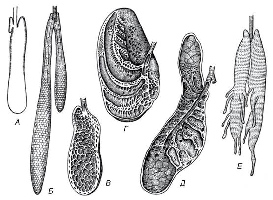
Fig. 5.26. Reptilian lungs (after C. Ritchel):
A - amphisbaenian (cross-section); B - anaconda (dorsal view); C - tuatara (cross-section); D - monitor lizard (cross-section);
E - alligator (cross-section); F - chameleon (ventral view); processes resemble air sacs
The complex lung structure in turtles, which are capable of absorbing oxygen even with weak pulmonary ventilation, is associated with the presence of their shell.
This new mode of respiration necessitated a restructuring of the respiratory tracts: a non-collapsible breathing tube—the Trachea—formed, supported by elastic cartilaginous rings. The entrance to the trachea (from the laryngeal chamber) is bordered by the cricoid and paired arytenoid cartilages; the chamber opens into the oral cavity via the glottis. At its posterior end, the trachea divides into two Bronchi that enter the lungs and branch into finer tubes within; the walls of the bronchi are also reinforced with rings. The breathing rhythm varies depending on the External Temperature and the animal's physiological state. For instance, in the lizard Sceloporus, the respiratory rate at 15°C is 26 breathing movements per minute, whereas at 25–35°C it rises to 37.
The Circulatory system. Reptiles possess a three-chambered heart. The atrium is divided by a complete septum, with each opening into the ventricle via an independent aperture equipped with a valve formed of semilunar folds. The ventricle features an incomplete septum that divides it into two compartments: during systole, this septum extends to the dorsal wall of the ventricle, briefly separating it completely and thereby segregating blood flows with different oxygen content. The sinus venosus is fused with the right atrium.
The conus arteriosus is reduced, and three distinct vessels emerge independently from various Regions of the ventricle: the pulmonary artery, divided into right and left branches, originates from the right region of the ventricle, which carries venous blood; the right aortic arch, which branches into the carotid and subclavian arteries, originates from the left region of the ventricle, carrying arterial blood; and the left aortic arch emerges from the middle of the ventricle (Fig. 5.27). The left and right aortic arches loop around The Heart and merge into the dorsal aorta. Venous blood flows through the pulmonary artery, whereas arterial blood flows through the right aortic arch and its branching carotid and subclavian arteries. The left aortic arch receives mixed blood. Consequently, the dorsal aorta carries mixed blood with a predominance of the arterial fraction.

Fig. 5.27. Diagram of the CIRCULATORY SYSTEM OF a lizard (after S. Ognyov, modified)
The Venous system of reptiles closely resembles that of amphibians. In the pelvic region, the caudal vein (Fig. 5.27) bifurcates into two iliac, or pelvic, veins, which receive blood from the hind limbs. The iliac veins branch into the renal portal veins and merge to form the abdominal vein. The abdominal vein, together with the HEPATIC PORTAL VEIN carrying blood from the intestine, breaks down into capillaries within the liver. The liver detoxifies protein breakdown products, initiates the synthesis of excretory waste, stores Glycogen reserves, and serves as a site for hematopoiesis. The capillaries of the hepatic portal system converge into the hepatic veins, which empty into the posterior vena cava. Blood from the head is drained by paired jugular veins that join the subclavian veins to form the right and left anterior vena cava, emptying into the right atrium. The left atrium receives the pulmonary vein, which is formed by the confluence of the right and left Pulmonary veins (carrying arterial blood).
The total blood volume and Hemoglobin content in the reptilian body are only slightly higher than those in amphibians.
The excretory system and water-salt balance. Among the adaptations that enabled reptiles to transition to a terrestrial lifestyle, the replacement of the mesonephric (trunk) kidney—typical of cyclostomes, Fishes, and amphibians—with the metanephric (pelvic) kidney, alongside a corresponding restructuring of the Water-Salt METABOLISM, is of paramount importance. Consequently, the composition of nitrogenous waste products excreted from the body has shifted. The end products include several substances: ammonia, uric acid, urea, creatine, creatinine, and others. The metanephric pelvic kidney differs not only in its anatomical position (situated in the pelvic region) but also in its microstructure. It features a simplified glomerular apparatus and a more complex structure of the renal (nephron) tubules. The glomeruli of the metanephros possess only 2–3 capillary loops, rendering them less capable of filtration. Accordingly, greater significance is placed on the renal tubules, where not only the reabsorption of water and sodium salts occurs, but also the active secretion of urea and uric acid by secretory cells. As a result, the concentration of these substances at the outlet of the tubule becomes twenty times higher than in the blood. The tubules are elongated and subdivided into a convoluted proximal segment, an intermediate segment, a convoluted distal segment, and a collecting duct. The first three segments facilitate secretory processes and the reabsorption of water and low-molecular-weight compounds vital to the Organism (such as sugars, Vitamins, and Amino Acids), whereas the final segment acts as a simple conduit for The excretion of the fluid. The final urine, enriched with metabolic waste, flows from the Kidneys via the Ureters into the cloaca and the urinary bladder, where The final stage of water reabsorption takes place, after which the highly concentrated urine is expelled from the body. The intensity of filtration and the reabsorption of substances within the tubules, cloaca, and urinary bladder depend on the permeability of their walls. Reabsorption is regulated by antidiuretic and several other Pituitary Hormones.
The embryonic pelvic kidneys develop posterior to the trunk kidneys from primordia that subsequently degenerate. In reptiles, the metanephric kidneys appear as two compact bodies located near the dorsal wall of the pelvic region. During their development, a pair of independent ureters buds off from the posterior section of the Wolffian ducts (mesonephric ducts). These ureters open into the DORSAL SIDE OF the cloaca. Species in which urea and other soluble substances predominate in excretion possess a well-developed urinary bladder that opens into the ventral side of the cloaca. Species whose urine is dominated by uric acid have a less developed urinary bladder (e.g., crocodiles, snakes, and certain lizards). Sea turtles and other reptiles forced to ingest saltwater are equipped with specialized salt glands that expel excess salts from the body. In turtles, these glands are located in the orbital cavity. The secretion of the salt glands in marine reptiles contains a seven-percent sodium chloride solution, causing them to literally "shed bitter tears" as they rid themselves of excess salt. Marine iguanas, which feed on salt-saturated Algae, possess salt glands in the form of so-called nasal glands, the ducts of which open into the Nasal cavity. Salts are excreted as droplets dripping from the nostrils.
The Reproductive System and reproduction. The Gonads are located within the body cavity on either side of the vertebral column. The Testes are paired, oval organs connected to the vas deferens. Both the right and left vas deferens open into their respective ureters. Males of all reptiles, except the tuatara, possess specialized copulatory organs: crocodiles and turtles feature a single median organ, whereas lizards and snakes possess paired outgrowths of the posterior cloacal wall that evert outward during copulation (Fig. 5.28).
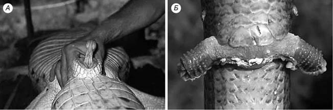
Fig. 5.28. Everted male copulatory organs: A - crocodile; B - lizard Pseudopus apodus
The paired Ovaries appear as granular, oval bodies. They give rise to the Müllerian ducts, which begin with ciliated funnels situated near the ovaries and terminate in apertures within the cloaca. Fertilization occurs in the upper section of the oviduct. Secretions from the glands in the middle section of the oviduct form an albumen layer around the egg Cell (yolk), which is poorly developed in snakes and lizards but well developed in turtles and crocodiles. The outer egg membranes are formed from secretions produced by the cells lining the walls of the lower section of the oviduct (the Uterus).
The majority of reptiles bury their laid eggs in the soil in well-warmed locations; some species deposit their eggs in mounds of decaying vegetation or beneath rotting stumps, utilizing the heat generated by decomposition. Certain crocodiles dig pits and cover their eggs with plant debris, with females remaining near the nest to guard the clutch. Some small lizards also guard their clutches (such as monitor lizards and others). Python females coil their bodies around the clutch of eggs, guarding and incubating them. In such a "nest," the temperature is 6–12°C higher than the ambient environment. Female crocodiles unearth the clutch after the hatchlings emerge, facilitating their journey to the surface; females of certain species also protect the young during the Early stages of their independent life.
A small number of extant squamate species exhibit ovoviviparity or, rarely, true viviparity (e.g., the common adder, the slowworm).
The Nervous system and Sense Organs. The reptilian brain differs markedly from that of amphibians; notably, the Forebrain is larger in reptiles, a feature associated with the Development of the cerebral roof (Pallium) and the enlargement of the corpora striata, which constitute the bulk of the forebrain mass (Fig. 5.29). The cerebral pallium clearly exhibits a primary cortex, or archipallium, which occupies a major portion of the hemispheric roof, alongside a rudiment of the neopallium. The olfactory center of reptiles is more complex and differentiated compared to that of amphibians. The Diencephalon is dorsally covered by the hemispheres of the forebrain. Its roof houses the Pineal Gland (an endocrine structure) and the parietal organ, which is capable of detecting light signals.

Fig. 5.29. Brain of a lizard, after W. Parker:
A - dorsal view; B - ventral view; C - lateral view; II-XII - Cranial Nerves
The optic tectum of the Midbrain is more highly developed than in amphibians. The reptilian Cerebellum is large, corresponding to the significant complexity and vigor of their locomotion. The Medulla Oblongata forms a vertical flexure characteristic of all amniotes. It coordinates vital autonomic Functions (such as respiration, Circulation, and digestion). Eleven pairs of cranial nerves originate from the brain.
The spinal cord clearly displays a distinction between White matter (nerve tracts) and Gray matter (neuronal cell bodies). This indicates an enhancement in the control exerted by higher brain centers over spinal reflex mechanisms.
The visual organ is adapted for functioning in an aerial medium. The eye is protected by external eyelids and a nictitating membrane. Snakes and certain lizards (such as geckos, skinks, and some legless lizards) possess fused eyelids that form a transparent spectacle. In nocturnal species, the eyes are enlarged and feature a vertical pupil. Lacrimal glands protect the eye from desiccation. Visual accommodation is achieved by displacing the crystalline lens and altering its curvature via striated ciliary muscles. Color Vision in most reptiles is shifted toward the yellow-orange region of the spectrum. Unlike in amphibians, the analysis and synthesis of visual images occur not in the retina, but primarily within the optic tectum of the midbrain.
Vision plays a crucial role in spatial orientation and social communication among reptiles. Pit vipers (Crotalidae), pythons (Pythonidae), and African vipers (Bitis) possess specialized thermal-sensing organs—thermoreceptors, and even thermographic locators. The thermolocators of pit vipers are paired pits located on the sides of the snout between the nostrils and eyes; in pythons, similar shallow pits are found on the labial scales.
The auditory organ of reptiles is structurally similar to that of frogs. It consists of a middle ear featuring a tympanic membrane and a single auditory ossicle—the stapes—which transmits vibrations from the membrane to the oval window separating the Inner ear cavity. Within the inner ear, a distinct cochlea (lagena) serves as the apparatus for analyzing and encoding acoustic signals. This cochlea is simple in structure and, in most species, forms a sac-like pouch. Reptiles perceive sounds within a frequency range of 20–6,000 Hz, though most have optimal Hearing between 60 and 200 Hz. Crocodiles are more sensitive to frequencies of 100–3,000 Hz. Snakes possess poor hearing; lacking a tympanic membrane, they primarily perceive vibrations transmitted through the substrate or water (so-called seismic hearing). Snake-like lizards perceive sound in a similar manner.
The majority of reptiles are voiceless. The sounds produced by snakes (such as hissing, wheezing, and tail-rattle buzzing) and certain lizards (such as scale-friction scraping) predominantly serve a warning function. Loud, bellowing vocalizations are characteristic of crocodiles (during territorial defense or mate seeking).
Chemoreceptors play a vital role in reptilian orientation and communication. The olfactory organs open externally via paired nostrils and into the oral cavity through slit-like choanae. Many lizards rely on Olfaction to locate food, digging it out from sand at depths of up to 6–8 cm. Monitor lizards, rat snakes, and vipers can use scent to distinguish between conspecifics and individuals of other species. Turtles, lizards, and crocodiles possess specialized scent glands, the secretions of which are used to mark their territory and deter intruders.
Last update: 13/08/2026
Editorial and Educational Adaptation: This material has been compiled based on the primary/original source text. The project team performed an editorial review, corrected technical inaccuracies, structured sections, and adapted the content for an educational format.
What was processed:
- elimination of formatting defects (OCR errors, structural breaks, corrupted characters);
- editorial organization of content;
- standardization of terminology in accordance with academic sources;
- verification of factual statements against the original source text.
All mentions of the author, publication year, and origin of the primary text have been preserved in accordance with the source.