MODERN BOTANY - P. RAVEN - 1990
SECTION V. STRUCTURE AND DEVELOPMENT OF THE ANGIOSPERM BODY
CHAPTER 19. EARLY STAGES OF PLANT DEVELOPMENT
In the previous section, we traced the long evolutionary journey of angiosperms from their hypothetical ancestor (a relatively complex, multicellular green alga) through a series of early vascular plants whose branched axes were the precursors to the leaves and roots of most modern forms.
This section focuses primarily on the result of this evolution: the flowering plant. This chapter begins where the Structure/133.html">Discussion of the angiosperm life cycle left off in Section 4—that is, with the seed, consisting of a seed coat, stored nutrients, and an embryo. We will trace The Development of the latter, because it is during this process, known as Embryogenesis, that the vegetative PARTS OF THE plant—ROOT, stem, and leaf—are established with their characteristic tissue Organization.
Mature Embryo and Seed
The mature angiosperm embryo consists of a stem-like axis bearing one or two cotyledons (Figs. 19-2 and 19-3), which are the first leaves of the young sporophyte. As the names of the angiosperm classes imply, dicotyledonous embryos have two, whereas monocotyledonous embryos have one.
At opposite ends of the embryonic axis are located the SHOOT and root apical Meristems. As mentioned earlier (see Ch. 1), such meristems are found at the tips of all shoots and roots. They consist of physiologically young, actively dividing Cells. In the embryo, the shoot apical meristem is located at the tip of the epicotyl, the stem-like axis above the cotyledons (epi- being Greek for "above"). In some cases, the epicotyl is represented almost exclusively by this meristem (Figs. 19-2, B and 19-3, B), whereas in others it bears one or more young leaves (Figs. 19-2, A and 19-3, A). Together with these leaves, it is called the plumule.
The stem-like axis below the cotyledons is called the hypocotyl (from the Greek hypo- meaning "under"). The lower end of the hypocotyl may transition into an embryonic root with well-defined root characteristics (Fig. 19-4). However, in many plants, this end consists almost entirely of an apical meristem covered by a root cap. If the root is difficult to distinguish in the embryo, the portion of the embryo below the cotyledons may be termed the hypocotyl-root axis.
Class="center">Fig. 19-2. Seeds and germination stages of some dicots. A. Bean (Phaseolus vulgaris); seed shown opened and from the edge view. B. Castor bean (Ricinus communis); seed opened to show the embryo in the cotyledonary plane and the plane perpendicular to it. C. Pea (Pisum sativum); external view of the seed. Germination of bean and castor bean seeds is epigeous, meaning that during germination the cotyledons are lifted above the soil surface by the elongating hypocotyl. In the seedlings of these plants, the elongating hypocotyl bends to form a hook and then straightens, pulling the cotyledons and plumule out of the soil. In contrast, germination of the pea seed (C) is hypogeous, meaning the cotyledons remain underground. In pea seedlings, the epicotyl forms a hook which then straightens to lift the plumule above the ground.


In the discussion of angiosperm seed development in Chapter 18, it was noted that in many dicots, most or all of the endosperm, along with the perisperm (when present), is absorbed by the developing embryo, which consequently forms fleshy cotyledons with stored nutrients that occupy the bulk of the seed. This is typical of most representatives of this class, such as sunflower, walnut, bean, and pea (Figs. 19-2, A, C). In dicots with abundant endosperm (e.g., castor bean), the cotyledons are thin and leaflike (Fig. 19-2, B); they function in absorbing nutrients from the endosperm.
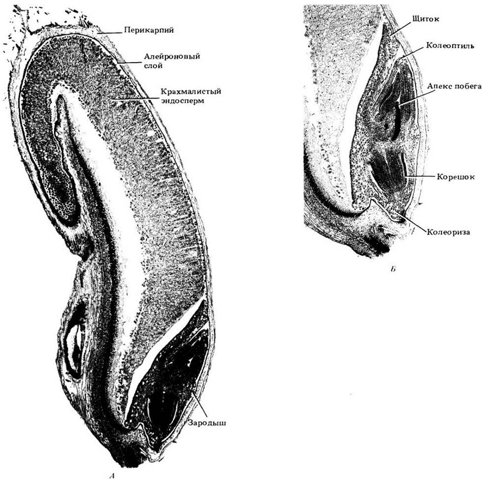
In monocots, the single cotyledon, although somewhat fleshy, typically performs an absorptive rather than a storage function (Fig. 19-3). Immersed in the endosperm, it absorbs nutrients previously broken down by Enzymes, which are then transported from the cotyledon to the growing Regions of the embryo. Among the most highly differentiated monocots is the embryo of grasses (Figs. 19-3, A and 19-4). In its fully formed state, it possesses a massive cotyledon (the scutellum) adjacent to the endosperm. The scutellum, like the cotyledons of most monocots, absorbs reserve Materials from the endosperm. It is attached to one side of the embryonic axis, at the lower end of which is the root and at the upper end the plumule, surrounded by sheath-like protective structures called the coleorhiza and coleoptile, respectively (Figs. 19-3, A and 19-4, B).
Fig. 19-3. Seeds and germination stages of some monocots: A — corn (Zea mays); B — onion (Allium cepa). Both seeds are shown in longitudinal section.

Fig. 19-4. A. Cytology/practical/54.html">Longitudinal section of a mature grain (caryopsis) of wheat (Triticum aestivum). Its seed coats are formed mainly by the pericarp. The seed coat, fused with it, disintegrates during grain development. B. Detail of a mature wheat embryo.
All seeds are covered by a seed coat (testa), which develops from the integuments of the ovule and provides protection for the embryo. It is usually much thinner than the original integuments. The thin, dry seed coat may have a papery texture, but in many species it is very hard and poorly permeable to Water. The micropyle (a channel in the integuments) often persists on the seed coat as a small opening. It is usually adjacent to the hilum, the scar remaining on the seed coat after the Separation of the seed from the funiculus or stalk (see the seed on the left in Figs. 19-2, A, C).
Embryo Formation
The Early stages of embryo development in dicots and monocots are very similar (Figs. 19-5 and 19-6). It begins with the division of the fertilized egg Cell (zygote) within the embryo sac of the ovule. In most flowering plants, the plane of the First Division is transverse (or nearly transverse) to its longitudinal axis (Figs. 19-5, A and 19-6, A). This establishes the polarity of the embryo: the upper (chalazal) pole is its main growth zone, while the lower (micropylar) pole forms a distinctive stalk-like structure called the suspensor, which anchors the embryo at the micropyle.
After several divisions, final differentiation occurs, separating the nearly spherical embryo proper from the suspensor (Figs. 19-5, B–G and 19-6, B–F). Up to this stage, the developing embryo is often referred to as a proembryo.
Suspensors were mentioned in Chapters 17 and 18 during the description of embryogenesis in clubmosses, spike mosses, and pines as structures that simply push the developing embryo into nutritive Tissues. Until recently, it was believed that they played only this limited role in angiosperms as well. However, it is now clear that angiosperm suspensors actively participate in the absorption of nutrients from the endosperm. Furthermore, in some cases, protein substances synthesized in the suspensor are utilized by the embryo proper during its rapid growth phase.
The newly formed embryo proper consists of a mass of relatively undifferentiated cells. However, changes in its internal structure soon lead to the initiation of plant tissue systems. The prospective epidermis (protoderm) is formed during periclinal Divisions of the outer Cells of the embryo proper (Figs. 19-5, D and 19-6, D) (periclinal divisions are those in which The Cell plates between two daughter cells are parallel to The surface of the plant part where the divisions occur). Subsequently, differences in the degree of vacuolation and cell density within the embryo lead to the segregation of the procambium and ground meristem. The latter, being more heavily vacuolated and less dense, gives rise to the ground tissue surrounding the less vacuolated and denser procambium, the precursor to the Vascular Tissues—xylem and phloem. The protoderm, ground meristem, and procambium (the so-called primary meristems) extend continuously from the cotyledons into the embryonic axis (Figs. 19-5, E, G and 19-6, D, E).
The formation of cotyledons may begin either during or after the initiation of the primary meristems (the developmental stage of the embryo prior to cotyledon emergence is often referred to as the globular stage). At this point, the globular embryo of dicotyledons gradually adopts a bilobed shape (this developmental phase is frequently called The Heart stage, Fig. 19-5, D). Because the monocotyledon embryo forms only a single cotyledon, it lacks a heart stage (Fig. 19-6, D).
Next, the cotyledons and the embryonic axis elongate (the so-called torpedo stage), and the primary meristems become distributed along them (Fig. 19-5, E and 19-6, E). As it extends, the embryo either remains straight or becomes curved. The single cotyledon of monocots often enlarges to such an extent that it becomes the largest embryonic structure (Fig. 19-3, B). With the continued growth of the embryo, the suspensor cells gradually degenerate.
Fig. 19-5. Stages of embryonic development in shepherd's purse (Capsella bursa-pastoris), a dicot. A. Two-celled stage resulting from the transverse division of the zygote. B. Three-celled proembryo. C. Six-celled proembryo. The suspensor is already distinct from the two terminal cells that develop into the embryo proper proper. D. Globular embryo proper with protoderm. At its base is the large basal cell of the suspensor. E. Embryo at the torpedo stage. In this species, it is curved. F. Mature embryo. The dark layer of cells lining the embryo sac (see A – E) is the endothelium, i.e., the inner layer of the integument.
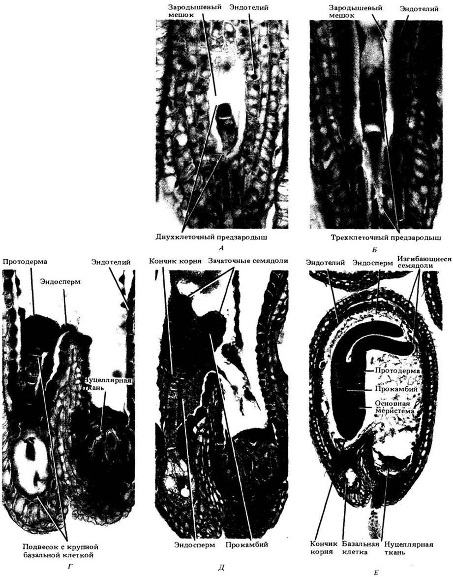
Fig. 19-6. Selected Developmental Stages of the arrowhead (Sagittaria) embryo, a monocot. A – D. Early stages. A. Two-celled stage—the result of the transverse division of the zygote. B. Three-celled proembryo. C. Excluding the large basal cell, the proembryo is now at the four-celled stage. All four cells participate in forming the embryo proper following a series of divisions. D. The protoderm is laid down terminally in the embryo proper. At this stage, the suspensor consists of only two cells, one of which is a large basal cell (continued on p. 14).
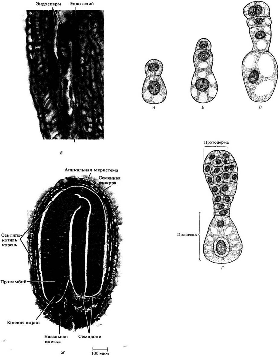
Fig. 19-6. D, E — Late stages of embryogenesis. D. A depression forms at the Base of the initiating cotyledon (the site of the future shoot apex). E. Curved cotyledon; the embryo approaches maturity.

In the early stages of embryogenesis, cell divisions occur throughout the entire mass of the young sporophyte. However, as embryonic development proceeds, the appearance of new cells gradually becomes restricted to the shoot and root apical meristems. In dicots, the shoot apical meristem is initiated between the two cotyledons (Fig. 19-5, G), whereas in monocots it forms on one side of the cotyledon and is entirely enclosed by a sheath-like outgrowth of its base (Fig. 19-6, E). Apical meristems are of paramount importance because they ultimately serve as the source of all new cells that drive the Development of the seedling and the adult plant from the embryo.
Throughout the entire period of embryogenesis, nutrients continuously flow from the parent plant to the Tissues of the ovule, resulting in the accumulation of substantial reserves in the endosperm, perisperm, or the cotyledons of the developing seed. Eventually, the ovule detaches from the funiculus—which connects it to the Ovary wall—becoming a nutritionally closed system. Ultimately, the seed dries out, releasing water into the environment, and the seed coat hardens, enveloping the embryo and nutrient reserves like a protective armor.
Seed Germination Requirements
During seed maturation and dispersal, embryonic growth typically ceases. Its resumption, or seed germination, depends on numerous external as well as internal factors. Among the external (environmental) factors, three are paramount: water, oxygen, and Temperature. In addition, the germination of small seeds—such as those of lettuce (Lactuca sativa) and many weeds—typically requires exposure to light (see p. 123).
Mature seeds are, in most cases, extremely dry (typically with a moisture content of 5–20%), making germination impossible until they absorb a specific amount of water required for metabolic activity. This process activates enzymes already present in the seed and synthesizes new ones involved in breaking down and utilizing the nutrient reserves accumulated during embryogenesis. Cells that previously synthesized massive amounts of storage compounds now completely reverse their metabolic pathways. Cell Division and elongation resume within the embryo, proceeding in accordance with the Specific characteristics of the given species. Further growth requires a continuous supply of water and nutrients. As the seed imbibes water, it swells, and considerable pressure can build up inside it (see Appendix to Chapter 4).
In the early stages of germination, Respiration may be entirely anaerobic, but as soon as the seed coat ruptures, it shifts to aerobic respiration and requires oxygen. If the soil is waterlogged, The amount of O2 available to the seed may prove insufficient for such respiration, rendering germination impossible.
Although many seeds germinate across a fairly wide temperature range, upper and lower limits specific to each species typically apply. The minimum for many plants is 0 – 5°C, the maximum is 45 – 48°C, and the optimum is 25 – 30°C.
Some seeds fail to germinate even under favorable environmental conditions. These are known as dormant seeds. The most common causes of such dormancy are physiological immaturity of the embryo and impermeability of the seed coat to water and, occasionally, to oxygen. Certain physiologically immature seeds must undergo a series of complex enzymatic and biochemical changes—collectively termed after-ripening—before they can germinate. In temperate zones, this process is stimulated by low winter temperatures. Thus, the requirement for after-ripening helps prevent germination during the cold season, when seedling survival is practically impossible.
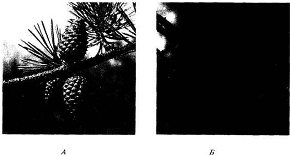
Dormancy is vital for plant survival. As demonstrated by after-ripening, it acts as a mechanism ensuring that germination occurs only under conditions favorable for subsequent development. Some seeds must pass through the digestive tract of birds or mammals, which facilitates wider dispersal of the species. In A number of desert plants, seeds germinate only after rainfall leaches out inhibitors contained in their seed coats; this adaptation ensures that seedlings develop during the rare periods when sufficient moisture is available for growth. The seed coat may also be mechanically scarified, for example, as seeds are tumbled by flowing water along a rocky streambed. Occasionally, dormancy is maintained within cones or fruits until high temperatures release the seeds. In certain pines, for instance, this occurs when heat melts the resin gluing the cone scales shut, ensuring the species' survival in fire-prone regions (Fig. 19-7). Finally, the germination of gap-phase species depends on the death of canopy trees or other disturbances that eliminate overhead shading. Thus, germination strategies are closely tied to the ecological characteristics of each plant's specific habitat (see also Chapter 25, p. 128).
Fig. 19-7. Resin-sealed cones of pitch pine (Pinus rigida) before (A) and after (B) fire exposure. They remain closed and attached to the tree long after seed maturation. The immediate cause of this is apparently a genetically controlled high melting point of the resin that seals the scales. Fire melts the resin, opening the cone and releasing seeds onto the fire-devastated areas. The same species, when bearing normally opening cones, favors habitats where vegetation is thinned primarily by factors other than fire.
1Gap phases refer to clearings in the forest canopy created by the fall of large trees, for example, due to windthrow. — Translator's Note.
From Embryo to Adult Plant
In most seeds, the radicle, or embryonic root, emerges first during germination (see Fig. 19-1); this allows the developing seedling to anchor itself in the soil and begin absorbing water. As it grows, the radicle develops into the primary root, which produces branch roots known as lateral roots, which in turn may produce lateral roots of higher orders. This gives rise to an extensively branching root system. In monocots, the primary root is generally short-lived, and The Root System of the adult plant develops from adventitious roots that originate at the nodes (the points of leaf attachment on the stem) and subsequently form lateral roots.
The shoot emerges from the seed in various ways depending on the species. In the common bean (Phaseolus vulgaris), for example, following root emergence, the hypocotyl elongates and arches into a loop (see Fig. 19-2, A). As a result, the delicate shoot tip is pulled rather than pushed through the soil, preventing damage. When this curved hypocotyl, commonly known as the hypocotyl loop or crook, reaches the soil surface, it straightens out, lifting the cotyledons and plumule into the air. This type of germination, in which the cotyledons are elevated above ground, is called epigeous.
During germination and subsequent development, the nutrients stored in the cotyledons are converted into a soluble form and transported to the growing parts of the seedling. The cotyledons gradually shrink, wither, and eventually drop off. By that time, the seedling has established roots, and its Nutrition is no longer dependent on the seed's food reserves. It becomes a photosynthesizing, autotrophic Organism.
The germination of castor bean seeds (Ricinus communis, see Fig. 19-2, B) is very similar to that described for the bean, except that the nutrient reserves are stored in the endosperm. As the hypocotyl loop straightens, the endosperm—and frequently the seed coat as well—is carried to the surface along with the cotyledons and plumule. During this stage, the broken-down nutrients of the endosperm are absorbed by the cotyledons and translocated to the growing regions of the seedling. Like those of the bean, the cotyledons of the castor bean turn green in the light, though they play a less significant role in Photosynthesis.
In peas (Pisum sativum), it is not the hypocotyl that elongates to form a loop, but the epicotyl. As soon as it straightens out, the plumule is lifted above the soil surface (see Fig. 19-2, B), while the cotyledons remain below ground, where they eventually break down. This type of germination, without the cotyledons being brought above ground, is called hypogeal germination.
In the vast majority of monocot seeds, nutrient reserves are stored in the endosperm. In relatively simple seeds, such as those of the onion (Allium cepa), a single tubular cotyledon emerges from the seed, forming a loop (see Fig. 19-3, B). As it straightens, it carries the seed coat, with the endosperm enclosed inside, up into the air. Throughout this period and for some time afterward, the embryo derives a significant portion of its nourishment from the endosperm via the cotyledon. Furthermore, the green onion cotyledon Functions as a photosynthetic leaf, substantially supplementing the nutritional resources of the developing seedling. Soon the plumule, initially protected by the sheath-like base of the cotyledon, elongates and emerges from it.
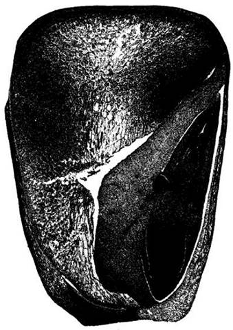
The final example is the development of a corn (Zea mays) seedling, a monocot with a highly differentiated embryo (Fig. 19-8). Both the radicle and the plumule are protected here by sheath-like structures—the coleorhiza and coleoptile, respectively. The coleorhiza is the first to pierce the pericarp (the wall of the mature ovary) during grain germination (in corn, the integuments break down during Seed and Fruit development, so the pericarp functions as the "seed coat"). The coleorhiza is followed by the radicle, or primary root, which elongates very rapidly and soon emerges from it. Following The Emergence of the primary root, the coleoptile is pushed out of the grain by the elongation of the mesocotyl (i.e., the first "internode," the stem section between the "nodes" of the scutellum and the coleoptile). When the base of the coleoptile reaches the soil surface, its tip parts, and the first leaves of the plumule begin to emerge from the earth. In addition to the primary root, two or more adventitious roots, initiated at the cotyledonary node, break through the pericarp and then bend downward.
Fig. 19-8. Longitudinal section of a mature corn grain (Zea mays). Comparing this photograph with the diagram in Fig. 19-3, A and the photomicrograph of the wheat grain and embryo in Fig. 19-4, one can distinguish the pericarp, endosperm, and various parts of the embryo. In grasses, the embryo typically includes two or more adventitious roots that initially point upward before bending and growing downward.
Regardless of how precisely the shoot emerges from the seed, The activity of its apical meristem results in the formation of an orderly sequence of leaves, nodes, and internodes. Apical meristems developing in the leaf axils (the upper angles between leaves and stems) give rise to axillary shoots, which in turn can form axillary shoots of subsequent orders.
The period from germination until the seedling becomes an independent organism is the most critical stage in a plant's ontogeny. During this time, it is most vulnerable to damage by insect pests and parasitic Fungi, and a lack of moisture can lead to its death very quickly.
The described type of root and shoot growth is called vegetative. Eventually, one or more shoot apical meristems transform into a reproductive apical meristem, which produces a flower or inflorescence. Once the flowers are formed, the plant is ready for sexual reproduction.
Summary
Flowers' seeds consist of an embryo, a seed coat, and stored nutrients. A fully formed embryo is composed primarily of a hypocotyl-root axis bearing one or two cotyledons and the shoot and root apical meristems. The cotyledons of most dicots are fleshy and contain the seed's nutrient reserves. In other cases, as in most monocots, these reserves are stored in the endosperm, and the cotyledons absorb pre-digested nutrients from it, which are then transported to the growing parts of the embryo.
During embryogenesis, the shoot and root are laid down as a single structure developing from the zygote. As a result of orderly divisions, the embryo differentiates into a suspensor and the embryo proper, in which the so-called primary meristems—the precursors of the epidermis, ground tissue, and vascular tissue—are formed. The development of cotyledons may begin either during or after the ESTABLISHMENT OF THE primary meristems.
As the embryo develops, the appearance of new cells gradually becomes restricted to the Zones of the apical meristems. Seed germination, i.e., the resumption of embryo growth, depends on environmental factors, including water, oxygen, and temperature. Many seeds are capable of germinating only after a period of dormancy.
Following the period of vegetative growth, one or more shoot apical meristems transform into reproductive apical meristems, which develop into flowers or inflorescences.
Appendix: Wheat
Like all grasses, common wheat (Triticum aestivum) is a monocotyledonous plant with a single-seeded fruit known as a caryopsis (grain). The protective layers of a wheat grain consist of the pericarp and remnants of the seed coat. Beneath these lie the endosperm and the embryo. More than 80% of the grain's volume is occupied by the endosperm. Its outer layer, called the aleurone layer, contains Reserve Proteins and Lipids and surrounds the starchy endosperm and embryo.
Wheat flour is milled from the endosperm. Bran, which consists of the seed coat remnants and the aleurone layer, is removed during milling. It accounts for about 14% of the total grain mass and slightly reduces its nutritional value, since it is composed mainly of Cellulose, cannot be digested in the human Stomach, and helps speed up the passage of food through the digestive tract, thereby reducing nutrient absorption. The embryo (about 3% of the grain mass) is also removed due to its high oil content, which shortens the shelf life of the flour. However, both bran and germ, which contain the bulk of the wheat's Vitamins, are now increasingly used in human diets and livestock feed.
Last update: 07/08/2026
Editorial and Educational Adaptation: This material has been compiled based on the primary/original source text. The project team performed an editorial review, corrected technical inaccuracies, structured sections, and adapted the content for an educational format.
What was processed:
- elimination of formatting defects (OCR errors, structural breaks, corrupted characters);
- editorial organization of content;
- standardization of terminology in accordance with academic sources;
- verification of factual statements against the original source text.
All mentions of the author, publication year, and origin of the primary text have been preserved in accordance with the source.