MODERN BOTANY - P. RAVEN - 1990
SECTION V. STRUCTURE AND DEVELOPMENT OF THE ANGIOSPERM BODY
CHAPTER 22. THE SHOOT: PRIMARY STRUCTURE AND DEVELOPMENT
The shoot, consisting of the stem and leaves, is laid down during embryo development, where it is represented by a plumule consisting of a stem axis (epicotyl), one or more leaf primordia, and an apical meristem (the plumule may be considered the plant's first bud). As the embryo resumes growth upon seed germination, new leaves develop from the apical meristem, while the stem elongates and differentiates into nodes and internodes. Gradually, bud primordia form in the leaf axils (Figs. 22-1 and 22-2), eventually undergoing successive stages of growth and differentiation more or less similar to those observed in the first bud. This developmental pattern is repeated many times during The formation of the plant's shoot system.
Class="center">Fig. 22-1. Part of a croton shoot (Croton). The leaves of this dicotyledonous plant are variegated and arranged in a spiral along the stem. Near the shoot apex, they are so congested that it is impossible to distinguish nodes and internodes as separate regions. Elongation of the stem between the leaves attached to it at the nodes results in the formation of internodes

Fig. 22-2. Cytology/practical/54.html">Longitudinal section of the shoot apex of the common houseplant Coleus blumei, a dicot. The leaves are arranged oppositely at the nodes, with each successive pair perpendicular to the previous one; thus, the leaves of the second node from the bottom are at a right angle to the plane of the section

The growing terminal (apical) bud of a shoot usually inhibits The Development of lateral buds. This is known as apical dominance (see Ch. 24). When the distance between the shoot apex and the lateral buds increases, this inhibitory effect diminishes, and the latter may begin their development. Pinching, i.e., the removal of shoot tips—a common horticultural practice used to promote bushiness in plants—is based on this phenomenon.
The two Main Functions of the stem are conduction and support. Substances synthesized in the leaves are transported via the stem phloem to sites of utilization, including growing leaves, the stems themselves, roots, developing flowers, seeds, and fruits. Many nutrient compounds accumulate in the parenchymal Cells of roots, seeds, and fruits, but stems are also important storage Organs, and some of them, such as the underground stems of potatoes, are specially adapted for this purpose. The primary photosynthetic Organs of the plant—the leaves—are supported by stems, which position them favorably with respect to light. In addition, most of the Water lost by the plant passes through the leaves (Ch. 27), reaching them from the roots via the stem through the xylem.
Origin and Growth of Primary Stem Tissues
The Structure of the shoot apical meristem is more complex than that of the ROOT. Here, it not only produces the cells of primary tissues but also participates in the formation of leaf primordia and axillary buds (Fig. 22-2), which subsequently develop into lateral branches. This meristem is not protected by a rootcap-like structure.
The vegetative shoot apex of most flowering plants is constructed According to the so-called tunica-corpus model (Fig. 22-3), consisting of two zones that generally differ in the planes of Cell Division. The tunica comprises an outer layer (or layers) of cells that divide anticlinally, primarily ensuring surface growth. The corpus is formed by a mass of cells lying beneath the tunica, where they divide in various planes, providing the bulk of the shoot volume. The corpus and each tunica layer have their own initials.
Fig. 22-3. Shoot apex of Coleus blumei showing tunica-corpus meristem Organization. The zone of central mother cells roughly corresponds to the corpus

In many angiosperms, the corpus corresponds to a region of distinctly vacuolated cells called the central mother cell zone (Fig. 22-3). It is surrounded by a peripheral meristem derived partly from the tunica and partly from the corpus, while the pith meristem lies deeper within. Cell division in the central mother cell zone is relatively infrequent, whereas the peripheral zone is mitotically very active. The protoderm always originates from the outermost layer of the tunica, whereas the procambium and part of the ground meristem (cortex and sometimes part of the pith) derive from the peripheral meristem. The remainder of the ground meristem (all or most of the pith) is formed by the pith meristem.
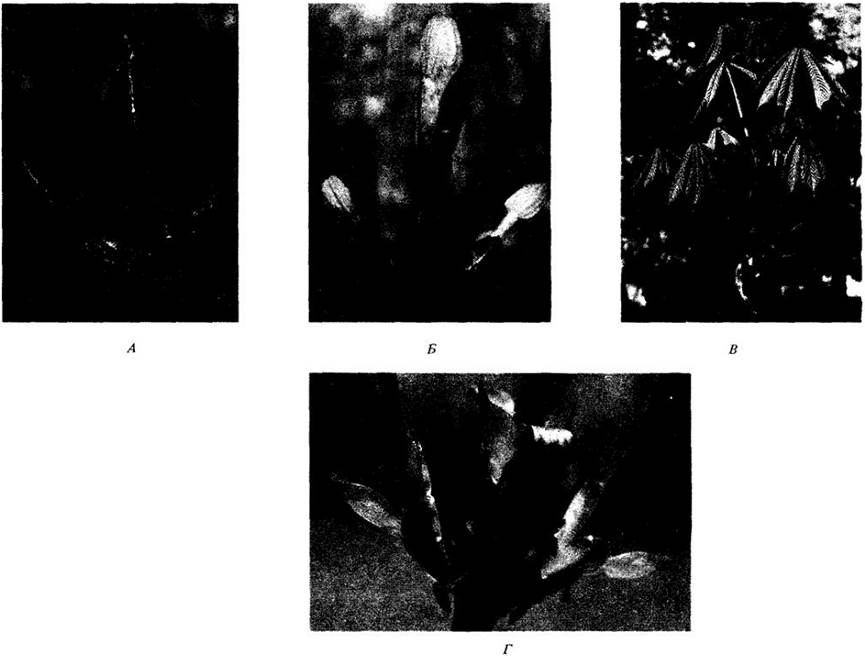
Although the growth periods of primary stem tissues are similar to those distinguished for the root, the stem cannot be divided along its axis into zones of cell division, elongation, and differentiation. During periods of active growth, the shoot apical meristem produces leaf primordia in such rapid succession that nodes and internodes are initially indistinguishable. Gradually, the regions between the leaf tiers begin to grow and elongate, taking on the appearance of internodes, while the areas of leaf attachment become defined as nodes (Fig. 22-4). Thus, longitudinal stem growth occurs primarily through internodal elongation.
Fig. 22-4. Developmental Stages of the terminal bud and two lateral buds of horse chestnut (Aesculus hippocastanum). A. Young shoots are tightly packed within the buds and protected by bud scales. B. The buds open, exposing the oldest rudimentary leaves. C. Internodal elongation has separated the nodes from one another. The terminal bud of the horse chestnut is mixed, meaning it produces both leaves and flowers (the latter are not visible in the figure). Lateral buds produce only leaves. D. Lower parts of young shoots; bud scales are separated and reflexed
Typically, the meristematic activity leading to their elongation is most intense at the base of these developing structures. If internodal elongation occurs over a relatively long period, such a zone at its base may be termed an intercalary meristem (i.e., located between more differentiated regions). Certain elements of the primary phloem and xylem, notably protophloem and protoxylem, differentiate here, connecting the more differentiated tissues located above and below.
Primary radial growth of the stem is accomplished through both longitudinal cell divisions and the elongation of cortex and pith cells. In plants with Secondary Growth, this is moderate. However, in many monocots, such as palms, substantial primary thickening occurs. This growth typically takes place near the apical meristem (often located in a depression of the apex) and is restricted to a relatively narrow subperipheral zone called the primary thickening meristem (Fig. 22-5).
Fig. 22-5. Diagram of the Anatomical Structure of the apex, or rosette, of a thick-stemmed monocot lacking secondary growth. Radial expansion of such stems occurs through The activity of the primary thickening meristem. The apical meristem and the youngest leaf primordia appear submerged within the surrounding stem tissues. Near the apex, rows of cells are arranged like a stack of saucers

As in the root, the shoot apical meristem gives rise to primary Meristems—the protoderm, ground meristem, and procambium (see Fig. 22-2). These, in turn, develop into the epidermis, ground tissue, and primary vascular tissue, respectively.
Introduction/19.html">Primary Structure of the Stem
This structure in seed plants is quite diverse, but three main types can be distinguished. 1. In some conifers and dicots, narrow, elongated procambial cells and the primary Vascular Tissues developing from them form a more or less continuous hollow cylinder embedded in the ground tissue (Fig. 22-6). Its outer zone is called the cortex, and the inner one, the pith. 2. In other conifers and dicots, primary vascular tissues form a cylinder composed of separate strands separated by ground tissue (Fig. 22-7). This tissue between the procambial strands (and later the mature vascular bundles) is continuous with the cortex and pith and is referred to as interfascicular parenchyma. Its interfascicular regions are frequently called medullary rays or pith rays. Such narrow regions connect the cortex to the pith in the first type of structure as well, though they are less pronounced there. 3. In the stems of most monocots and some herbaceous dicots, the arrangement of vascular bundles is more complex. Instead of forming a single ring between the cortex and pith, they typically form several rings or a system of strands scattered throughout the ground tissue, which in this case can often not be divided into cortex and pith (Fig. 22-8).
Stem of Linden
The stem of linden (Tilia americana) is structured according to the first type discussed above (Fig. 22-6). As in most cases, its epidermis consists of a single layer of cells covered by a cuticle. It contains significantly fewer Stomata here than the leaves do.
Fig. 22-6. A. Cross section of a linden (Tilia americana) stem at the primary growth stage. Vascular tissues form a continuous hollow cylinder that divides the ground tissue into an inner pith and an outer cortex. B. A fragment of the same stem at a higher magnification

The cortex includes parenchyma and collenchyma cells. Several layers of the latter, which provide support for the young stem, form a continuous cylinder beneath the epidermis. The rest of the cortex consists of parenchymal cells containing METABOLISM/14.html">Chloroplasts in their mature state. Due to its dark coloration, the innermost layer of the cortex sharply delimits it from the primary vascular tissues.
In the vast majority of stems, including that of linden, primary phloem develops from the outer Cells of the procambium, and primary xylem from the inner ones. However, not all procambial cells differentiate into these tissues. A single layer of them between the primary xylem and phloem remains meristematic, becoming the cambium. The linden stem is woody, meaning it produces a large amount of secondary xylem (secondary stem growth is discussed in Chapter 23). Once internodal elongation is complete, fibers known as primary phloem fibers form within the primary phloem here (see Fig. 23-10).
The inner boundary of the primary xylem in linden is sharply delineated by one or two layers of darker pith cells, which consist primarily of parenchyma and contain numerous large mucilage ducts or canals (specialized CARBOHYDRATES). Similar ducts form in the cortex. As cortical and pith cells increase in size, numerous intercellular spaces develop between them. These air-filled spaces are crucial for gas exchange with the atmosphere. The parenchymal cells of the pith and cortex serve for storage of various substances.
Stem of Elderberry
In the stem of elderberry (Sambucus canadensis), the procambium and primary vascular tissues are arranged in discrete strands around the pith. The latter, as well as the epidermis and cortex, are very similar in structure to those discussed for linden; therefore, only the Development of the primary vascular Tissues of the stem will be examined in detail below.
Fig. 22-9, A shows three procambial strands in which primary vascular tissues are just beginning to differentiate. The leftmost strand is slightly older than the other two and contains at least one mature sieve element and one mature tracheal element. As can be seen, the mature sieve element is located in the outer part of the strand (near the cortex), while the first mature tracheal element is in its inner part (at the border with the pith). Comparing Fig. 22-9, A and 22-9, C reveals the appearance of younger sieve elements closer to the center of the stem, whereas the xylem differentiates in the opposite direction.
The earliest elements of the primary xylem and phloem (protoxylem and protophloem, respectively) are stretched during internode elongation and are frequently destroyed. Like linden stems, elderberry stems undergo secondary growth resulting in woodiness. In the former case, almost all the cambium originates from procambial cells between the primary xylem and phloem, and the interfascicular zones are very narrow. In elderberry, these zones are relatively wide, As a result of which a substantial portion of the cambium develops from the interfascicular parenchyma.
Stems of Alfalfa and Buttercup
The stems of many dicots with little or no secondary growth are herbaceous, or non-woody (see Chapter 23). Examples include the stems of alfalfa (Medicago) and buttercup (Ranunculus).
Alfalfa is an herbaceous dicot with some secondary growth (see Fig. 22-7). The structure and development of primary stem tissues in this plant are largely similar to those known for elderberry and other woody members of its class. The vascular bundles are separated by wide interfascicular zones and surround a prominent pith. The cambium is partly procambial and partly interfascicular in origin, but secondary vascular tissues form mainly within the bundles. The interfascicular cambium deposits predominantly sclerenchymatous cells toward the xylem side. In the herbaceous stem of buttercup, the vascular bundles resemble those found in many monocots. Following the differentiation of primary vascular tissues, no procambium remains within them; consequently, cambium never forms, and the capacity for further growth is lost. Such bundles (Fig. 22-10) are termed "closed," in contrast to "open" bundles, in which a cambium is established. In most dicots, vascular bundles of the second type produce at least some secondary vascular tissues.
Fig. 22-7. A. Cross section of a stem of alfalfa (Medicago sativa), a dicotyledonous plant with separate vascular bundles. B. A fragment of the same stem at a higher magnification

Stem of Corn
In the herbaceous stem of corn (Zea mays) and many other monocots, closed vascular bundles form a system of strands scattered throughout the ground tissue (see Fig. 22-8).
Fig. 22-8. Stem of corn (Zea mays). A. Cross section of an internode, showing numerous vascular bundles scattered among the ground tissue. B. Cross section of a node of a young corn stem, showing horizontal procambial strands connected to vertical bundles. C. A mature stem split longitudinally; the ground tissue has been removed to reveal The Vascular System

Fig. 22-9. Cross sections of an elderberry (Sambucus canadensis) stem at the primary growth stage. A. A very young stem with protoderm, ground meristem, and separated procambial strands. The leftmost strand contains one mature sieve element (upper arrow) and one tracheal element (lower arrow). B. More developed primary tissues. C. The stem prior to the completion of primary growth. Fascicular and interfascicular cambia have not yet formed. Further stages of elderberry stem growth are shown in Figs. 23-8, 23-9, and 23-11

Fig. 22-10. Cross section of a vascular bundle of buttercup (Ranunculus), an herbaceous dicot. The vascular bundles of this plant are closed, meaning all procambial cells within them are differentiated and no secondary tissues develop. The primary phloem and xylem are surrounded by a bundle sheath composed of thick-walled sclerenchyma cells. Compare this bundle with the mature corn vascular bundle shown in Fig. 22-11, C

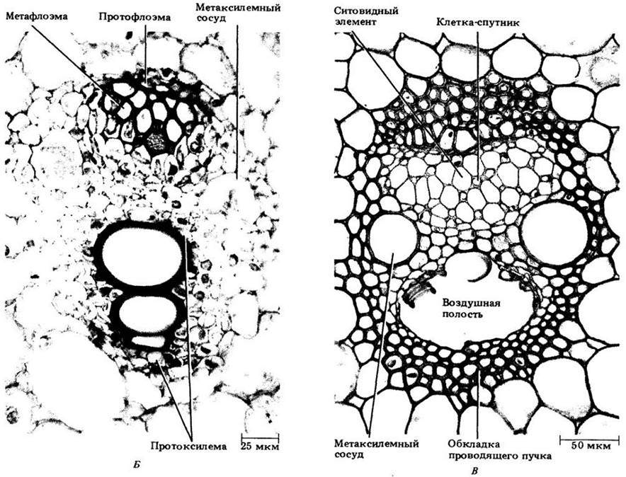
Fig. 22-11 shows three developmental stages of such a bundle. Just like in the stem of dicots, the phloem develops from the outer cells of the procambial strand, whereas the xylem develops from the inner cells, with the differentiation of phloem and xylem proceeding in opposite directions. Cross-sections reveal that the former differentiates centripetally, while the latter differentiates centrifugally. The earliest elements of the phloem and xylem (protophloem and protoxylem) are stretched and destroyed during internode elongation, resulting in the formation of a large air cavity on the xylem side of the bundle (Fig. 22-11, C). The mature vascular bundle, comprising two wide vessel members (metaxylem vessels) and phloem (metaphloem) consisting of sieve tube members and companion cells, is enclosed within a sheath of sclerenchymatous cells.
Fig. 22-11. Three stages of vascular bundle differentiation in corn stems, shown in cross-section. A. Mature elements of the protophloem and (two) protoxylems. B. Sieve elements of the protophloem are destroyed; a significant portion of the metaphloem is mature. The protoxylem contains three mature elements, and two metaxylem vessel members have almost completed elongation. C. The mature vascular bundle is surrounded by a sheath of thick-walled sclerenchymatous cells. The metaphloem consists entirely of sieve tube members and companion cells. The region of the vascular bundle previously occupied by protoxylem elements has now transformed into a large air cavity. The Cell wall thickenings of the destroyed protoxylem elements can be seen along its border.

Relationship Between the Vascular Tissues of the Stem and Leaf
The arrangement of vascular bundles in the stem indicates a close structural and developmental relationship between the stem and its lateral appendages, namely the leaves. The umbrella term "shoot" serves not only as a convenient designation for the complex of above-ground vegetative organs, but also reflects this interconnectedness.
The procambial strands of the stem originate immediately behind the apical meristem beneath developing leaf primordia, and sometimes even beneath the site of their future initiation prior to the onset of this development. As the primordia elongate, the strands differentiate toward the leaf tip. Thus, from the very beginning, the procambial System of the leaf is continuously connected with the procambium of the stem.
In each node, one or more vascular bundles extend from the central cylinder of the stem through the cortex into the leaf (or leaves) of that node (Figs. 22-12 and 22-13). Within the stem, the bundle extending from the longitudinal stem bundle to the Base of the leaf, where it connects with the leaf's vascular system, is termed a leaf trace. The wide gap—that is, the region of ground tissue located in the central cylinder directly above the point where the leaf trace departs—is called a leaf gap. A single leaf may receive one or more leaf traces from the stem. The number of internodes they traverse varies; i.e., the lengths of the leaf traces differ.
Fig. 22-12. Diagrammatic representations of the primary vascular system structure in the stem of Chenopodium glaucum, whose discrete bundles form a cylinder of interconnected strands around the pith. A. Three-dimensional diagram of bundle arrangement in the stem. B. The vascular system unfolded in a single plane. Leaf traces departing outward from the vascular cylinder and their connection with the stem vascular bundles are clearly visible. In this species, three leaf traces enter each leaf.
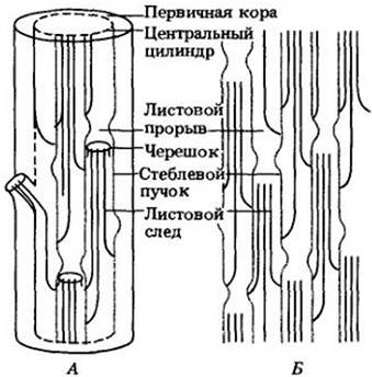
Fig. 22-13. Diagrams of longitudinal (A) and transverse (B) sections of a tobacco shoot (Nicotiana tabacum), illustrating the interrelationships between the leaf and stem vascular systems. The "continuous" central cylinder here consists of regions varying in width. In each node, a single leaf trace departs into the leaf, accompanied by branch traces that are often closely associated with the leaf traces. In tobacco, two branch traces extend from the stem vascular system toward the bud, and the branch gap is not separated from the leaf gap.

Tracing the course of a vascular bundle up and down the stem reveals that it connects with several leaf traces, forming what is known as a sympodium. In some stems, certain or all sympodia are interconnected, whereas in others, each represents an independent vascular complex. In either case, the STRUCTURE OF THE stem's vascular system reflects its leaf arrangement (phyllotaxy).
Leaf Morphology
Leaves exhibit great diversity in shape and internal structure. In dicots, they typically consist of a flat, expanded portion, the blade (lamina), and a stem-like petiole (Fig. 22-14). At the base of certain leaves, small scale-like or leaf-like structures called stipules are formed (Fig. 22-15). Many leaves lack petioles and are termed sessile (Fig. 22-16). In most monocots and some dicots, the leaf base is expanded into a sheath that clasps the stem (Fig. 22-16, B). In A number of grasses, the sheath covers the entire internode. Leaf arrangement on the stem may be spiral (alternate), opposite (paired), or whorled (three or more leaves per node). For instance, in white mulberry (Morus alba) and oak (Quercus), phyllotaxy is spiral; in maple (Acer), it is opposite; and in Veronicastrum virginicum, it is whorled (Fig. 22-14).
Fig. 22-14. Several examples of simple leaves. A. White mulberry (Morus alba). B. Veronicastrum virginicum. C. Sugar maple (Acer saccharum). D. Silver maple (Acer saccharinum). E. Red oak (Quercus rubra). Spiral or alternate phyllotaxy is visible in the mulberry, and whorled phyllotaxy in Veronicastrum. Phyllotaxy is opposite in the maple and spiral in the oak, though only individual leaves of these trees are shown here.

Dicot leaves are either simple or compound. In the former, the blade is not divided into segments, although it may be deeply lobed (Fig. 22-14); in the latter, it is divided into separate leaflets, each usually provided with its own small stalk (petiolule). Pinnately and palmately compound leaves can be distinguished (Fig. 22-17). In pinnate leaves, the leaflets are arranged in two rows on either side of the axis, or rachis, which represents an extended petiole. In palmate leaves, the leaflets diverge from the tip of the petiole, and the rachis is absent.
Because leaflets outwardly resemble simple leaves, these structures can sometimes be difficult to distinguish. Two criteria are useful: (1) a bud is located in the axil of both simple and compound leaves, but no bud is found in the axil of a leaflet; and (2) leaves diverge from the stem in different planes, whereas leaflets diverge from the rachis in a single plane.
Leaf Structure
Differences in the leaf structure of angiosperms are largely related to the plants' environmental conditions; a particularly crucial factor influencing their shape and anatomy is water availability. Based on their water requirements and corresponding adaptations, species are generally classified into mesophytes (requiring high soil moisture and a relatively humid atmosphere), hydrophytes (growing in waterlogged habitats, often fully or partially submerged), and xerophytes (adapted to arid conditions). These ecological categories lack sharp boundaries, and leaves frequently exhibit mixed characteristics. Regardless of their form, angiosperm leaves are specialized organs of Photosynthesis and, like roots and stems, consist of dermal, ground, and vascular tissue systems.
Epidermis
The bulk of the leaf epidermal cells, much like those of the stem, are densely arranged and covered by a cuticle that reduces water loss (see Chapter 3 and p. 25). Stomata may occur on both surfaces, but are typically more numerous on the lower surface (Fig. 22-18). In floating leaves of hydrophytes, they may be restricted to the upper epidermis (Fig. 22-19), while in submerged leaves they are generally entirely absent. Xerophytic leaves typically possess more stomata than those of other plants, which is thought to facilitate more intensive gas exchange during relatively brief periods of favorable water supply. In many xerophytes, stomata are sunken in depressions on the lower leaf surface (Fig. 22-20), which may be lined with numerous epidermal hairs. Both of these features apparently help minimize water loss. Epidermal hairs, or trichomes, may occur on either leaf surface or on both simultaneously, and their dense coverage also curtails moisture loss.
In dicot leaves, stomata are often scattered across the surface in a random pattern (Fig. 22-21). Their development is mixed; i.e., developing and fully differentiated stomata occur side by side on the same developing leaf. In monocots, stomata are arranged in rows parallel to the leaf axis (Fig. 22-22) and develop in a basipetal direction from the tip to the base.
Mesophyll
The mesophyll—the primary leaf tissue characterized by large intercellular spaces and numerous chloroplasts—is the tissue best adapted for photosynthesis. The intercellular spaces connect with atmospheric air via stomata, ensuring the rapid gas exchange necessary for this process to proceed efficiently. In mesophytes, the mesophyll is differentiated into palisade and spongy parenchyma. In the former, cells are columnar in shape with their long axes oriented at right angles to the epidermis. In spongy parenchyma, cell shapes are irregular (see Fig. 22-18, B, D). Although the palisade tissue appears denser than the spongy tissue, a greater portion of the vertical surface of its palisade cells borders intercellular spaces, and in some leaves the total surface area of these cells may be two to four times greater than that of the spongy parenchyma. Chloroplasts are also more abundant in palisade cells, which presumably concentrates the primary photosynthetic activity within this region.
Usually, the palisade parenchyma is located on the upper side of the leaf, and the spongy parenchyma on the lower side (see Fig. 22-18). In xerophytes, the former is frequently found on both sides of the leaf, whereas in some plants, such as corn (see Fig. 7-19) and other grasses (see Figs. 22-25 — 22-27), the mesophyll cells are more or less uniform in shape, and there is no clear distinction between the Two Types of parenchyma.
Fig. 22-15. Pinnately compound leaf of the pea (Pisum sativum). Note the stipules at its base (in this genus, they are often larger than the leaflets) and the slender tendrils at the apex

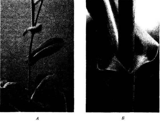
Fig. 22-16. Sessile leaves are common in dicotyledons, such as Moricandia of the mustard family (A), but are especially characteristic of grasses and other monocotyledons. B. In corn (a monocotyledonous plant), the leaf base forms a sheath around the stem, from which projects the ligule, a small outgrowth of leaf tissue

Fig. 22-17. Some examples of compound leaves. A. Palmately compound leaf of the red horse chestnut (Aesculus pavia). The remaining leaves are pinnately compound. B. Shagbark hickory (Carya ovata). C. Green ash (Fraxinus pennsylvanica var. subintegerrima). D. Black locust (Robinia pseudo-acacia). E. Honeylocust (Gleditsia triacanthos). In this species, each leaflet is further subdivided into smaller leaflets

Vascular Bundles
The leaf mesophyll is densely permeated by numerous vascular bundles, or Veins, which are directly connected with the vascular system of the stem. In most dicotyledons, they form a branched system in which progressively smaller veins diverge from larger ones. Such venation is termed reticulate (Fig. 22-23). Often, the largest (midrib) vein extends along the leaf axis and, together with the adjacent ground tissue, projects from the lower surface as a ridge (Fig. 22-18, A). In contrast, in most monocotyledons, numerous veins of roughly equal size run parallel to one another along the leaf. This type of venation is called parallel (Fig. 2-16, B). Additionally, these longitudinal veins are interconnected by smaller ones, forming an intricate network (Fig. 22-24).
The veins contain xylem and phloem, which are typically of primary origin (the midribs, and occasionally other major veins in some dicotyledons, are capable of secondary growth). The vein endings in dicotyledons frequently contain only tracheary elements, although both xylem and phloem elements may reach their tips. Generally, the xylem is located on the upper side of the leaf and the phloem on the lower side (Fig. 22-18, A, B).
Vascular bundles that are more or less completely embedded within the mesophyll are referred to as minor veins, whereas those accompanied by projections on the lower side of the leaf are called major veins. The former play the primary role in exporting assimilates from the mesophyll cells. As veins increase in size, their connection with the mesophyll diminishes, and they become increasingly surrounded by the non-photosynthetic tissues of the Ribs. Consequently, the function of assimilate collection is gradually superseded primarily by transport.
The vascular tissues of the veins rarely border directly on the intercellular spaces of the mesophyll. Major veins are surrounded by parenchyma containing few chloroplasts, whereas minor veins are enclosed by one or more layers of compactly arranged cells that form a bundle sheath (see Figs. 22-18 — 22-20) and often resemble the mesophyll cells in which these veins are embedded. The bundle sheath accompanies the bundle all the way to its termination, so that the vascular tissues nowhere come into contact with intercellular spaces, and all substances entering or leaving the vascular tissues must inevitably pass through its cells (see Fig. 22-18, D). Thus, the bundle sheath functions analogously to the root endodermis.
Fig. 22-18. Sections of a lilac leaf (Syringa vulgaris). A. Cross section through the midrib. B. Cross section through a portion of the blade, showing two minor veins. C. Paradermal section, i.e., approximately parallel to the leaf surface. From the top to the bottom of this photomicrograph, the section extends progressively deeper into the leaf tissues, so that the upper part shows a portion of the upper epidermis and the lower part shows a portion of the lower epidermis. It is clearly visible that stomata are more numerous on the latter. Lilac exhibits reticulate venation. D and E. Enlarged details of Fig. C. D. Palisade parenchyma (top) and spongy parenchyma (bottom) are visible. At the upper right of this same photomicrograph is a vein ending; the section has passed through several tracheary elements surrounded by the bundle sheath. E. A region of the lower epidermis with two trichomes (epidermal hairs), several stomata, and numerous ordinary epidermal cells


Fig. 22-19. Cross section of a floating leaf of the water lily (Nymphaea odorata) with stomata restricted to the upper epidermis. As is typical of hydrophytes, the vascular tissue is strongly reduced, especially the xylem. The palisade parenchyma consists of several cell layers overlying the spongy parenchyma

In many leaves, the bundle sheaths are connected to the upper and/or lower epidermis by cells resembling those of the sheath itself (Fig. 22-20). Such extensions are termed bundle sheath extensions. They provide mechanical support to the leaf and, in dicotyledons, apparently conduct water to the epidermis as well.
Fig. 22-20. Cross section of an oleander leaf (Nerium oleander). This species is a xerophyte, which is reflected in its leaf structure. A very thick cuticle can be seen covering the multilayered epidermis on both its upper and lower surfaces. Stomata and trichomes are confined to depressions in the lower epidermis known as stomatal crypts

The epidermis itself largely provides leaf rigidity owing to the tight arrangement of its cells and the presence of a cuticle. Major veins in dicotyledons are often surrounded by collenchymatous or sclerenchymatous cells, which also perform a supportive function. In monocotyledons, veins may be bordered by fibers. Collenchyma cells and fibers are sometimes also located along the margin of dicot and monocot leaves, respectively.
Grass Leaves
Following the discovery of C4 photosynthesis in sugarcane (Fig. 7-18), numerous studies have been devoted to the comparative anatomy of grass leaves and its relationship to the various types of this process. It has been established that the leaves of C3 and C4 grasses differ quite distinctly in their anatomy. For example, in C4 species, the mesophyll and bundle sheath cells typically form two concentric layers around the vascular bundles (see cross section, Fig. 22-25). The densely packed, very large parenchymatous bundle sheath cells of C4 grasses contain numerous voluminous, prominent chloroplasts. This concentric arrangement of mesophyll and bundle sheath cell layers is termed Kranz anatomy (derived from the German word Kranz, meaning "wreath").
Fig. 22-21. Scanning electron micrograph of a potato leaf (Solanum tuberosum), showing the irregular distribution of stomata characteristic of dicotyledons. The guard cells in potato are Kidney-shaped and lack associated subsidiary cells

Fig. 22-22. Scanning electron micrograph of a corn leaf. The parallel arrangement of stomata is typical of monocots. In corn, each pair of narrow guard cells is associated with two subsidiary cells, one on each side of the stoma

Fig. 22-23. Cleared leaf (chlorophyll removed) of the eastern cottonwood (Populus deltoides) at two magnifications. This species exhibits reticulate venation, which is characteristic of dicots. Small regions of mesophyll enclosed by veins are called areoles. All mesophyll landmarks are located near the veins. Water with dissolved minerals is delivered here via the xylem, while synthesized organic molecules are transported away via the phloem
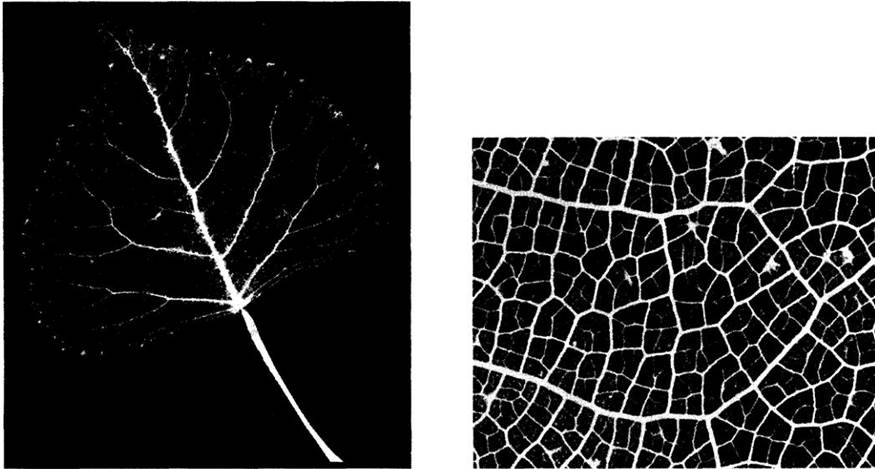
Fig. 22-24. Paradermal section of a corn leaf demonstrating parallel venation, characteristic of monocots. Numerous transverse veins connecting the longitudinal (parallel) vein system are clearly visible

Fig. 22-25. Cross section of a sugarcane leaf (Saccharum officinarum). As is typical for C4 grasses, the mesophyll cells are arranged radially around the bundle sheaths, which consist of large cells containing chloroplasts

In C3 plants, the mesophyll and bundle-sheath cells do not exhibit this concentric arrangement. Furthermore, the relatively small parenchymatous bundle-sheath cells contain smaller chloroplasts and appear pale and empty under low magnification. C3 species also typically possess an inner, so-called mestome sheath composed of more or less thick-walled cells (Fig. 22-26).
Fig. 22-26. Cross section of a wheat leaf (Triticum aestivum), a typical C3 grass. The mesophyll cells are not arranged radially around the vascular bundle sheath. The sheath itself consists of two layers of parenchyma cells: an outer layer of relatively thin-walled cells and an inner (mestome) layer of thick-walled cells

Another consistent structural difference between the leaves of C3 and C4 grasses lies in the interveinal distance, i.e., the distance between the sheaths of adjacent vascular bundles. In C4 grasses, this distance spans only two to four mesophyll cells, whereas in C3 grasses it always encompasses a greater number (averaging 12 in one study).
Photosynthetic products are generally exported from the leaves of C4 plants more rapidly and completely than in C3 species. The reasons for this remain unclear, but it has been suggested that the distance between the mesophyll cells and the phloem of the vascular bundles may influence the rate at which substances enter the sieve tubes.
The grass epidermis is composed of various cell types. Most of them are narrow and elongated. Some cells, notably larger and termed bulliform cells, are arranged in longitudinal rows and are thought to be involved in the folding (rolling) and unfolding (unrolling) of leaves (Fig. 22-27). Thick-walled stomatal guard cells are associated with subsidiary cells (see Fig. 22-22 and also Fig. 20-20).
Fig. 22-27. Cross section of a leaf of the C3 grass annual bluegrass (Poa annua). B. Enlarged portion of the section showing the midrib. In the grass leaf, the mesophyll is not differentiated into palisade and spongy parenchyma. Sclerenchyma cell strands are typically located above and below the veins. The epidermis contains large bulliform cells, which are believed to participate in leaf folding and unfolding. In the bluegrass specimen shown, these cells have partially lost turgor, causing the leaf to fold. Apparently, an increase in the turgor of these cells would cause the leaf to unfold

Leaf Development
The initial structural indication of leaf initiation in most angiosperms is the onset of periclinal divisions beneath the protoderm in the peripheral zone of the shoot apex. The combination of cell expansion and subsequent divisions soon results in the formation of a protrusion, or leaf buttress (Fig. 22-28, A), while new centers of mitotic activity arise at the apex at specific times and positions relative to previously initiated leaves. Before or during the Formation of the buttress, a procambial strand appears beneath the young leaf primordium.
Fig. 22-28. Early stages of leaf development in Coleus blumei shown in longitudinal sections of the shoot apex. In this species, leaves are arranged in opposite pairs at the nodes (see Fig. 22-2). A. Two small opposite protrusions—leaf buttresses—are visible at the margins of the apical meristem. Below, in the axils of the two young leaves, bud primordia can be seen. B. Two upright wedge-shaped leaf primordia that have developed from the leaf buttresses. The entering procambial strands are clearly visible. The bud primordia (below) are more advanced than those in Fig. A. As the leaf primordia grow (C), procambial strands originating from the stem vascular bundles continue to develop within them. Trichomes, or epidermal hairs, form from certain protodermal cells very early, long before the Differentiation of the protoderm into the epidermis is complete

As it grows in length, the leaf buttress develops into an upright, wedge-shaped leaf primordium (Fig. 22-28, B). In dicots, zones of meristematic activity soon appear on the primordium, roughly on opposite sides of its axis. These regions, which initiate the formation of the leaf blade, are called marginal meristems (Figs. 22-29 and 22-30).
Apical growth of the primordium is relatively brief. The leaf increases in length primarily through intercalary growth, i.e., cell divisions and (principally) cell expansion throughout the blade. As a result of marginal meristem activity, a specific number of mesophyll cell layers in the blade is established fairly early, although this number may increase during subsequent development. Differences in the rates of cell division and expansion among various layers of the blade lead to the formation of numerous intercellular spaces and the typical mesophyll structure. As a rule, the leaf tip ceases growth first, and the base ceases last. Compared to the stem, most leaves grow for a relatively short time. Unlimited or prolonged growth of the vegetative apical meristem is termed indeterminate, whereas restricted growth (characteristic of leaves and floral apices) is termed determinate.
As the wedge-shaped leaf primordium elongates, the procambial strand penetrates further into it (Fig. 22-28, C), forming a system of major veins branching off from the main, or mid-, vein (Fig. 22-29). Smaller veins are initiated near the leaf tip and develop downward toward the base, merging continuously with the larger veins. Thus, the leaf tip is the first to acquire a complete venation system, reflecting the overall "top-to-bottom" developmental sequence of this organ.
Fig. 22-29. Diagrams of longitudinal and cross sections illustrating early stages of tobacco leaf development (Nicotiana tabacum). A. Young wedge-shaped leaf primordium without a blade. B. Marginal meristem activity has begun on opposite sides of the primordium, leading to blade formation. The existence of such a meristem in tobacco leaves was recently questioned. Like the apical growth of the leaf primordium, its activity may be relatively short-lived. C. As the blade grows, major lateral veins (narrow pale regions) branching from the midrib become visible. D. The primordium has elongated. With further growth of the blade, parenchymal tissue outgrowths (ridges) appear along the major veins. Minor veins begin to develop near the leaf tip. The lowest diagrams on the right depict cross sections of a winged petiole. Compare these diagrams with the sections in Fig. 22-30

Fig. 22-30. Cross section below the apical meristem of developing tobacco leaves clustered around the shoot apex. The younger the leaves, the closer they are to the axis. The primordium is initially undifferentiated into midrib and lamina. Some early stages of their development can be observed (compare these sections with the diagrams in Fig. 22-29). Numerous trichome fragments are visible around the developing leaves.

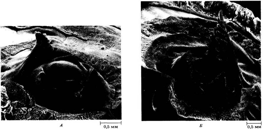
Figure 22-31 shows a three-dimensional view of a shoot apex and some early stages of leaf development in celery (Apium graveolens). The leaf primordia and young leaves are arranged in a spiral here, with the younger leaves located closer to the center of the apex. In Fig. 22-31, A, two leaf bulges can be seen on the right and left near its tip. Developing from the bulge, the leaf primordium expands in a shell-like fashion and, through rapid and relatively brief apical growth, forms the petiole-rachis portion of the future pinnately compound leaf. Below the apex on both sides of the rachis—first at its base and then progressively closer to the apex—outgrowths arise from which the leaflets subsequently form. Over time, secondary outgrowths appear on the initial outgrowths, becoming either the lobes of the primary leaflets or separate secondary leaflets. These can be seen in the relatively large leaf primordium in Fig. 22-31, B. Each leaflet develops similarly to a simple leaf.
Fig. 22-31. Scanning electron micrograph of early stages of leaf development on a celery (Apium graveolens) shoot apex. A. The closer the developing leaf is to the center of the apex, the younger it is. B. Two leaves (right and left) are more advanced in development than any of those shown in Fig. A.
Sun and Shade Leaves
Environmental factors, especially light, acting on developing leaves can have a substantial impact on their final size and thickness. In many species, leaves grown under high light intensity (sun leaves) are smaller and thicker than shade leaves formed under lower light levels. The increased thickness of sun leaves is primarily associated with the enhanced development of palisade parenchyma. Their vascular system is also more extensive, epidermal cell walls are thicker, and The ratio of internal mesophyll surface area to leaf blade area is much higher. The Effect of these differences is that while both leaf types exhibit similar photosynthetic rates under low light, shade leaves are not adapted to bright light and consequently photosynthesize much less efficiently than sun leaves under such conditions.
Because light levels vary considerably across different parts of a tree canopy, extreme forms of both leaf types can be found there. Sun and shade leaves also occur in shrubs and herbaceous plants. The formation of either type can be induced by growing plants under specific light intensities.
Leaf Abscission
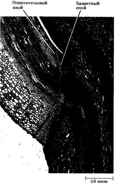
In many species, the normal detachment of a leaf from the stem—that is, the abscission process—is preceded by structural and chemical changes near the base of the petiole (see p. 102) leading to the formation of an abscission zone (Fig. 22-32). In woody dicots, two distinct layers can be distinguished here: the abscission layer and the protective layer. The former consists of relatively short cells with poorly developed cell wall thickenings, making it mechanically weak. The latter is formed by the deposition of suberin in The Cell walls and intercellular spaces beneath the abscission layer. Following leaf fall, the protective layer remains visible on The Stem as a leaf scar (see Fig. 23-18). Hormonal factors associated with abscission are discussed in Chapter 24.
Fig. 22-32. Abscission zone in a maple (Acer) leaf. Longitudinal section through the petiole base.
Transition Between Root and Shoot Vascular Systems
As noted in the previous chapter, differences between plant organs lie primarily in the relative arrangement of the vascular and ground tissues. For example, in dicot roots, vascular tissues typically form a solid cylinder surrounded by the cortex. Furthermore, strands of primary phloem alternate with radial rays of primary xylem. In the stem, however, the vascular tissues form a cylinder of discrete bundles arranged around the pith, with phloem on the outside and xylem on the inside. Clearly, somewhere within the primary plant body, one type of structure must transition into the other. This occurs gradually in a specialized region known as the transition zone.
As discussed in Chapter 19, the shoot and root are laid down during Embryogenesis as a single continuous structure. Consequently, the reorientation of vascular tissues occurs within the axis of the young plant itself. It begins with the initiation of procambium in the embryo and is completed alongside the differentiation of its variously arranged tissues in the seedling. Continuous connection between the shoot and root vascular systems is maintained throughout the plant's life.
The structure of the transition zone can be very complex and varies widely among species. In most gymnosperms and dicots, the vascular rearrangement is observed between the root and cotyledons. Figure 22-33 depicts a transition zone typical of dicots. One can observe the diarch structure of the root (with two protoxylem poles), the branching and reorientation of the primary xylem and phloem leading to the formation of a pith in the upper part of the axis, as well as the traces of the first epicotyl leaves.
Fig. 22-33. Transition zone connecting the root and cotyledons in dicot seedlings with a diarch root. The primary vascular system is represented within it by a single cylinder of vascular tissue. In the hypocotyl-root axis, this system branches toward the cotyledons, while the xylem and phloem within this axis are reoriented.

Flower Development
The meristematic activity of the vegetative shoot apex culminates in the development of a flower or inflorescence.
During the transition to flowering, successive physiological and structural changes occur in this apex, transforming it into a reproductive apex. Consequently, flowering can be viewed as a developmental stage of this shoot region and the plant as a whole. Because the reproductive apex exhibits determinate growth, flowering in annuals signals the approaching end of their life cycle. In perennials, it may occur repeatedly. Environmental factors, including day length and Temperature, are known to be involved in the induction of flowering (see Chapter 25).
The transition from vegetative to floral apex is frequently preceded by internodal elongation and the Early Development of lateral buds below it. The apex itself exhibits a marked increase in mitotic activity, accompanied by changes in its Size and Structure: initially relatively small and organized according to the tunica-corpus model, it becomes broad and dome-shaped.
The initiation and early Selection/3.html">Stages of development of sepals, petals, stamens, and carpels proceed in much the same way as in leaves. Typically, sepals are initiated first, followed by petals, stamens, and finally carpels (Fig. 22-34). This typical order of floral part appearance may be altered in some cases, but their spatial interrelationship remains consistently the same (Figs. 22-35 and 22-36).
Fig. 22-34. Scanning ELECTRON MICROGRAPHS OF developmental stages of a radially symmetrical, cyclic bisexual flower of Neptunia pubescens (Fabaceae). The tips of opposing bracts in figs. B–I have been removed. In figs. E–I, most sepals and petals have also been removed. A. Floral apex (a) in the axil of a bract (b). B. Five sepal primordia (se) surrounding the floral apex. C. Five petal primordia (p) surrounding the floral apex, alternating with the sepals (se). As they develop, the sepals form a calyx tube. D. Five stamen primordia (arrows) surrounding the floral apex, alternating with the petals (p). E. Surrounding the floral apex and alternating with the members of the first (outer) whorl of stamens (st1), a second (inner) whorl (st2) has been initiated (arrow). A carpel (c) has differentiated in the center of the floral apex. All floral parts are now present. F. A longitudinal furrow forms on the carpel, which will develop into the locule of the Ovary. The stamens of the outer whorl (st1) begin to differentiate into anthers and filaments. G. The carpel begins to differentiate into style and ovary (o). H. Flower at a later stage with two whorls of stamens. I. Flower at an even later stage; several stamens have been removed to reveal the carpel differentiated into ovary (o), style, and stigma (arrow).

Fig. 22-35. Longitudinal section of a young capitulum (flower HEAD) of fleabane (Erigeron) of the Asteraceae family. The inflorescence contains numerous floral primordia. The more developed ones are located closer to the periphery of the inflorescence, while the younger ones are in the center

Fig. 22-36. Stages of Flower Development in fleabane shown in longitudinal sections. A. Floral primordium with a developing corolla. B. Initiation of stamens fused to the corolla. C. Flower with developing corolla, stamens, and carpels (for the continuation of Fig. 22-36, see p. 66)

Fig. 22-36. D, E. Flowers with developing ovules and styles. Two carpels form a dome over the cavity enclosing the ovule, and subsequently elongate into a solid style with a bilobed stigma. The calyx, or pappus, is initiated at approximately the same time as the anthers, but develops slowly. F. Flowers almost fully developed. Pollen grains can be distinguished within the anthers. The fleabane flower has an inferior ovary

They may remain separate throughout development or unite within individual whorls (connation) and between whorls (adnation).
Typical flower structure and some of its variations were discussed in Chapter 18.
Leaf and Stem Modifications
Stems and leaves can become modified to perform non-typical functions. One of the most common modifications is the formation of tendrils used for support. In some cases, these represent modified stems. For example, in ivy (Hedera), tendrils form expanded cup-like adhesive discs at their tips. In grapes (Vitis) (Fig. 22-37) and Virginia creeper (Parthenocissus quinquefolia), they are also modified stems that twine around a support. Small leaves and flowers sometimes develop on grape tendrils.
Fig. 22-37. Grape tendrils are modified shoots

In most cases, however, tendrils are leaf modifications. In legumes, particularly the garden pea (Pisum sativum), they correspond to the terminal part of a pinnately compound leaf (see Fig. 22-15). Nevertheless, only a few plants in this family form them. In the peanut (Arachis hypogea), for instance, another interesting adaptation is known. After Fertilization, the stamens and corolla drop off, and the internode between the ovary and the receptacle begins to elongate, curves downward as it stretches, and drives the developing fruit several centimeters into the soil, where it matures. If the ovary remains above the soil surface, it withers and no ripe seeds are formed.
Branches that take on the shape of leaves are called cladophylls or cladodes. The thread-like, leaf-like branches of asparagus (Asparagus officinalis) are a typical example (Fig. 22-38). Its thick and fleshy above-ground shoots (“spears”) are edible. The scales on them are true leaves. As growth continues, cladodes develop in the axils of these tiny, inconspicuous scales, functioning as photosynthetic organs. In some cacti, the branches resemble leaves (Fig. 22-39).
Fig. 22-38. The thread-like branches of common edible asparagus (Asparagus officinalis) resemble leaves. Such modified stems are called cladodes

Fig. 22-39. The Branches of the spineless cactus Epiphyllum resemble leaves, but are actually modified stems, or cladodes

Sometimes leaves are modified into dry, hard spines incapable of photosynthesis. The terms “spine” and “thorn” are often used interchangeably, but in origin, the latter represent modified branches developing in leaf axils (Fig. 22-40). “Prickle” is also commonly considered a synonym for these terms. However, it is not a stem or leaf modification, but rather a small, more or less elongated sharp outgrowth of the bark and epidermis (an example being the prickles on a rose stem). Thorns, spines, and prickles are protective structures that prevent plants from being eaten by herbivorous animals. Highly specialized relationships between trees and herbivores are known in so-called “ant-acacias,” whose spines1 serve as shelter for ants that kill other insects attempting to feed on these plants (see p. 256).
1 The strict distinction between terms based on structural origin proposed here is not generally adopted in Soviet literature; therefore, the “spines” of acacias (modified stipules) or cacti are also referred to as thorns, and sometimes as prickles. In morphology, a distinction is specified as “spine of leaf origin,” etc. — Ed. note
Fig. 22-40. A. The spines of this cactus (Ferocactus melocactiformis) are modified leaves. B. Thorns are modified branches formed, as seen in this photograph of hawthorn (Crataegus sp.), in the leaf axils

The modified or specialized leaves of carnivorous plants, such as pitcher plants (Sarracenia), sundew (Drosera), and Venus flytrap, are highly striking and serve to capture and digest insects. The resulting nutrients are absorbed by the plant (see Chapter 26).
Nutrient Storage
Stems, much like roots, serve for nutrient storage. Perhaps the most widespread type of specialized storage stem is the tuber, as seen in the potato (Solarium tuberosum). In this plant, when grown from seed, tubers develop at the ends of stolons (slender stems growing parallel to the soil surface). However, if cut tubers are used for propagation, new structures of the same kind form at the ends of long, thin rhizomes, i.e., underground stems (Fig. 22-41). With the exception of vascular tissues, almost the entire mass of the tuber beneath the periderm (Skin) consists of storage parenchyma. The so-called “eyes” of the potato are depressions containing bud clusters, which represent the axils of scale-like leaves.
A bulb is essentially a large bud containing a small conical stem with numerous modified leaves attached to it. These leaves are scale-like, with thickened bases where nutrients are stored. Adventitious roots emerge from the base of the stem (the basal plate). Examples of plants with such structures include onions (Fig. 22-42, B) and lilies.
Fig. 22-41. Potato (Solanum tuberosum) with tubers attached to a rhizome, i.e., an underground stem

Fig. 22-42. Examples of modified leaves and stems. A. Fleshy storage stem of kohlrabi (Brassica oleracea var. caulorapa). B. Bulb of the common onion (Allium cepa) consisting of a conical stem and attached scale-like leaves containing nutrient reserves and forming the edible part of this modified shoot. C. Corm of a gladiolus (Gladiolus grandiflorus) representing a fleshy stem with small membranous leaves

Corms, although resembling bulbs in external appearance, consist primarily of stem tissues. Their leaves are typically thin and much smaller than those of bulbs, meaning that reserves are stored here in the fleshy stem portion. Corms are produced by such well-known plants as gladioli (Fig. 22-42, C), crocuses, and cyclamens.
Kohlrabi (Brassica oleracea var. caulorapa) is an example of an edible plant with a fleshy storage stem. Short and thick, it rises above the ground and bears several leaves with very broad bases (Fig. 22-42, A). Head cabbage (Brassica oleracea var. capitata) is closely related to kohlrabi. Its so-called "head" consists of a short stem with numerous thick, overlapping leaves. In addition to the terminal bud, several well-developed axillary buds can be found here.
In some plants, the leaf petioles become thick and fleshy. Well-known examples include celery (Apium graveolens) and rhubarb (Rheum rhaponticum).
Water Storage: Succulence
Succulents are plants with juicy tissues specialized for water storage. Most of them—such as American cacti, the superficially similar African spurges (Euphorbia) (see Appendix 3 to this chapter), and agaves (Agave)—typically grow in arid regions, where The ability to store water is essential for survival. The green, fleshy stems of cacti serve simultaneously as photosynthetic and storage organs. Water is accumulated in large, thin-walled parenchyma cells lacking chloroplasts.
In agaves, the succulent parts are the leaves. In these, as in succulent stems, the water-storing tissue consists of non-photosynthetic parenchyma cells. Other examples of plants with succulent leaves include ice plant (Mesembryanthemum crystallinum), stonecrop (Sedum), and certain Peperomia species. In the ice plant, the water-storage function is performed by large epidermal cells with appendages (trichomes) called bladder cells, which bear a striking resemblance to crystal beads (see Fig. 20-22, B). The water-storage cells of Peperomia leaves form part of a multi-layered epidermis (Fig. 22-43).
Fig. 22-43. Cross-section of a Peperomia leaf blade. The exceptionally thick, multi-layered epidermis on the upper side of the leaf presumably functions as water-storage tissue

The vegetative shoot apices of most flowering plants are organized according to the tunica-corpus model, where the tunica comprises one or more peripheral cell layers, while the corpus is represented by the mass of tissue beneath it. Although the primary tissues of the stem undergo the same developmental stages as those in the root, the stem cannot be similarly divided into zones of cell division, elongation, and differentiation (maturation). It grows in length primarily through internodal elongation.
As in the root, the shoot apical meristem gives rise to the protoderm, ground meristem, and procambium, which develop into primary tissues. There are Three types of distribution for the ground and primary vascular tissues here: (1) as a more or менее continuous hollow cylinder, (2) as a cylinder composed of discrete strands, and (3) as a system of strands scattered throughout the ground tissue. Regardless of the structural type, the phloem is typically located exterior to the xylem.
In most dicots, leaves consist of a blade and a petiole. Sometimes the blades are divided into leaflets. Stomata are generally more numerous on the lower surface of the leaf. The ground tissue, or mesophyll, of the leaf is specialized for photosynthesis and, in mesophytes, is differentiated into palisade and spongy parenchyma. It is thoroughly permeated by intercellular spaces and veins—that is, vascular bundles consisting of phloem and xylem surrounded by parenchymatous bundle sheaths. The xylem is usually located on the upper side of the vein, and the phloem on the lower.
In most monocots, including grasses, the leaf consists of a blade and a sheath that encloses the stem. The leaves of C3 and C4 grasses exhibit significant anatomical differences. The most prominent of these is the presence in C4 species and absence in C3 species of Kranz anatomy—a structural arrangement in which mesophyll and bundle sheath cells are organized in two concentric layers around the vascular bundles.
Leaves originate in the peripheral zone of the shoot apex, and their position on the stem is reflected in the arrangement of the vascular system within it. Their growth is determinate, meaning it is relatively short-lived, whereas growth at vegetative shoot apices can be indefinite, or indeterminate. In many species, leaves grown under high light intensity are smaller and thicker than those developed under relative shade. The former are termed sun leaves, and the latter shade leaves.
In many plants, leaf fall is preceded by the formation of an abscission zone at the base of the petiole.
The transition from the structural pattern characteristic of the root to that typical of the shoot occurs in a specific region of the embryo axis and young seedling known as the transition zone.
During flowering, the vegetative apex directly transforms into a reproductive one.
Shoots, like roots, can store nutrients. Examples of fleshy shoots specially adapted for this purpose include tubers, bulbs, and corms. Plants that store water are called succulents. Their water-storing tissue consists of large parenchyma cells. Succulence may occur in stems, leaves, or both.
Appendix 1. Plants, Air Pollution, and Acid Rain
A plant leaf, much like a human lung, can only function properly when capable of gas exchange with the surrounding air. Consequently, like the lung, it is extremely sensitive to atmospheric pollution.
This pollution takes various forms. Sometimes it is caused by particulate matter, which can be organic (such as in smoke from burning fossil fuels and refuse) or inorganic (including dust from cement plants and metallurgical works, as well as lead compounds released from burning leaded gasoline). As a major component of "smog," these particles reduce The amount of sunlight reaching the Earth's surface and exert a direct damaging effect on plants. They can clog stomata, hindering their functioning, or act as poisons (especially metal-based particles).
Fluorine compounds released into the air as waste from phosphate, steel, aluminum, and other industries act as cumulative poisons. They penetrate leaves through the stomata and destroy leaf tissues, apparently by inhibiting Enzymes involved in Cellulose synthesis. Thousands of hectares of citrus groves in Florida have been damaged by fluoride emissions from phosphate fertilizer plants.
Processing ore that contains sulfur produces sulfur dioxide:
2СuS + 3O2 —> 2СuO + 2SO2.
This compound has an unpleasant, sweetish-tart taste (an unusual property for an air pollutant is that its taste is perceived at lower atmospheric concentrations than its smell). Sulfur oxides are also formed during the combustion of fossil fuels containing this element. In humid air, they react with water to form droplets of sulfuric acid, which is highly corrosive and a component of acid rain. In some areas of the United States, the landscape has been literally turned into a desert under The Influence of atmospheric emissions of SO2 and metals. Efforts to combat air pollution around the copper smelters in Tennessee began as early as 1905, but even today, the lush forests that once covered the area are entirely absent. All vegetation has perished, and soil nutrients have been leached away by acid. Around the smelters of the Sacramento Valley in California, vegetation has been completely destroyed over an area of 260 km2, and its growth is severely stunted over an additional 320 km2.
The most widespread form of air pollution in California is photochemical smog, which forms when sunlight acts on automobile exhaust. The Los Angeles area is an "ideal" Location for its development, as life there is heavily dependent on automobiles, and the mountains to the north and east form a barrier that impedes air Circulation. Many plant species cannot survive not only within the city itself (which is also typical of many other large cities), but even 160 km away, where smog drifting from the basin damages crops and destroys pine forests in the mountains.
A. Damage to a blackberry (Rubus) leaf by sulfur dioxide; the affected areas are surrounded by healthy tissue. B. Ozone damage to a tobacco (Nicotiana tabacum) leaf, manifested as streaks or specks of dead tissue on its upper surface. Under severe ozone stress, these specks merge into larger patches visible on both surfaces. C. When sulfur and nitrogen oxides react with water in the atmosphere, they form sulfuric and nitric acids, which fall to the ground as acid rain.

One of the Main Components of photochemical smog is nitrogen dioxide (NO2), which is produced during any combustion processes in the air (dry air contains 77% nitrogen) and is therefore also present in automobile exhaust. Under the influence of light, NO2 dissociates into NO and atomic oxygen. The latter is extremely reactive and combines with molecular oxygen to form ozone.
Similar reactions occur under the influence of ultraviolet radiation in the upper atmosphere, forming the ozone layer described on p. 13, vol. 1. Ozone can also be generated by electrical discharges, producing the "fresh" smell after a thunderstorm. This substance is highly toxic. In plants, it damages thin-walled palisade cells, apparently altering the permeability of cell and chloroplast membranes. Another component of photochemical smog is PAN (peroxyacetyl nitrate — C2H3O5). It is several times more toxic than ozone, but is usually present in the air at much lower concentrations. The presence of 0.25 ppm of photochemical smog in the atmosphere reduces photosynthesis by 66%.
Currently, the environmental impact of acid rain is a major concern. "Normal" rain falling in unpolluted areas typically has a pH of 5.6. The combustion of fossil fuels and the processing of sulfide ores release large amounts of sulfur and nitrogen oxides into the atmosphere, where they react with water to form strong acids (sulfuric and nitric). Rain and snow under these conditions have a pH of less than
5.6, meaning they are acidic by definition. Such precipitation is now widespread, particularly in Western Europe, the eastern United States, and southeastern Canada, where the average annual pH is A — 4.5. Moreover, individual downpours can be significantly more acidic here. In Scotland, Norway, and Iceland, rains with pH levels of 2.4, 2.7, and 3.5, respectively, have been recorded. Attempts to reduce local pollution by increasing the height of industrial smokestacks have created regional problems. Pollutants emitted from tall stacks are carried long distances by the air currents. For example, it is estimated that more than 75% of the sulfur in rain falling in Scandinavia originates in the British Isles and Central Europe. The impact of acid rain on plants is not fully understood, but it is being studied extensively throughout the Northern Hemisphere. It has been shown to slow the growth of forest tree species in Sweden. Experimental acid rain damages leaves and inhibits seed germination. Over the past few years, the area of damaged forests in Germany has increased from a few percent to over 50%.
The impact of acid rain is particularly noticeable on fish populations, which have effectively perished in acidified lakes in various PARTS OF THE world. This is believed to be caused primarily not by a direct drop in water pH, but by an increase in aluminum concentrations. This metal, which makes up about 5% of the Earth's crust, is almost insoluble in neutral or alkaline environments and is consequently biologically inactive. However, as a result of acid rain, dissolved aluminum concentrations in some lakes can rise to levels toxic to fish and other aquatic organisms. The solubility of other toxic metals, notably lead, cadmium, and mercury, also increases sharply as the pH drops.
Appendix 2. Leaf Dimorphism in Aquatic Plants
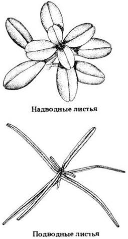
In their natural environment, aquatic flowering plants sometimes develop two different leaf forms: narrow and often deeply dissected leaves underwater (submersed leaves), and leaves of normal appearance above the surface (emersed leaves). Various treatments can stimulate developing leaves that have not yet fully formed to develop into a shape atypical for their normal living conditions.
Recent studies on the aquatic plant Callitriche heterophylla revealed that the plant hormone gibberellic acid induces its aerial shoots (those projecting above the water) to form submersed leaves. Another plant hormone, Abscisic acid (see Chapter 24), leads to the formation of emersed leaves on submersed shoots. Elevated temperatures or The addition of the sugar alcohol mannitol to the water produce the exact same result.
In nature, the cellular turgor pressure of submersed leaves is relatively high, whereas in emersed leaves it is relatively low. The latter can be partly explained by transpirational water loss through numerous stomata on the leaf surface. High turgor in developing submersed leaves is associated with the development of long epidermal cells upon maturity.
Gibberellic acid causes the cells of emersed leaves to elongate, increasing Water uptake and, consequently, turgor pressure. Once fully formed, such leaves possess all the characteristics of typical submersed leaves, including long epidermal cells. The limited cell elongation in submersed shoots treated with abscisic acid or high temperatures does not appear to result from reduced turgor; the treated cells become less extensible, so high turgor does not promote an increase in their size. Culturing submersed shoots in a mannitol solution results in turgor pressure similar to that observed in control emersed shoots, leading to the formation of leaves with short epidermal cells.
The results of these experiments demonstrate that the relative magnitude of cellular turgor pressure determines the ultimate size and shape of the leaf in Callitriche heterophylla. Thus, the mere presence or absence of surrounding water during its development leads to the appropriate adaptation to aerial or aquatic conditions.
Appendix 3. Convergent Evolution
Similar selection pressures acting on plants in comparable environments in different parts of the world often lead entirely unrelated species to acquire a similar appearance. The process responsible for this is called convergent evolution.
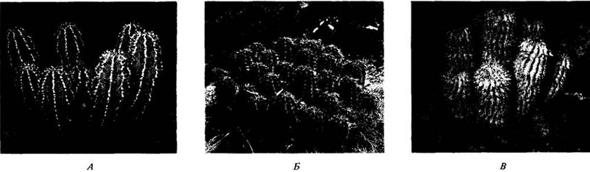
Let us examine some adaptive features of desert plants: succulent columnar stems (which store water), protective spines, and reduced leaves. Representatives possessing all these traits are found in three quite distinct flowering plant families: the Euphorbiaceae (spurges), Cactaceae (cacti), and Asclepiadaceae (milkweeds). The cactus-like spurges and milkweeds shown in the illustrations evolved from leafy plants that bore no resemblance to one another.
With a single exception, the natural range of cacti is restricted to the New World. Relatively succulent Representatives of the other two families are found primarily in the desert regions of Asia and especially Africa, where they occupy the same ecological niche as cacti do in the Americas.
Although the plants shown here (A — Euphorbia from the Euphorbiaceae; B — the cactus Echinocereus; C — Hoodia, a succulent from the Asclepiadaceae) exhibit CAM photosynthesis, they are all closely related to and descend from C3 plants. It can therefore be concluded that the physiological adaptations associated with CAM photosynthesis also arose as a result of convergent evolution (see Chapter 7).
Vascular bundles that are more or less completely embedded in the mesophyll are referred to as minor veins, whereas those accompanied by ribs on the lower side of the leaf are called major veins. The former play the primary role in collecting assimilates from the mesophyll cells. As the veins increase in size, their connection with the mesophyll weakens, and they become increasingly surrounded by non-photosynthetic bundle sheath tissues. Consequently, the function of assimilate collection is gradually superseded primarily by transport.
Fig. 22-16. Sessile leaves are common in dicotyledons, such as Moricandia of the Brassicaceae family (A), but are especially characteristic of grasses and other monocotyledons. B. In maize (a monocot), the leaf base forms a sheath around the stem, from which a ligule projects as a small outgrowth of leaf tissue
Last update: 07/08/2026
Editorial and Educational Adaptation: This material has been compiled based on the primary/original source text. The project team performed an editorial review, corrected technical inaccuracies, structured sections, and adapted the content for an educational format.
What was processed:
- elimination of formatting defects (OCR errors, structural breaks, corrupted characters);
- editorial organization of content;
- standardization of terminology in accordance with academic sources;
- verification of factual statements against the original source text.
All mentions of the author, publication year, and origin of the primary text have been preserved in accordance with the source.