BOTANY WITH BASICS OF HYDROBOTANY (AQUATIC PLANTS OF UKRAINE) - B.Ye. Yakubenko - 2011
VIII. BASICS OF PLANT MORPHOLOGY AND ANATOMY
Leaf Morphology and Anatomy
Anatomical Structure of the Leaf
The Internal Structure of a leaf is closely related to its function and habitat conditions. A typical dicotyledonous leaf has a dorsiventral structure (upper and lower sides are structurally distinct). Some plants possess an isobilateral leaf structure.
Externally, the flat leaf is covered by the epidermis—a primary dermal tissue that regulates Transpiration and gas exchange, protecting the leaf from desiccation and damage. The upper epidermis is single-layered, occasionally two- or three-layered (as in Ficus). Epidermal Cells are parenchymatous, tightly interlocking with wavy contours, or feature projections that ensure a tight connection between them. These cells contain a dense protoplast, typically lacking METABOLISM/14.html">Chloroplasts, though leucoplasts frequently occur. The Cell walls of the epidermis are unevenly thickened, with the outer and radial walls being the most thickened. The cells may contain anthocyanin pigments, which color the leaf blade in various shades. A significant number of Stomata are formed within the epidermis (their guard cells contain chloroplasts), ensuring gas exchange and transpiration. They are predominantly formed on the lower side of the leaf, whereas in aquatic plants, the reverse is true—they occur on the upper side.
The epidermal surface is frequently covered with various formations, such as prickles and hairs. Their shape, structure, and dimensions serve as a characteristic feature for specific systematic plant groups. Hairs play an important physiological role by protecting the leaf's dermal tissue. In all plants, the epidermis is covered by a cuticular layer on the outside.
The lower epidermis features a weakly developed cuticle, numerous stomata, and is covered with hairs. Typically, the Cells of the lower epidermis are smaller and possess more sinuous cell walls compared to those of the upper epidermis.
The epidermis performs a protective function, while the presence of stomata facilitates metabolic exchange with the environment. To give an idea of their density, 1 mm2 of wheat epidermal surface contains 50-70 stomata, apple features 250, potato 263, lemon 623, and Tradescantia 7 (on the upper leaf surface). In aquatic plants whose leaves float on the Water surface, stomata are located exclusively on the upper side, while the lower side, submerged in water, lacks them entirely. Submerged leaves possess no stomata.
The fundamental tissue of the leaf is called the mesophyll. It is situated between the upper and lower epidermis and consists of parenchymatous cells with thin, delicate walls containing a protoplast and green Plastids. These cells constitute the chlorophyll-bearing parenchyma or chlorenchyma. Within the mesophyll cells, palisade (columnar) and spongy parenchyma are distinguished.
Cells of the palisade parenchyma are elongated, cylindrical, tightly appressed to one another, and rich in chloroplasts. They are arranged in one or several regular, compact rows. Palisade tissue serves as the primary assimilation tissue, which explains its Location on the upper side of the leaf.
Cells of the spongy parenchyma are irregular in shape and loosely arranged. Spongy parenchyma predominates throughout the thickness of the mesophyll, yet it contains fewer chloroplasts compared to the palisade layer. A major portion of the spongy cells' surface borders intercellular spaces. The structure of typical spongy parenchyma is dictated by its primary function—transpiration. The number of layers varies from 2 to 7, depending on environmental conditions. The shape of spongy parenchyma cells can be rounded, sinuous, lobed, or stellate. Large intercellular spaces traverse this tissue in various directions. The uppermost cells of the spongy parenchyma form a specialized layer of collecting cells that are funnel-shaped and contact multiple palisade cells simultaneously. They accumulate Photosynthesis products synthesized within the columnar parenchyma cells.
The leaf mesophyll is permeated by a system of Veins. The vascular tissue of the leaf blade and petiole forms a continuous unit with the stem's Vascular System. All parts of this system originate from the procambium. During The formation of the leaf vascular system, identical cell types develop. The central vein consists of xylem oriented upwards and phloem facing downwards. This arrangement of xylem and phloem results from their emergence from the stem and a 90-degree rotation within the leaf blade. The xylem, which occupied a central position in the stem, ends up facing outward, whereas the phloem, positioned peripherally in the stem, shifts downward. The xylem consists of vessels or tracheids, xylem parenchyma, and radial rays. Adjacent to the xylem in dicots is the cambium, which ensures the thickening of veins. Below the cambium lies the phloem, composed of sieve tubes, companion cells, and phloem parenchyma. The vascular bundle is surrounded by a sclerenchymatous sheath. Lamellar or angular collenchyma develops both above and below the vascular bundle.
Lateral vascular bundles (veins) and their branches have a simplified structure, frequently lacking mechanical tissue and phloem. Only xylem vascular bundles—or merely their elements, such as vessels or tracheids—reach the margins of the leaf blade.
Thus, collateral-type vascular bundles are formed in the leaves, which are closed in the majority of plants.
A classic object for studying leaf Anatomical Structure is the leaf of Japanese camellia (*Camellia japonica* L.) (Fig. 77). This is a typical leaf characteristic of dicotyledonous representatives. The upper surface of the leaf is covered by the epidermis. Epidermal cells are large, flat, and nearly transparent. Stomata are clearly visible on the lower side of the leaf, subtended by an air cavity. The epidermis is covered by a cuticular film. Beneath the upper epidermis lies the assimilation tissue—the palisade parenchyma. This consists of two rows of elongated cells that fit tightly together without forming intercellular spaces. The cells are packed with chloroplasts concentrated in a stationary layer of Cytoplasm. Beneath the palisade cells lie funnel-shaped collecting cells, through which accumulated photosynthetic products are transferred into the leaf's vascular system. The lower portion is filled with spongy parenchyma, whose cells are irregularly shaped with large intercellular spaces. Large druses and crystals of calcium oxalate occur within the camellia leaf mesophyll.
Class="center">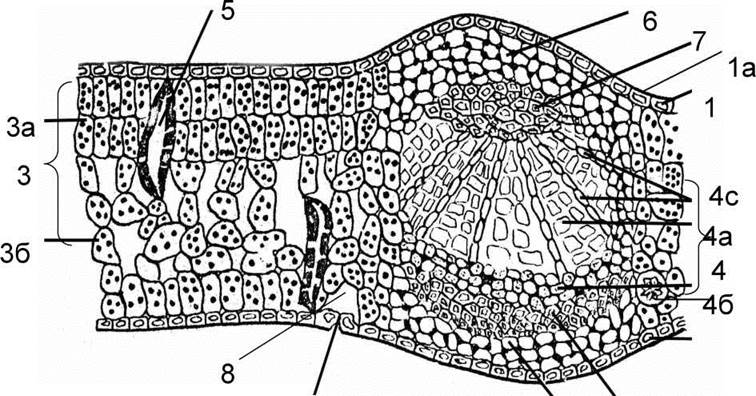
Fig. 77. Cytology/practical/72.html">Cross section of a Japanese camellia leaf:
1 - upper epidermis, 1a - cuticle; 2 - stoma; 3 - leaf mesophyll;
3a - palisade parenchyma, 3b - spongy parenchyma; 4 - vascular bundle:
4a - xylem, 4b - phloem; 4c - radial ray; 5 - supportive cells;
6 - collenchyma cells; 7 - sclerenchymatous sheath cells; 8 - air cavity.
The vascular bundle is protected by several layers of lamellar-angular collenchyma. It is formed by living parenchymatous cells featuring angular thickenings and thickened tangential walls. The collenchyma adjoins the central vascular bundle beneath both the upper and lower epidermis.
STRUCTURE OF THE white water-lily leaf (*Nymphaea alba* L.). This is an aquatic plant with large leaves floating on the water's surface. The leaf structure is dorsiventral, yet it exhibits specific adaptive structures.
The palisade parenchyma is fine-celled and multi-layered. Its cells are relatively tightly appressed, though narrow intercellular spaces run parallel to the length of the palisade cells. The latter contain a high concentration of chloroplasts and facilitate photosynthesis. The spongy parenchyma consists of large cells interspersed with large air cavities, containing only a sparse amount of chloroplasts. The upper epidermis is covered by a thick cuticular layer containing stomata. Beneath the stomatal guard cells lie air chambers extending across three to five layers of palisade cells and resting against the apex of one of them. The cell walls of the upper epidermis are sinuous, whereas those of the lower epidermis are somewhat thickened and conical, with the cuticle absent. Interspersed among the living epidermal cells are dead cells that support the adjacent living cells and, together with the air cavities, help keep the heavy, fleshy water-lily leaves afloat on the water surface. Vascular-fibrous bundles are weakly developed; phloem and xylem elements are small and few in number, and mechanical fibers are virtually absent. A bundle sheath develops around them. Within the leaf mesophyll, a system of branched solitary sclereids—supportive cells with thickened and cutinized walls—can be observed.
The Anatomical Structure of the submerged and floating leaves of the yellow water-lily (Nuphar luteum (L.) Smith). The leaves of this plant are distinguished by their unique Anatomical Features. Specifically, the submerged leaf lacks palisade parenchyma; its mesophyll is uniform, formed by spongy parenchyma with large air cavities, and stomata are absent. The floating leaf is dorsiventral, featuring a multi-layered, small-celled palisade parenchyma containing a significant number of chloroplasts. Large, thick-walled supporting cells are interspersed among the parenchymal cells. The spongy parenchyma also contains chloroplasts and large air cavities. Stomata are located in the upper epidermis. Beneath the substomatal aperture, narrow respiratory air chambers extend deep into the mesophyll, reaching down to the spongy parenchyma. The vascular bundles consist of small xylem and phloem elements and are poorly developed.
The anatomical structure of monocot leaves is exceptionally diverse, which is associated with adaptation to various living conditions. The leaves of many monocots receive uniform sunlight from both sides, resulting in an isolateral structure. The leaf blades of such plants are typically narrow, with parallel venation. The upper and lower sides of the leaf are similar and show little differentiation. The upper and lower epidermis are identical in structure and stomatal density. The leaf mesophyll is not differentiated into palisade and spongy parenchyma, or this differentiation is very weakly expressed. When palisade parenchyma is present, it is poorly developed and represented by a single row of elongated cells.
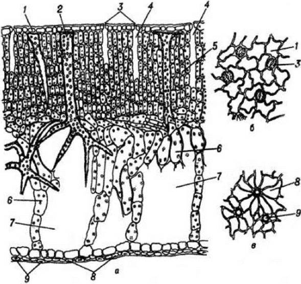
Fig. 78. Cross-section of a white water-lily leaf: a — part of the leaf blade, b - surface view of the upper epidermis, c - surface view of the lower epidermis, 1 - upper epidermis, 2 - supporting branched sclereids,
3 - stomata, 4 - air cavities, 7 - air cavities, 8 - lower epidermis, 9 - suberized cells of the lower epidermis.
Anatomical structure of the leaf of the yellow iris (Iris pseudacorus L.). The leaf surface is covered by a single-layered epidermis (Fig. 79). Stomata are found among the cells of the lower epidermis, each equipped with two large guard cells containing chloroplasts. The palisade parenchyma consists of a single row of cells located directly beneath the epidermis. In the central part of the leaf, there is an optimally developed system of intercellular spaces functioning as aeration channels. Closed collateral vascular bundles, comprising phloem and xylem surrounded by a sclerenchymatous sheath, are arranged in a single row at equal intervals from both the lower and upper sides of the leaf.
The leaves of aquatic plants exhibit several anatomical adaptations related to their habitat. The leaf blade is thin, transparent, and often dissected into narrow linear segments. Assimilatory tissue is poorly developed due to low light conditions. Palisade parenchyma is absent, while in the spongy parenchyma, trabeculae (cross-bridges) form between the upper and lower epidermis. Large air cavities develop between these trabeculae. The epidermis resembles a delicate film, lacking any thickening of the cell walls. The cuticle is extremely thin or entirely absent. Stomata are completely absent from the epidermis of fully submerged leaf blades; however, some aquatic plants possess specialized structures that differ from ordinary cells in size, shape, and greater wall permeability to water, known as hydropotes.
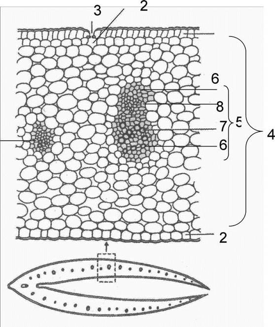
Fig. 79. Cross-section of an iris leaf: 1) upper epidermis; 2) lower epidermis; 3) stoma; 4) leaf mesophyll; 5) vascular bundle; 6) sclerenchyma; 7) xylem; 8) phloem
In the leaf blades of submerged plants, the xylem is poorly developed, vascular bundles are scarce, and vessels are few in number. Often, a narrow intercellular canal is present in the xylem instead of vessels. The phloem is better developed. The system of air intercellular spaces is highly developed, supplying the plant with oxygen and carbon dioxide.
In plants with floating leaves, the leaf blade is typically dense and leathery. A significant number of stoma develop on the upper surface, positioned directly above well-developed air cavities.
Last update: 07/08/2026
Editorial and Educational Adaptation: This material has been compiled based on the primary/original source text. The project team performed an editorial review, corrected technical inaccuracies, structured sections, and adapted the content for an educational format.
What was processed:
- elimination of formatting defects (OCR errors, structural breaks, corrupted characters);
- editorial organization of content;
- standardization of terminology in accordance with academic sources;
- verification of factual statements against the original source text.
All mentions of the author, publication year, and origin of the primary text have been preserved in accordance with the source.