BIOCHEMISTRY AND MOLECULAR BIOLOGY - W. ELLIOTT - 2002
CHAPTER 14. PHOTOSYNTHESIS - THE MECHANISM OF ENERGY TRANSFER TO WATER ELECTRONS
The generation of ATP in aerobic Cells relies on The transfer of electrons from high-energy donor molecules down an energy gradient to oxygen. Because the Earth's food supply is finite, sustaining life requires a mechanism to "lift" electrons back to the top of the energy scale. A minor exception can be made for recently discovered deep-sea organisms. They inhabit hydrothermal vent regions in the Earth's crust, fed by hot springs rich in hydrogen sulfide (H2S). Hydrogen sulfide is a powerful reducing agent, allowing its electrons to flow down an energy gradient and release energy that can be coupled to cellular ATP production, provided these organisms possess the appropriate biochemical machinery. However, such life forms can persist only as long as hydrogen sulfide and similar compounds continue to be emitted by crustal vents. For the vast majority of other organisms, an electron cycle is essential. This holds true for any early life forms in the primordial ocean as well. Regardless of the ready-to-use organic nutrients available there, exponentially multiplying organisms would have exhausted them long ago. Consequently, electron sinks would have been depleted even within the anaerobic atmosphere believed to have existed prior to the advent of Photosynthesis.
Photosynthesis - a biological process that drives the electron cycle. It offers several distinct advantages:
✵ an inexhaustible energy source – the sun;
✵ an inexhaustible electron donor – Water;
✵ the released oxygen serves as an inexhaustible electron sink, enabling the release of energy from high-energy electrons in food molecules.
Together, these points indicate that The Emergence of photosynthesis was the single most pivotal event in The history of life. The global energy cycle sustained by photosynthesis is illustrated in Fig. 14.1.
Class="center">Fig. 14.1. Schematic diagram of The electron transport cycle linking photosynthesis and Oxidative Phosphorylation in nature
CO2 assimilation is a secondary process relative to the elevation of water electrons to a high energy level

As you may know, photosynthesis converts CO2 and H2O into CARBOHYDRATES (typically Monosaccharides or starch). The overall equation describing this process can be written as:
6СО2 + 6Н2О —> С6Н12О6 + 6О2
∆G°′ = 2872 kJ • mol-1.
Synthesizing glucose from CO2 and H2O requires two conditions to be met. First, a reducing agent with a sufficiently low redox potential (high energy) must be available. In photosynthesis, this role is fulfilled by NADPH; in animal Gluconeogenesis, the reducing agent is NADH rather than NADPH (see Chapter 11). Second, ATP must be available as the driving force for synthesis. Light energy absorbed during photosynthesis is specifically expended to satisfy these two requirements: transferring electrons from water to NADP+ and synthesizing ATP.
The Site of Photosynthesis: The Chloroplast
Photosynthetic reactions take place within the METABOLISM/14.html">Chloroplasts of green plant cells. Much like Cell/35.html">Mitochondria, chloroplasts are intracellular Organelles whose outer
membrane is permeable to protons, whereas the inner membrane is not. Like mitochondria, chloroplasts contain their own DNA, which encodes a portion of their Proteins, as well as their own protein-synthesizing machinery with prokaryote-like properties (see p. 296). It is widely accepted that chloroplasts are distant descendants of photosynthetic prokaryotic unicellular organisms that once established a symbiotic relationship with Eukaryotic cells.
Unlike mitochondria, the internal membrane structures of chloroplasts – thylakoids – consist of closed, flattened membrane sacs stacked into grana and interconnected by membrane extensions (lamellae). The fluid-filled space inside the chloroplast (excluding the grana) is called the stroma (Fig. 14.2).
Fig. 14.2. Structure of a chloroplast. Grana are formed by stacks of thylakoids
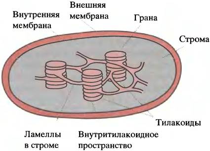
Chlorophyll (the light-harvesting pigment) and all electron carriers reside in the thylakoid membrane, whereas the synthesis of carbohydrates from CO2 and H2O occurs in the stroma. Synthetic processes in the chloroplast are referred to as dark reactions, implying not that they occur in the dark, but that they are not directly driven by light; rather, they are a consequence of NADPH and ATP generation in the thylakoid membrane. The division of labor between thylakoids and the stroma is illustrated in Fig. 14.3.
Fig. 14.3. The photosynthetic process (schematic diagram)

The Photosynthetic Apparatus and Its Organization in the Thylakoid Membrane
It is worth recalling how the Electron Transport Chain in mitochondria is organized, as it shares much in common with the photosynthetic apparatus. The inner mitochondrial membrane houses four types of Protein Complexes between which electrons move, mediated by mobile carriers: ubiquinone, a low-molecular-weight lipid-soluble substance, and cytochrome c, a small water-soluble protein.
The thylakoid membrane contains Three types of complexes (Fig. 14.4). The first two interact with a diffusing electron carrier—plastoquinone, which is structurally similar to ubiquinone—while the second and third interact with plastocyanin, a small water-soluble protein that also participates in electron transport. Plastocyanin contains a copper atom that alternately serves as an electron donor and acceptor, thus cycling between the Cu+ and Cu2+ states.
Fig. 14.4. The light-dependent reactions of photosynthesis (schematic). The primary function of the system is to transfer electrons from water to NADP+ and establish a proton gradient across the thylakoid membrane sufficient for chemiosmotic ATP synthesis. This gradient is generated through the splitting of water by Photosystem II and The Active Transport of protons by the cytochrome bf complex. Electrons are delivered from photosystem II to the cytochrome bf complex via plastoquinone (Q). All of this is analogous to the electron transport chain in mitochondria (where Q is ubiquinone). Plastocyanin (Pc), much like cytochrome c in mitochondria, acts as a protein electron carrier, while ferredoxin is located on the opposite side of the membrane.

These three complexes are designated respectively as photosystem II (PSII), the cytochrome bf complex, and Photosystem I (PSI). Do not let it confuse you that photosystem II precedes photosystem I in the chain—the numbering simply reflects the chronological order of their discovery. The overall function of this apparatus is to carry out the reaction:
2NADP+ + 2Н2O —> 2NADPH + 2Н+ + O2.
This reaction is accompanied by a very large increase in Free energy. Up to this point, we have considered only endoergic reactions driven by energy stored in chemical bonds. Photosynthesis, however, harnesses light energy: The formation of each NADPH molecule requires the energy of two absorbed photons. This energy is sufficient to reduce NADP+ molecules using water, with the surplus being utilized for ATP synthesis. This setup fully satisfies the two fundamental requirements for carbohydrate synthesis from CO2 and H2O formulated above: the presence of a reducing agent and ATP. We now turn to the mechanisms of light energy capture by Photosystems II and I.
What Is Chlorophyll?
In green plants, the light receptor is chlorophyll. Although Bacteria and Algae possess other light-harvesting pigments, our Discussion here will be limited to higher plants.

Chlorophyll is a tetrapyrrole structurally similar to heme (see Chapter 27). It differs from heme in the identity of its central atom (magnesium instead of iron) as well as The structure of its side chains. One of these side chains in chlorophyll contains a long hydrophobic hydrocarbon tail that acts as an anchor, holding chlorophyll within The Lipid Bilayer. Like heme, chlorophyll possesses a system of conjugated double bonds, which accounts for its intense coloration. Plants contain Two Types of chlorophyll (a and b), which differ in their side groups: in chlorophyll b, an aldehyde group replaces one of the methyl groups. Both chlorophylls absorb light in the blue and red Regions of the visible spectrum (although their absorption maxima differ slightly) and reflect green light, which imparts their characteristic color to the pigment and plant leaves.
Upon absorbing a photon, a chlorophyll molecule transitions to an excited state accompanied by the promotion of one of its electrons to a higher energy level (jumping to a new atomic orbital). However, the excited molecule is unstable and tends to return to its ground state. When a photon is absorbed by an isolated chlorophyll molecule, this energy is subsequently dissipated as heat and fluorescence, with no further consequence. In contrast, when chlorophyll molecules are tightly and orderly packed, as in the thylakoid membrane, excitation is transferred to neighboring molecules via so-called Resonance energy transfer. In green plants, chlorophyll molecules are organized into functional complexes termed photosystems, thereby restricting the spread of energy to the dimensions of these complexes (Fig. 14.5).
Fig. 14.5. Activation of a chlorophyll molecule in the reaction center. The reaction center is represented by the light circle. The chlorophyll molecule in this center is excited via resonance energy transfer from antenna chlorophyll molecules that capture light quanta (dark circles).

In each photosystem, alongside A large number of "ordinary" chlorophyll molecules, there exist so-called reaction centers presumably comprising
two chlorophyll molecules complexed with proteins. Within these centers, the excitation energy of chlorophyll is relatively low, so that resonance transfer back to free chlorophyll molecules does not occur. Thus, the reaction center acts as a sort of trap into which the energy of any photon absorbed by any "ordinary" chlorophyll molecule within that photosystem eventually falls. Once this happens, the high-energy electron is transferred to an acceptor possessing the appropriate redox potential. This acceptor initiates the photosynthetic electron transport chain.
Thus, photosystems are sophisticated complexes consisting of light-harvesting chlorophyll molecules, a reaction center, and an electron transport chain. The chlorophyll within the reaction center of photosystem II is designated as P680, and that of photosystem I as P700 (derived from "pigment"; the numbers correspond to the wavelength of maximum Light absorption in nm). The chlorophyll molecules that funnel captured energy into these centers are referred to as antenna pigments.
Mechanism of Light-Dependent NADP+ Reduction
Figure 14.6 illustrates the so-called Z-scheme of the Two Photosystems, demonstrating not only the ORGANIZATION OF THE electron transport chain but also the redox potentials of its individual components. Why are there two photosystems? The following analogy helps answer this question: powering a three-volt bulb requires two 1.5-volt batteries connected in series. In photosynthesis, the reduction of NADP+ serves as the "load," while photosystems II and I act as the "batteries."
Fig. 14.6. Photosynthesis: Electron transfer from water to NADP+ (Z-scheme). Analysis of this diagram should begin with the electron transfer from the excited P680 molecule to P700. P680 subsequently acquires a replacement electron from water and awaits another excitation event. The electron delivered to P700 is, upon molecular excitation, transferred to NADP+. Ph, pheophytin; Q, plastoquinone; Pc, plastocyanin; Fd, ferredoxin; Fp, ferredoxin-NADP reductase.

In reality, these "batteries" generate a greater redox potential difference than is strictly necessary for reducing NADP+, with the excess being utilized for ATP synthesis.
Let us begin with chlorophyll P680 in the reaction centers of photosystem II. In the dark, it resides primarily in the ground (unexcited) state, exhibiting no reducing properties. When P680 receives photon energy from antenna chlorophyll, it transitions to an excited state and tends to release the high-energy electron occupying its outer orbital. Consequently, this electron is captured by the electron carrier of photosystem II—pheophytin. Pheophytin is a pigment structurally similar to chlorophyll but lacking Mg2+.
Two reduced pheophytin molecules sequentially pass the acquired electrons to reduce plastoquinone, a lipid-soluble electron carrier that transports electrons from photosystem II to the cytochrome bf complex.

The latter consists of two Cytochromes and an iron-sulfur center, mediating electron transfer from reduced plastoquinone to plastocyanin.
What happens to photosystem I? Chlorophyll P700 in the reaction center of this photosystem also receives the energy of a photon harvested by antenna chlorophylls, converting it into a powerful reducing agent. An electron from the excited P700 chlorophyll is passed along a short chain of carriers (omitted in Fig. 14.6 for simplicity) to ferredoxin, a water-soluble stromal protein containing an electron-accepting iron atom cluster. Ferredoxin, with the help of the FAD-dependent enzyme ferredoxin-NADP+ reductase, reduces NADP+ to NADPH.
2Ferredoxinred + NADP+ + 2H+ —> 2Ferredoxinox + NADPH + H+.
Clearly, both photosystems must regain the electrons they lost in order to return to their initial state.
In other words, they shift from reducing agents to oxidizing agents. P700+ acquires an electron from reduced plastocyanin:
P700+ + PcCu+ —> P700 + PcCu2+.
As for photosystem II, P680+ returns to its ground state in an unusual way: it gets its electron from water!
Water Splitting by Photosystem II
P680+ is an extremely strong oxidizing agent; its electron affinity is even higher than that of oxygen, enabling it to extract an electron from water. To convert 2 molecules of water into 4 protons (H+) and O2, 4 electrons must be removed. Just as during O2 reduction in mitochondria, this process must avoid the formation of reactive intermediates like hydrogen peroxide, which are extremely hazardous to biological systems. In photosystem II, this task is performed by an Mn2+-containing protein complex (Fig. 14.8a), known as the water-splitting center, which extracts electrons from water by splitting it into oxygen and protons, and transfers them to P680+, preparing the latter for subsequent reactions.
How is ATP Generated?
The cytochrome bf complex, which uses plastohydroquinone to reduce plastocyanin, is analogous to mitochondrial complex III (see Fig. 8.19) in that its electron transfer is coupled to the Transmembrane Transport of protons from the outer side of the thylakoid membrane to the lumen. By analogy with the mitochondrial Q-cycle, one might expect four protons to enter the thylakoid lumen for every oxidized plastohydroquinone molecule (see p. 127). However, this stoichiometry is not supported by experimental data. Protons are also generated inside the thylakoids during water splitting. Both of these processes (proton translocation across the membrane and their generation within the thylakoid) drive the thylakoid lumen pH down to 4.5, establishing a proton gradient across the thylakoid membrane. Proton uptake during NADP+ reduction by ferredoxin in the stroma helps maintain this transmembrane gradient.
Thus, the photosynthetic apparatus incorporates a proton pump. This pump establishes an H+ gradient across the thylakoid membrane sufficient for the chemiosmotic synthesis of ATP from ADP and Pi. The operation of this proton pump is not rigidly coupled to NADP+ reduction. If all available NADP+ has been reduced to NADPH, ferredoxin passes its electrons to the cytochrome bf complex, which in turn transfers them to plastocyanin. This transfer is coupled to proton pumping. The electrons thus move in a cyclic pathway: photosystem I —> ferredoxin —> cytochrome bf complex —> plastocyanin —> photosystem I (Fig. 14.7). Consequently, this mode of ATP synthesis is termed cyclic Photophosphorylation, driven by a cyclic flow of electrons.
Fig. 14.7. Cyclic electron transport. When all NADP+ molecules are reduced, ferredoxin transfers electrons to the cytochrome bf complex. This enhances proton transport and thereby promotes ATP synthesis

The complete scheme of electron and proton pathways during photosynthesis in chloroplasts is shown in Fig. 14.8.
Fig. 14.8. Electron and proton pathways occurring within the thylakoid (schematic). a — Overall reaction at the Mn2+-containing center; b — movement of protons and electrons

Why does the proton pump in mitochondria pump protons out of the matrix into the surrounding medium, whereas the thylakoid pump pumps them inward? According to one hypothesis, both Mitochondria and chloroplasts are descendants of prokaryotic organisms that took up residence inside eukaryotic cells. Therefore, the direction of active proton transport and the orientation of the proton ATP synthase in the membranes of chloroplasts and mitochondria should be identical. However, the thylakoid membrane is formed by the pinching off of invaginations of the inner chloroplast membrane; consequently, its polarity is reversed, which explains why the proton gradients and ATP synthases of mitochondria and thylakoids have opposite orientations.
How is CO2 Converted into Carbohydrates?
Among all the biochemical processes discussed so far, photosynthesis is unique in that NADP+ reduction and ATP synthesis are driven by light energy.
All subsequent Chemical Reactions leading to the formation of glucose and other carbohydrates are exclusive to plants and do not differ fundamentally from standard enzymatic reactions.
The key metabolite here is 3-phosphoglycerate, from which carbohydrates are subsequently synthesized in the same manner as in the Liver (see Fig. 11.5), with the sole exception that NADPH, rather than NADH, serves as the reducing agent in these pathways (Fig. 14.9). Thus, the problem reduces to elucidating The Mechanism of 3-phosphoglycerate formation during photosynthesis.
Fig. 14.9. Formation of starch from 3-phosphoglycerate during photosynthesis. This process resembles gluconeogenesis, differing only in that NADPH, rather than NADH, serves as the reducing agent. Unlike Glycogen synthesis, the activated form of glucose used in starch production is ADP-glucose rather than UDP-glucose

How is 3-phosphoglycerate formed?
Of all Enzymes, the most abundant on Earth is ribulose-1,5-bisphosphate carboxylase, commonly referred to simply as "Rubisco." It is this very enzyme that utilizes CO2 to synthesize 3-phosphoglycerate.

Recall that ribose is an aldopentose, whereas ribulose (its optical isomer) is a ketopentose. The carboxylase cleaves ribulose-1,5-bisphosphate into 2 molecules of 3-phosphoglycerate while fixing 1 molecule of CO2. Subsequently, 3-phosphoglycerate is converted into various monosaccharides.
What is ribulose-1,5-bisphosphate synthesized from?
The answer is, in principle, very simple. From 6 molecules of ribulose bisphosphate (totaling 30 C-atoms) and 6 molecules of CO2 (6 C-atoms), 12 molecules of 3-phosphoglycerate (36 C-atoms) are formed. Two of these (2C3) are directed toward the synthesis of carbohydrates (C6), which proceeds According to the scheme shown in Fig. 14.9.
The remaining 10 molecules of phosphoglycerate (totaling 30 C-atoms) are converted into 6 molecules of ribulose bisphosphate (totaling 30 C-atoms). This process is quite complex and boils down to various interconversions of C3, C4, C5, C6, and C7 sugars mediated by aldolase and transketolase (see p. 181; recall that transketolase transfers C2 units). In a simplified form, this can be described by the following scheme:

Net result: 5 C3 —> 3 C5.
Thus, 6 molecules of ribulose bisphosphate, 6 molecules of CO2, and 6 molecules of H2O are converted into 12 molecules of phosphoglycerate; these are transformed into 6 molecules of ribulose bisphosphate and 1 molecule of fructose-6-phosphate, which can then be used to form reserve carbohydrates such as starch. The overall process is known as The Calvin Cycle (Fig. 14.10). The overall reaction is described by a rather cumbersome equation:
6CO2 + 18ATP + 12NADPH + 12H+ + 12H2O —> C6H12O6 + 18ADP + 18Pi + 12NADP+ + 6H+.
Fig. 14.10. Reactions of the Calvin cycle (simplified scheme). Only the Stoichiometry of the transformations is shown here, not their mechanism.

Where did evolution stumble?
The chemical processes occurring within Living organisms are so sophisticated and efficient that it seems incredible for evolution to have stumbled on arguably the most crucial enzyme of all—the CO2-fixing ribulose-1,5-bisphosphate carboxylase.
At the dawn of photosynthesis, the primordial atmosphere was entirely devoid of oxygen, while the CO2 content vastly exceeded current levels. However, As a result of photosynthesis, oxygen levels gradually rose. The carboxylase reacts not only with CO2 but also with oxygen. These two reactions are competitive, so the enzyme would be more accurately called ribulose-1,5-bisphosphate carboxylase/oxygenase. The reaction of the carboxylase with oxygen is entirely futile, leading to a senseless expenditure of ATP (a process known as Photorespiration).
In bright light and at sufficiently high air temperatures, photosynthesis in a green leaf is so intense that a drop in CO2 levels and an increase in oxygen content can be detected in its immediate microenvironment. Under such conditions, oxygenation reactions take place, which can reduce CO2 assimilation by as much as one-third. In ancient times, when atmospheric CO2 levels were significantly higher than O2 levels, oxygenation could not occur, but this is no longer the case today.
It is peculiar that carboxylase did not undergo evolutionary improvement. Perhaps there are reasons unknown to us, for it is hard to assume negligence in a process so vital to The Fate of all life. It is known, however, that certain plants possess special adaptations that maintain a high level of CO2 around the cells where carboxylase operates. As a result, losses due to oxygenation are minimized in these plants. Examples of such plants, which thrive in bright sunlight and warm climates, include corn and sugarcane.
The C4 pathway
The designation C3 plants indicates that the first detectable product of photosynthesis, determined using radiolabeled 14CO2, is a C3 compound—3-phosphoglycerate, formed through the action of ribulose-1,5-bisphosphate carboxylase. However, in some plants, the primary product of atmospheric CO2 fixation is oxaloacetate (Fig. 14.11). Oxaloacetate contains four carbon atoms, hence the alternative name: C4 plants.
Fig. 14.11. The C4 pathway, which ensures an elevated CO2 concentration in photosynthetic cells. The mechanism of oxaloacetate formation from Pyruvate here differs fundamentally from analogous processes in animal cells, where pyruvate is never converted to phosphoenolpyruvate, nor is the latter ever carboxylated. Furthermore, unlike animal systems, NADPH rather than NADH is used here to reduce oxaloacetate.

C3 and C4 plants differ in their leaf Anatomical Structure. In the latter, mesophyll cells on the leaf surface lack ribulose-1,5-bisphosphate carboxylase, and CO2 fixation occurs via the carboxylation of phosphoenolpyruvate. This reaction is catalyzed by phosphoenolpyruvate carboxylase, an enzyme absent in animals:
CO2 + H2O + Phosphoenolpyruvate —> Oxaloacetate + Pi.
This enzyme has a high affinity for CO2, so no oxygenation problems arise. Oxaloacetate is subsequently reduced to malate and transported to the cells where photosynthesis takes place. Within those cells, malate is decarboxylated by the so-called malic enzyme (see p. 141), thereby increasing the local CO2 concentration 10- to 60-fold:
Malate + NADP+ -> Pyruvate + СO2 + NADPH + Н+.
Clearly, this creates optimal conditions for the efficient functioning of ribulose-1,5-bisphosphate carboxylase. The resulting pyruvate is returned to mesophyll cells, where pyruvate phosphate dikinase converts it back into phosphoenolpyruvate. This reaction is particularly interesting because the ATP involved is converted not into ADP, but into AMP:

In animals, The conversion of pyruvate into phosphoenolpyruvate occurs through an entirely different pathway—via the formation of oxaloacetate (see p. 148). As soon as phosphoglycerate is formed in photosynthetic cells, it is immediately channeled into the Calvin cycle.
The С4 pathway can be viewed as a metabolic cost for efficient photosynthesis, since precious ATP is expended on the synthesis of phosphoenolpyruvate and The transport of acids.
Nevertheless, in hot climates, it offers significant advantages. In any case, corn and sugarcane are among the primary producers of carbohydrates.
Individual biochemical reactions in С4 plants can vary considerably. For instance, in some species, the primary С4 acid is aspartate rather than malate. Such plants contain high levels of aspartate aminotransferase instead of NADP+-dependent malate dehydrogenase (malic enzyme; see Fig. 14.11). The photosynthetic cells of С4 plants also contain additional С4 acid Decarboxylases. Two such decarboxylases are located not in the Cytoplasm, but within the chloroplasts. However, regardless of the mechanisms employed, the ultimate goal remains the same: to increase the concentration of СO2 in the cells where ribulose-1,5-bisphosphate carboxylase operates.
Questions for Chapter 14
1. Explain the differences between the "light" and "dark" reactions of photosynthesis.
2. What is meant by the term "antenna chlorophyll"?
3. What do the terms "photophosphorylation" and "cyclic photophosphorylation" mean?
4. The oxidation of water requires very strong oxidizing agents. What actually oxidizes water during photosynthesis?
5. The proton pump drives electron transport in photosystem II (PSII) and directs proton translocation from the exterior to the interior of the thylakoids. In mitochondria, the proton pump operates in the reverse direction. Explain why.
6. If a plant exposed to light is placed in an atmosphere containing radioactive 14СО2 for a very brief period, the label first appears in 3-phosphoglycerate. Why?
7. Explain what the Calvin cycle is.
8. Describe the biosynthetic reactions leading from 3-phosphoglycerate to starch.
9. The enzyme ribulose-1,5-bisphosphate carboxylase (Rubisco) can react with both oxygen and СO2. At low СO2 concentrations (under intense sunlight), the oxygenase reaction is intensified. How do plants prevent the oxygenase activity of ribulose-1,5-bisphosphate carboxylase?
10. In С4 plants, pyruvate is converted into phosphoenolpyruvate utilizing ATP. How would you comment on this?
Last update: 06/08/2026
Editorial and Educational Adaptation: This material has been compiled based on the primary/original source text. The project team performed an editorial review, corrected technical inaccuracies, structured sections, and adapted the content for an educational format.
What was processed:
- elimination of formatting defects (OCR errors, structural breaks, corrupted characters);
- editorial organization of content;
- standardization of terminology in accordance with academic sources;
- verification of factual statements against the original source text.
All mentions of the author, publication year, and origin of the primary text have been preserved in accordance with the source.