Biochemistry: The Chemical Reactions of Living Cells, Volume 2 - D. Metzler 1980
How electrons encounter oxygen, how ATP is generated in the process, and related phenomena.
Hemoproteins
Cytochromes
The Changes in the cellular absorption spectrum observed by Keilin result from The oxidation of the ferrous iron of cytochromes to the ferric state. Thus, these Proteins function as single-electron carriers with a heme group acting as their active center. Numerous cytochromes isolated from various sources have been described [12—14]. The Classification introduced by Keilin, which divides cytochromes into groups a, b, and c, is based on THE POSITION OF the a-band in the absorption spectrum (the band with the longest wavelength, Fig. 10-2). Currently, however, new cytochromes are conventionally designated by indicating the wavelength of the a-band, such as cytochrome 552 or cytochrome b-557.5. As for the designations a, b, c, and d, they are now associated with the specific type of heme (Fig. 10-1).
Class="center">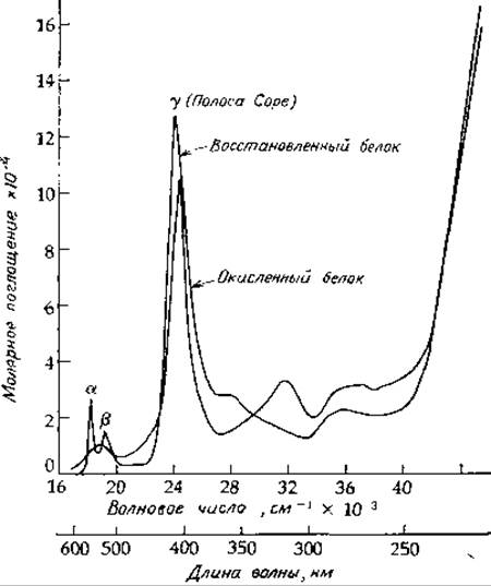
FIG. 10-2. Absorption spectra of oxidized and reduced cytochrome c from horse Heart Muscle at pH 6.8. Based on Margoliash E., Frohwirt N., BJ, 71, 570—572 (1959).
Type b cytochromes contain protoheme, which is also found in bacterial cytochrome o [13—16]. Typical b cytochromes do not react with O2; however, cytochrome o serves as a terminal electron acceptor (cytochrome oxidase) and is capable of autooxidation by molecular oxygen. Another protoheme-containing cytochrome involved in hydroxylation is designated as cytochrome P-450. Here, the number 450 indicates the position of the intense "Soret band" (also referred to as the y-band, Fig. 10-2). Other properties are also used to designate cytochromes. For example, cytochrome a3 is similar to cytochrome a in its absorption spectrum, but unlike the latter, it readily reacts with both CO and O2.
Cytochrome c is one of the few intracellular heme pigments that are soluble in Water and easily dissociated from the membrane. Mitochondrial cytochrome c was among the first cytochromes to be obtained in purified form and crystallized. The Structure of this protein in both the ferric and ferrous states has been determined by X-Ray Diffraction Analysis. Cytochrome c is a small protein with a Molecular Weight of ∼13,000, consisting of 104 amino acid residues; it has been isolated from plants, animals, and eukaryotic microorganisms [12, 17, 18]. The complete Amino Acid Sequence of this protein has been determined for more than 50 species, with 24 positions in the peptide chain remaining invariant. A number of other positions are characterized exclusively by conservative substitutions. Cytochrome c has served as one of the model proteins for tracing evolutionary relationships among species through observed sequence variations. For instance, humans and chimpanzees share an identical cytochrome c, whereas 12 amino acid differences are found between human and horse cytochrome c, and Neurospora cytochrome c differs from human cytochrome c by 44 residues [17]. The Cells of the photosynthetic bacterium Rhodospirillum rubrum contain a related cytochrome c2, which is believed to have diverged from the precursor of mammalian cytochrome c 2·109 years ago. Even in this case, however, 15 positions in the chain remain invariant, while other positions contain functionally equivalent residues [19].
Structural studies of cytochrome c and cytochrome c2 indicate [12, 17—19] that the heme group serves as a core around which the peptide chain is folded. The 104 residues of cytochrome c are just sufficient to form a shell enclosing the heme. In both the oxidized and reduced forms of the protein, the fifth and sixth coordination positions of the iron atom are occupied by Methionine-80 (Fig. 10-3, left) and Histidine-18 (Fig. 10-3, right). Consequently, the heme is tightly enclosed by the polypeptide chain and rendered almost inaccessible to the solvent. This structural feature raises a number of questions.
How does an electron enter ferricytochrome c to reduce it, and how does it leave the shielded heme group of ferrocytochrome c to reduce cytochrome oxidase? One possibility is The transfer of an electron from the sulfur atom of methionine-80 to the iron (Fe3+), resulting in The formation of an electron-deficient radical. As one can readily imagine, the resulting "hole" could be filled by an electron coming from the —OH group of the adjacent Tyrosine-67. This tyrosine might then shift toward the surface and, by coming into contact with tyrosine-74, accept an electron from it [17]. Thus, the reductase acting on ferricytochrome c would interact with tyrosine-74 located On the surface. Although such a mechanism, based on the transient formation of mobile tyrosine radicals, lacks direct precedent, it merits consideration. A similar hypothesis [20, 21] suggests that electrons enter individually or in pairs from the surface via a hydrogen-bonding network to tyrosine-67 and subsequently to methionine-80. An alternative pathway involving electron exchange through a surface-exposed heme crevice has also been proposed [22]. Regarding the oxidation of cytochrome c, Dickerson has hypothesized that a specific domain of cytochrome oxidase (the subsequent enzyme in the Electron Transport Chain) inserts into a cleft in ferrocytochrome c to accept an electron.
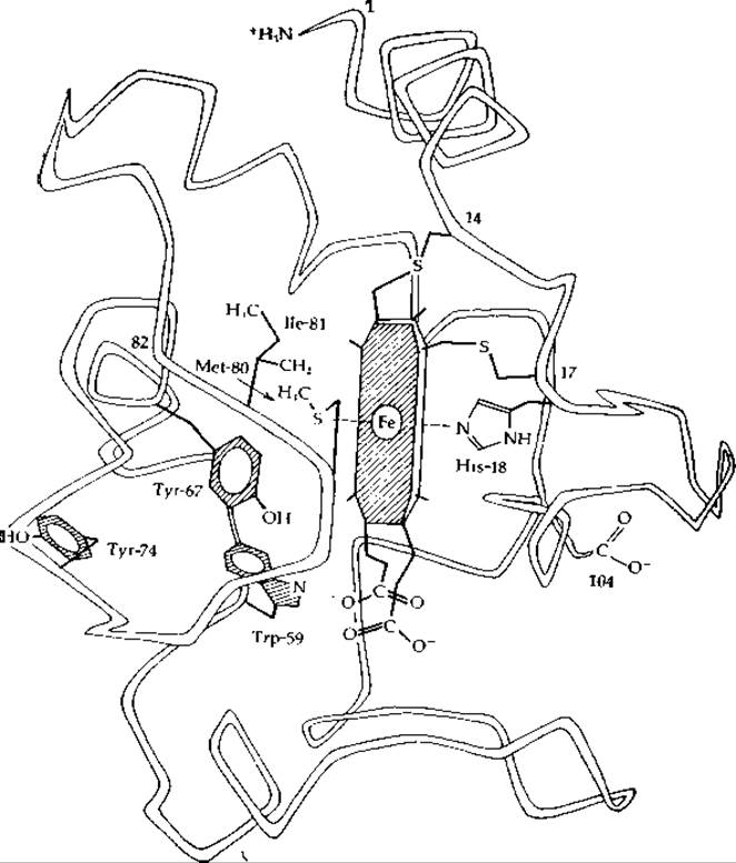
FIG. 10-3. Peptide backbone, heme group, and selected side chains of cytochrome c. From Dickerson [12].
The structure of solubilized cytochrome b5 from Liver microsomes has also been resolved. Although its precise function remains unknown, it is thought to play a role analogous to that of cytochrome c by interacting with The Endoplasmic reticulum enzyme system that catalyzes the Formation of Unsaturated Fatty acids. The protein contains 93 amino acid residues, with an additional 44 residues (predominantly hydrophobic) cleaved from the N-terminus during solubilization. This N-terminal segment likely Functions as a hydrophobic anchor embedded in the endoplasmic reticulum membrane. The heme in cytochrome b5 is not covalently bound to the protein but is firmly anchored between two histidine side chains. In terms of its folding pattern, this protein bears no resemblance to either cytochrome c or Myoglobin. Furthermore, The pathway of Electron transfer from the iron atom to the molecular surface remains obscure in this case [23].
The structures of other cytochromes functioning in The electron transport chain are currently unknown. Cytochrome c1 (whose a-band in the reduced state is at 554 nm, compared to 550 nm for cytochrome c) contains a heme bound to the protein in a manner very similar to that of cytochrome c, but the protein appears to have a higher molecular weight. Several forms of cytochrome b are known (Table 10-5); its molecular weight exceeds that of cytochrome c, and it is capable of forming a stable complex with ubiquinone [24].
Cytochrome oxidases perform a unique function in aerobic organisms: they bind O2 in a manner strikingly similar to Hemoglobin and subsequently rapidly reduce O2 to two molecules of H2O [24a]. This process involves the Cleavage of the O—O bond and requires four electrons. Naturally, this complex pathway is not yet fully understood. It is important to note that mammalian mitochondrial cytochrome oxidase contains two heme groups (cytochrome a) and two Cu(I) atoms per functional unit. Thus, upon the reduction of both cytochrome a molecules and the two copper atoms, four electrons can be stored for the subsequent reduction of a single O2 molecule. The chemistry of cytochrome oxidase remains poorly understood. As Keilin first discovered, only half of the cytochrome a molecules combine with CO; this fraction was designated as cytochrome a3. According to sodium dodecyl sulfate-Polyacrylamide gel Electrophoresis, Yeast cytochrome oxidase comprises six or seven subunits with molecular weights ranging from 5,000 to 42,000 [24b, c]. Interestingly, the three largest subunits appear to be encoded by Mitochondrial DNA genes, whereas the heme groups are attached to the smaller Peptides. It has been proposed that in the intact enzyme, the O2 molecule initially binds between the iron atom of cytochrome a3 and a cuprous copper ion: a2+3—O2—Cu+. The next stage involves a two-electron reduction of O2 to form a peroxide intermediate, followed by further reduction to two water molecules.
Additional cytochromes are discussed in Sections E.1 and E.2. Detailed reviews on bacterial cytochromes [13, 15] as well as broader discussions of cytochrome properties [12, 24] have been published.
Last update: 06/08/2026
Editorial and Educational Adaptation: This material has been compiled based on the primary/original source text. The project team performed an editorial review, corrected technical inaccuracies, structured sections, and adapted the content for an educational format.
What was processed:
- elimination of formatting defects (OCR errors, structural breaks, corrupted characters);
- editorial organization of content;
- standardization of terminology in accordance with academic sources;
- verification of factual statements against the original source text.
All mentions of the author, publication year, and origin of the primary text have been preserved in accordance with the source.