Biochemistry - The Chemical Reactions of Living Cells, Volume 2 - D. Metzler 1980
How electrons meet oxygen, how ATP is generated in the process, and related phenomena
Hemoproteins
Catalase and peroxidases
Another group of heme Enzymes catalyzes reactions that involve hydrogen peroxide, H2O2, rather than O2. Peroxidases are widely distributed in plant Tissues, where they are found predominantly in Peroxisomes; they also occur in small amounts in animal tissues. They catalyze the following reaction:
Class="center">Н2O2 + АН2 → 2Н2O + А. (10-4)
Catalase is an enzyme invariably present in aerobic Cells; sometimes its amount can reach up to 1% of the dry weight of Bacteria. This enzyme catalyzes The breakdown of H2O2 into Water and oxygen. The postulated mechanism is essentially identical to the mode of action of peroxidases. If in equation (10-4) we substitute H2O2 for AH2 and O2 for A, we obtain

Catalase Functions extremely rapidly, almost 104 times faster than peroxidases. Its molecular activity (per catalytic center) is approximately 2∙105 s-1.
It is generally believed that catalase plays a protective role by preventing the accumulation of H2O2, which exerts a damaging effect on cellular components. The lethal effect of O2 on obligate anaerobes is likely due to their lack of this enzyme. This view is supported by the existence of a hereditary disease known as acatalasemia [25]. Individuals with very low catalase activity are found in many regions, but they are particularly numerous in Korea. According to estimates, there are about 1,800 people lacking catalase in Japan. Since roughly half of these individuals exhibit no symptoms, catalase might be considered a non-essential enzyme. However, many affected persons develop ulcerative lesions around the Teeth, which can lead to serious complications. Apparently, hydrogen peroxide produced by bacteria accumulates and oxidizes Hemoglobin to methemoglobin (Supplementary Section 10-A), thereby depriving the affected tissues of oxygen.
The most extensively studied peroxidase is isolated from horseradish and contains a single heme per protein molecule with a Molecular Weight of 40,000. Catalases typically exist as tetramers with a molecular weight of 250,000. Both peroxidases and catalase contain high-spin Fe(III) iron and closely resemble metmyoglobin in their properties. These Enzymes can be reduced to the Fe(II) state, in which they can bind O2 (irreversibly). We can see that the very same Active Site found in hemoglobin is also present in peroxidases and catalase, yet the chemistry of this active site is heavily influenced by the protein environment. The affinity for O2 changes dramatically, and new types of catalytic activity emerge in these ferrihemoproteins.
The MECHANISM OF ACTION of catalases and peroxidases remains largely unclear, but particular attention has been drawn to The formation of a series of striking, distinctly colored intermediate forms in the presence of substrates [25a]. When a slight excess of H2O2 is added to a peroxidase solution, the dark-brown enzyme first turns olive-green (compound I) and then becomes pale pink as compound II is formed. Compound II reacts slowly with the substrate AH2 (or with another molecule of H2O2) to regenerate the native form of the enzyme. In equation (10-6), this sequence of reactions is represented by the upper horizontal line of arrows:
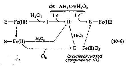
Titration demonstrates that compound I is converted into compound II by a one-electron reduction, and compound II is converted to free peroxidase via another one-electron reduction (for instance, using ferrocyanide). Thus, the iron in compound I can formally be represented as Fe(V), and in compound II as Fe(IV). However, this tells us very little about The Fate of H2O2 during its reaction with the enzyme when compounds I and II are formed. Do the oxygen atoms of H2O2 remain in compounds I and II or not? This question has sparked intense debate, yet a definitive answer has not been reached. Let us consider the following evidence, however. As mentioned earlier, the enzyme in its Fe(III) form can be reduced to the Fe(II) state. When the Fe(II) enzyme reacts with H2O2, it presumably converts into compound II, from which one can conclude that the latter represents an Fe(II) complex with a peroxide anion [26–28]:
Е—Fe+(II)—ООН
Clearly, the iron in this complex is in a low-spin state. Nevertheless, researchers prefer to view compound II as a ferryl-iron complex, which can be formed from the preceding Structure by The addition of a proton and the elimination of water:
Е—Fe2+(IV) О.
The addition of H2O2 apparently converts compound II into compound III, which is thought to be identical to oxyperoxidase. The latter is formed by the addition of O2 to the ferrous form of the free enzyme [equation (10-6)]. The H2O2 involved in The conversion of compound II into oxyperoxidase is apparently reduced to yield two molecules of water.
If The structure of compound II is uncertain, this is even more true for compound I. Initially, it was believed to be a complex of Fe(III) with H2O2 or its anion. However, such a structure fits poorly with the observed magnetic and spectral properties. There has been much speculation regarding the localization of the oxygen atoms belonging to the bound hydrogen peroxide. We shall introduce the reader to certain details that must be taken into account when pondering this problem. In equation (10-5), H2O2 can be replaced by an organic peroxide, ROOH; the conversion of form I into form II and the subsequent transformation into the free enzyme can be achieved using a one-electron donor, notably K2IrBr6. If the substrate AH2 serves as the electron donor, the product of the conversion of compound I into compound II will be the free radical AH2 (AH•). This free radical can give up a second electron required to generate the free enzyme; a second molecule of AH2 may be utilized, resulting in the formation of two AH radicals. The latter can subsequently disproportionate into A and AH2.
Chloroperoxidase [29], isolated from the mold Caldariomyces fumago, catalyzes the chlorination of Organic compounds using H2O2 and Cl- [equation (10-7)].
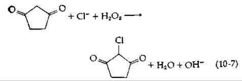
Chlorination is accompanied by the transient formation of a reaction intermediate of iron that is spectrally indistinguishable from compound I. An Fe—OCl structure has been proposed for it [29]. The protein has a molecular weight of ~42,000 and has been isolated in the low-spin ferri-state. The reduced ferroenzyme is a high-spin form; spectroscopically, it is nearly identical to cytochrome P-450 (Section G,2,e) [29, 30]. Milk lactoperoxidase, in the presence of I- and H2O2, catalyzes a completely analogous Iodination of Tyrosine and Histidine residues in Proteins. The method of introducing 125I- and 131I- isotopes into proteins at exposed membrane surfaces is widely used [31].
Myeloperoxidase from polymorphonuclear leukocytes (Chapter 1, Section D,2,b) utilizes H2O2 and a halide ion to attack phagocytized bacteria [32, 33]. Phagocytosis induces an enhancement of leukocyte Respiration and The production of H2O2, partly mediated by a membrane-bound NADPH oxidase. A portion of this H2O2 is utilized by myeloperoxidase to attack bacteria, apparently through the generation of HOCl [33a]. Other oxygen-dependent mechanisms of bacterial killing may also operate [34]. A hereditary deficiency of NADPH oxidase (an X-linked trait) can lead to a dangerous condition known as chronic granulomatous disease, characterized by a loss of the body's resistance to infections by many common bacteria.
Last update: 06/08/2026
Editorial and Educational Adaptation: This material has been compiled based on the primary/original source text. The project team performed an editorial review, corrected technical inaccuracies, structured sections, and adapted the content for an educational format.
What was processed:
- elimination of formatting defects (OCR errors, structural breaks, corrupted characters);
- editorial organization of content;
- standardization of terminology in accordance with academic sources;
- verification of factual statements against the original source text.
All mentions of the author, publication year, and origin of the primary text have been preserved in accordance with the source.