Biochemistry: The Chemical Reactions of Living Cells, Volume 2 - D. Metzler 1980
How electrons meet oxygen, how ATP is formed in the process, and some related phenomena
Non-heme iron proteins
Far from all the iron within Cells forms chelate complexes with porphyrin groups. Although hemerythrin had been known for many years (Sec. B,4), Structure/19.html">The Importance of non-heme iron Proteins only became apparent after Crane developed a method in 1945 for obtaining large quantities of Cell/35.html">Mitochondria. It was noted that the iron content of mitochondria far exceeds that of Hemoproteins. A major discovery was made in 1960 by H. Beinert, who was studying mitochondrial dehydrogenase systems specific for succinate and NADH. He observed that when the Electron Transport Chain was partially reduced by these substrates, solutions frozen at a low Temperature yielded a strong EPR signal with a value of g = 1.94. This signal appeared only after reduction by substrates, and fractionation results indicated that it was produced by non-heme iron proteins. Although the function of these proteins is still not fully understood, it has been suggested that at least six proteins of this type are present in The electron transport chain [35, 36a]. Three more such proteins are associated with the flavin-containing succinate dehydrogenase (Chap. 8, Sec. I,3) [36b].
The presence of non-heme iron proteins is even more evident in clostridia, which contain no heme at all. It was from these Bacteria that the first non-heme iron-containing protein, named ferredoxin, was isolated. This protein, possessing a strikingly low redox potential (E0 = —0.41 V), participates in the reaction catalyzed by Pyruvate:ferredoxin oxidoreductase (Chap. 8, Sec. K,3), in Nitrogen Fixation in certain species, and in H2 evolution. It is a small, greenish-brown protein containing only 54 amino acid residues, yet it forms a complex with eight iron atoms. If the pH is lowered to ~1, eight molecules of H2S are released. Thus, the protein contains eight atoms of "labile sulfur" somehow linked by iron-sulfide bonds. Ferredoxins turned out to be only the first representatives of a large family of iron-sulfur proteins discovered later [37–39]. Most of them contain iron and labile sulfur in a 1:1 ratio, but the number of iron atoms per protein molecule varies. Furthermore, one group of proteins contains no labile sulfur at all: the iron in them is held by the side chains of four Cysteine residues. The simplest iron-sulfur Proteins can be classified According to the table below. In addition to these, there are more complex iron-sulfur proteins, such as Nitrogenase (Chap. 14, Sec. A,2), which also contain molybdenum. The standard redox potentials of iron-sulfur proteins cover a strikingly wide range of values, from —0.42 V for spinach ferredoxin to +0.35 V for the so-called high-potential iron-sulfur protein from Chromatium.
|
Iron and labile sulfur content |
Protein designation |
|
1 Fe |
Rubredoxin |
|
2 Fe, 2 S2- |
Chloroplast ferredoxin, adrenodoxin, putidaredoxin, E. coli ferredoxin [40] |
|
4 Fe, 4 S2- |
High-potential iron-protein (Chromatium), certain bacterial ferredoxins, Hydrogenase |
|
Fe, nS2- |
Bacterial ferredoxins (typically n=8) |
The structure of several such proteins has been determined by X-ray crystallography [38, 41]. The simplest of these is rubredoxin from Clostridium pasteurianum, a small peptide with a Molecular Weight of ~6000 (Fig. 10-4). Among its 54 amino acid residues are four cysteine residues whose side chains form a distorted tetrahedron around a single iron atom [38]. Three Fe—S bonds have a "normal" length of about 0.23 nm, but the fourth, with the cysteine-41 residue, is unusually short (0.205 nm). The function of clostridial rubredoxin is not precisely known; it is believed to participate in electron transfer and may replace ferredoxin in certain reactions. There is also a larger rubredoxin with a molecular weight of ~19,000 that binds two iron ions; it participates in electron transfer as a component of the Pseudomonas cell hydroxylase system (Sec. G,2, e) [42].
Class="center">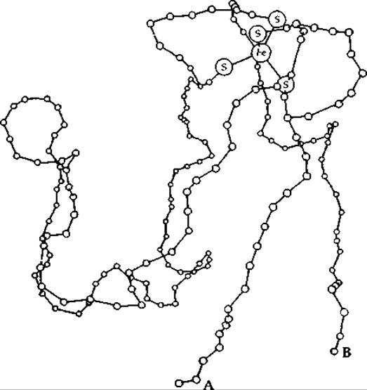
FIG. 10-4. Model of the polypeptide chain folding and iron coordination in rubredoxin from Clostridium pasteurianum. A and B are the N- and C-termini. From Herriott J. R., Sieker L. C., Jensen L. H., Lovenberg W., JMB, 50, 391–406 (1970).
X-ray structural studies have shown that in the high-potential iron-protein from Chromatium, the 86-amino-acid polypeptide chain is wrapped around an iron-sulfur cluster containing the side chains of four cysteine residues along with four iron atoms and four sulfur atoms (Fig. 10-5) [41]. Each of the four sulfur atoms in the cysteine residues is bonded to one Fe atom, and these four Fe atoms form an irregular tetrahedron with an Fe—Fe distance of 0.28 nm. Four labile sulfur atoms (S2-) form a tetrahedron with a side length of 0.35 nm, in which each sulfur atom is bonded to three iron atoms. Typically, the cluster can accept only one electron. The structure of such an iron-sulfur cluster came as somewhat of a surprise, but following its discovery, it was established that ions of the type [Fe4S4(S—CH2CH2COO-)4]6- form spontaneously from their components and possess a similar cluster structure [43, 44]. Thus, living organisms, as usual, merely perfected a structure that originated in the inanimate world.

FIG. 10-5. Fe4S4 cluster in the high-potential iron-protein from Chromatium.
Fig. 10-6 depicts the STRUCTURE OF THE bacterial ferredoxin from Peptococcus aerogenes [38, 45]. Note that this protein, with eight Fe atoms and eight labile sulfur atoms, contains two Fe4S4 clusters with practically the same structure as in the Chromatium protein. Each cluster apparently accepts one electron. The Amino Acid Sequence of the first half of the chain is largely repeated In the second half, suggesting that this half of the chain arose from Gene Duplication. The sequence contains many invariant positions, including the locations of the cysteine residues that form the Fe—S cluster.
Surprisingly, the redox potentials of bacterial ferredoxins (Fd) and the high-potential protein from Chromatium differ greatly despite the similarity in the structure of their active centers. It is believed that Fe4S4 clusters can exist in three oxidation states [equation (10-8)], successively differing from one another by one electron [46–49].
The Chromatium protein and ferredoxins probably have a similar average oxidation state1).
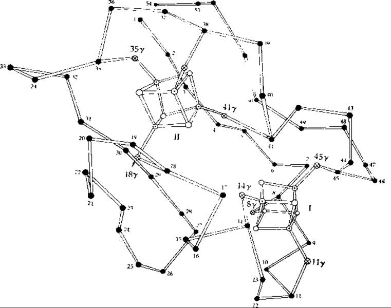
FIG. 10-6. Model of the spatial arrangement of alpha-carbon, iron, and sulfur atoms in ferredoxin from Peptococcus aerogenes [45]. Circles with a dot in the middle represent Fe, light circles represent S2-, crossed circles represent S (cysteine), and dark circles represent Cα.
In more oxidized states, the cluster size is slightly smaller (in the Chromatium protein, the Fe—Fe distance changes upon oxidation from 0.281 to 0.272 nm). Synthetic iron-sulfur clusters exhibit weakly basic properties [47] and act as proton acceptors with pKa values ranging from 3.9 to 7.4.

1) However, in the Azotobacter ferredoxin containing 8 iron atoms, one cluster has E0 = —0.42 V and the other has E0 = +0.34 V; EPR spectra indicate that both clusters, despite significant differences in their potentials, undergo a transition between the —2 and —1 oxidation states (oxidized and super-reduced states; Fig. 10-8 [49a]). Soluble mammalian succinate dehydrogenase contains three iron-sulfur clusters with E0 values of —0.40 V, —0.005 V, and +0.06 V. In this enzyme, the center with the highest potential apparently also undergoes a transition between the —2 and —1 states [equation (10-8)] [49b].
Iron and labile sulfur can be removed from certain iron-sulfur proteins and the active enzyme then reconstituted by appropriately adding sulfide and iron atoms. Similarly, the natural 56Fe isotope (with zero nuclear spin) can be exchanged for the 57Fe isotope, which possesses magnetic nuclei [50]. In the same way, 32S can be replaced by 77Se. The resulting proteins function without apparent impairment and display hyperfine structure in their EPR spectra arising from the interaction of nuclei with the unpaired electrons of the clusters (Fig. 10-7). These observations lead to the Conclusion that the electrons accepted by Fe4S4 clusters are not localized on atoms of any single type, but interact with both Fe and S nuclei.

FIG. 10-7. Electron paramagnetic Resonance spectrum of the Fe-S protein putidaredoxin in its native form (32S) and with labile sulfur replaced by selenium isotopes. At the low-field end of the spectrum for the 77Se-containing protein  , well-defined peak shoulders are visible. From Orme-Johnson W. H., Hansen R. E., Beinert H., Tsibris J. C. M., Bartholomaus R. C., Gunsalus I. C., PNAS, 60, 369–372 (1968).
Chloroplast ferredoxins contain two iron atoms and two labile sulfur atoms; their putative structure is as follows:

Although the three-dimensional structure of chloroplast ferredoxin has not been established, the structure of A number of related model compounds has been determined [51].
Last update: 06/08/2026
Editorial and Educational Adaptation: This material has been compiled based on the primary/original source text. The project team performed an editorial review, corrected technical inaccuracies, structured sections, and adapted the content for an educational format.
What was processed:
- elimination of formatting defects (OCR errors, structural breaks, corrupted characters);
- editorial organization of content;
- standardization of terminology in accordance with academic sources;
- verification of factual statements against the original source text.
All mentions of the author, publication year, and origin of the primary text have been preserved in accordance with the source.