Principles of Biochemistry, Volume 1 - A. Lehninger 1985
Biomolecules
Proteins: Covalent Structure and Biological Functions
Proteins can be isolated and subjected to purification
Cells contain hundreds, if not thousands, of different Proteins. To determine the Amino Acid Composition or sequence of a specific protein, it must first be isolated in pure form. How can a single protein, such as a specific enzyme, be separated from the hundreds of other proteins present in a Cell or tissue extract and purified to the desired degree?
First, proteins are separated from low-molecular-weight substances in cell or tissue extracts by dialysis (Fig. 6-3). Large molecules, such as proteins, remain inside a dialysis bag made of a material with ultramicroscopic pores, such as cellophane. When a dialysis bag containing a cell or tissue extract is immersed in Water, small molecules present in the extract, such as salts, pass through the pores, whereas the high-molecular-weight proteins remain inside the bag.
Once the protein mixture is freed of small molecules via dialysis, the proteins can be fractionated according to size by Gel filtration. In this Procedure, which is a type of Chromatography, a solution containing the protein mixture is passed through a Column packed with very small, porous beads of a highly hydrated polymer. Relatively small protein molecules penetrate the pores of these beads, which retards their passage through the column (Fig. 6-4), whereas larger protein molecules cannot enter the beads and move through the column much faster. Proteins of intermediate size pass through the column at intermediate speeds depending on their ability to enter the beads. Such a gel-filtration column acts essentially as a molecular sieve.
Class="center">
Fig. 6-3. Dialysis. The membrane enclosing the protein solution freely permeable to water and low-molecular-weight solutes, such as NaCl or glucose, but impermeable to large molecules such as proteins. Small molecules diffuse from the dialysis bag into the external medium because diffusion drives molecules toward regions of lower concentration. By periodically replacing the aqueous phase in the external vessel with distilled water, the concentration of low-molecular-weight solutes in the protein solution can be reduced to a negligibly small level.
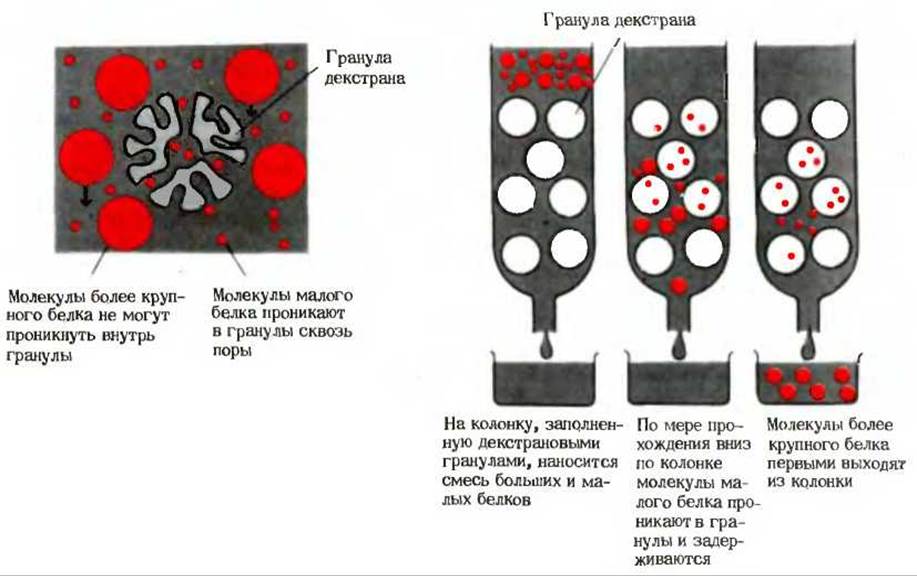
Fig. 6-4. Separation of proteins according to molecular size by gel filtration. A solution containing a protein mixture is passed through a column packed with very small, porous beads of a hydrophilic polymer; dextran derivatives are widely used for this purpose. Small protein molecules enter the pores of the beads, whereas larger molecules are excluded. The Molecular Weight of a protein can be determined by comparing its elution rate through the column with the elution rates of reference proteins of known molecular weights.
Proteins can also be separated from one another by Electrophoresis (Section 5.16). A decisive role in this method is played by the sign and number of electrical charges localized on the R groups, N-terminal amino groups, and C-terminal carboxyl groups of proteins. Like simple Peptides, polypeptide chains are characterized by a characteristic isoelectric point, which is determined by the relative number of acidic and basic R groups (Table 6-5). At a given pH, some proteins in a mixture will carry a net negative charge, others a net positive charge, and still others a net zero charge. When such a protein mixture is placed in an electric field, positively charged proteins will migrate toward the negative electrode (cathode), negatively charged proteins toward the positive electrode (anode), and electrically neutral proteins will remain stationary. Furthermore, protein molecules with a higher charge density will move toward the corresponding electrode faster than those with a lower charge density. Electrophoresis is frequently carried out not in free solution, but on a supporting medium such as a strip of paper, a Cellulose acetate membrane, or a hydrophilic gel plate, which effectively retards the diffusion of fractionated protein molecules in the aqueous phase (Fig. 6-5).
Table 6-5. Isoelectric Points (pHI) of Some Proteins
|
pHI |
|
|
< 1.0 |
|
|
Egg albumin |
4.6 |
|
Serum albumin |
4.9 |
|
Urease |
5.0 |
|
ß-Lactoglobulin |
5.2 |
|
γ1-Globulin |
6.6 |
|
6.8 |
|
|
7.0 |
|
|
Chymotrypsinogen |
9.5 |
|
Cytochrome c |
10.7 |
|
11.0 |
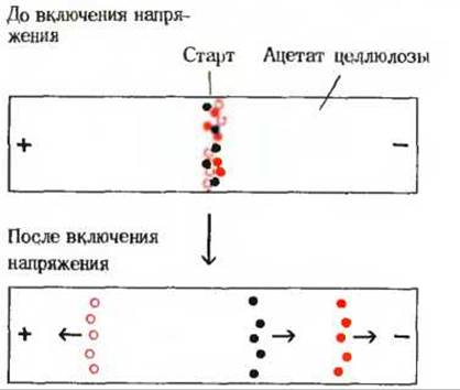
Fig. 6-5. Electrophoresis of a three-protein mixture. The protein mixture is applied to a cellulose acetate strip moistened with a buffer at a specified pH. The ends of the strip are immersed in electrode buffer compartments. The cellulose acetate serves as a support that prevents random migration of protein molecules via diffusion. After applying the protein mixture to the center of the strip, the proteins are subjected to an electric field generated by a potential difference between the electrodes. Each of the three proteins migrates toward the positive or negative electrode at a different rate, depending on the pH of the medium and the acid-base Properties of the individual protein. Upon completion of electrophoresis, the positions of the proteins can be visualized using protein-binding stains.
Another effective method for protein separation is Ion-exchange chromatography, which relies primarily on differences in the charge density and net charge of proteins at a given pH. Thus, ion-exchange chromatography can be used to separate not only Amino Acids (Section 5.18) and peptides (Section 5.21), but proteins as well.
To isolate a specific protein from a complex mixture, one must have a convenient assay to measure the concentration of that protein and monitor each stage of purification. For example, when purifying an enzyme, measuring its catalytic activity allows us to distinguish it from all other proteins.
Last update: 06/08/2026
Editorial and Educational Adaptation: This material has been compiled based on the primary/original source text. The project team performed an editorial review, corrected technical inaccuracies, structured sections, and adapted the content for an educational format.
What was processed:
- elimination of formatting defects (OCR errors, structural breaks, corrupted characters);
- editorial organization of content;
- standardization of terminology in accordance with academic sources;
- verification of factual statements against the original source text.
All mentions of the author, publication year, and origin of the primary text have been preserved in accordance with the source.