Biochemistry of Amino Acids - A. Majster 1961
Natural Amino Acids
Amino Acids Commonly Found in Proteins
Amino Acid Composition of Proteins
"...The properties of a protein are not merely a reflection of the sum of the properties of its constituent components, but rather are determined by The Structure of its molecule... We can gain some insight into the potential diversity of combinations of protein building blocks if we recall that their number corresponds to the number of letters in the English alphabet, which can be used to express an infinite range of thoughts." Kossel (1911).
Protein Hydrolysis is carried out using acids (hydrochloric, sulfuric, hydriodic), alkalis (sodium or barium hydroxide), or Proteolytic Enzymes. Sulfuric acid was used by Braconnot in 1820 [46] for the hydrolysis of gelatin. Hydrochloric acid [15] was subsequently introduced and has since become universally adopted. The standard hydrolysis Procedure involves treating the protein with a 5- to 20-fold excess by weight of 3–12 N hydrochloric acid at 100–120°C for 3–40 hours. Following hydrolysis, excess hydrochloric acid is removed by repeated evaporation. A variety of Methods exist for separating mixtures of amino acid hydrochlorides, including precipitation with relatively specific Reagents, the preparation and Separation of Amino Acid Derivatives (esters, silver or copper salts), Electrophoresis, ionophoresis, and Chromatography. The latter method has proven exceptionally valuable and will be discussed in detail below (p. 40).
Acid Hydrolysis of Proteins is typically accompanied by the destruction (via oxidation) of the majority of Tryptophan, The oxidation of Cysteine to cystine, and some decomposition of Serine and Threonine. Alkaline hydrolysis has the advantage over acid hydrolysis in that tryptophan remains considerably more stable under these conditions. However, alkaline hydrolysis results in extensive breakdown of serine, threonine, cystine, cysteine, and Arginine. Furthermore, racemization of Natural Amino Acids is observed during alkaline hydrolysis. Both acid and alkaline hydrolysis of proteins are accompanied by the deamidation of glutamine and asparagine. These amino acid amides, along with tryptophan, can be isolated from hydrolysates prepared using proteolytic enzymes. Nevertheless, the enzymatic method also suffers from certain drawbacks: specifically, hydrolysis may be incomplete, and the enzyme itself may undergo degradation, releasing free amino acids. The isolation of amino acids from proteins and their quantitative recovery present a formidable challenge that has engaged many researchers. This extensive field is comprehensively reviewed in the monograph by Block and Bolling [98].
Recently, The Use of ion-exchange resins has been recommended for protein hydrolysis. According to one procedure, casein is hydrolyzed by refluxing in the presence of a strong cation exchanger (Dowex-50); the resulting Amino acids are bound by the resin and can be recovered by Treatment with baryta [571, 572].
As is well known, proteins vary widely in their Amino acid composition; a substantial body of literature on this subject has already accumulated [98–101, 571, 572]. Investigating the Amino acid composition of proteins has proven to be a complex task due to various difficulties associated with preparing pure proteins and subsequently isolating their hydrolysis products quantitatively.
Despite the considerable efforts of numerous researchers, relatively little data has been obtained thus far that allows for a definitive interpretation of Cell/13.html">Protein Structure. It is abundantly clear that protein properties cannot be determined solely by the sum of the properties of their constituent amino acids. Nevertheless, knowledge of a protein's amino acid composition is a fundamental prerequisite for successfully elucidating The sequence of Amino Acids and other structural features. Data on amino acid composition are crucial for resolving practical nutritional issues and for understanding the physical and Chemical properties of purified proteins.
Although a detailed treatment of Protein Chemistry falls outside The Scope of this book, it is nonetheless of interest to discuss The amino acid composition of certain proteins. To date, over 50 proteins have been analyzed, yet the findings do not permit any broad generalizations. Even so, the investigation of proteins with specific Functions has revealed A number of characteristic features. Selected Examples from the literature are presented in Table 1.
In an attempt to identify General Principles underlying protein amino acid composition, Bailey [101] constructed a series of histograms reflecting the amino acid composition of two dozen proteins. On the whole, Bailey was unsatisfied with the results of this endeavor, yet he was still able to draw several Conclusions. For instance, he pointed out the widespread occurrence of valine, leucine, and isoleucine, noting that proteins generally contain less isoleucine than leucine. Phenylalanine, Proline, Tyrosine, aspartic acid, glutamic acid, and cystine are commonly present in proteins, whereas tryptophan, Methionine, Lysine, Histidine, arginine, Glycine, and Alanine are encountered less frequently.
Tristram [99] applied this approach to several other proteins and concluded that "certain groups of amino acids are distributed within proteins in such a way that the histograms of the latter resemble one or more partially overlapping normal distribution curves. If such a distribution reflects an underlying natural law, it may indicate either that The Mechanism of Protein Synthesis is more or less uniform across all cell types, or, more likely, that synthetic mechanisms are selective and preclude The formation of all stereochemically possible proteins."
The Development of chromatographic methods for protein research and novel techniques for determining amino acid sequencing suggests that data on protein amino acid composition will find much broader application in the future.
Certain proteins exhibit distinct peculiarities in their amino acid composition. For example, salmine, a protamine, is characterized by a high arginine content and the complete absence of such common protein components as glutamic acid and leucine, although isoleucine and valine residues are present (see Table 1). Wool keratin is distinguished by a high cystine content, Silk Fibroin contains relatively large amounts of glycine, alanine, and serine, while Collagen is unique among proteins in containing hydroxyproline and hydroxylysine. It has long been known that thyroglobulin contains thyroxine and diiodotyrosine, and it has recently been discovered that other iodine-containing amino acids may also be constituents of thyroglobulin. These examples provide An Overview of the characteristic amino acid composition of several well-known proteins.
Class="center">Table 1. Amino acid composition of certain proteins [99]
|
Amino acids |
Salmine * |
Bovine Insulin * |
Bovine thyroglobulin ** (0.54% diiodotyrosine and 0.21% thyroxine) |
Horse hemo globin * |
Human serum albumin * |
Human y-globulin ** |
a-Casein ** |
Pepsin * |
Wool keratin ** |
Silk fibroin ** |
Collagen ** |
|
Alanine |
1 |
3 |
83.1 |
54 |
42.8 |
46.4 |
334.0 |
106.9 |
|||
|
Arginine |
40 |
1 |
73.2 |
14 |
25 |
27.9 |
24.7 |
2 |
59.7 |
6.3 |
49.4 |
|
Aspartic acid |
0 |
3 |
— |
51 |
46 |
66.2 |
63.2 |
41 |
54.1 |
20.8 |
47.3 |
|
Valine |
2 |
5 |
12.4 |
50 |
45 |
83.0 |
53.9 |
21 |
39.7 |
30.8 |
29.1 |
|
Histidine |
0 |
2 |
14.4 |
36 |
16 |
16.1 |
18.7 |
2 |
6.8 |
2.3 |
4.8 |
|
Glycine |
3 |
4 |
50.7 |
48 |
15 |
56.0 |
30.2 |
29 |
87.0 |
581.0 |
363.0 |
|
Glutamic acid |
0 |
7 |
— |
38 |
80 |
80.4 |
153.1 |
28 |
96.0 |
14.7 |
77.0 |
|
Isoleucine |
1 |
1 |
_ |
0 |
9 |
20.6 |
28 |
8.4 |
|||
|
Leucine |
0 |
6 |
97.8 |
75 |
58 |
71.0 |
109.2 |
27 |
86.3 |
7.0 |
42.8 |
|
Lysine |
0 |
1 |
23.5 |
38 |
58 |
55.5 |
61.0 |
2 |
18.9 |
4.7 |
30.7 |
|
Methionine |
0 |
8.7 |
4.5 |
6 |
7.3 |
16.8 |
4 |
4.7 |
5 4 |
||
|
Hydroxylysine |
0 |
||||||||||
|
Hydroxyproline |
0 |
107 |
|||||||||
|
Proline |
4 |
1 |
— |
22 |
31 |
70.5 |
65.1 |
15 |
82.6 |
6.4 |
131.3 |
|
Serine |
7 |
3 |
10.3 |
35 |
22 |
108.8 |
60.0 |
40 |
95.4 |
154.3 |
32.3 |
|
Tyrosine |
0 |
4 |
17.2 |
11 |
18 |
37.6 |
44.8 |
16 |
25.7 |
70.7 |
5.5 |
|
Threonine |
0 |
1 |
— |
24 |
27 |
70.6 |
41.1 |
28 |
53.9 |
13.5 |
19.2 |
|
Tryptophan |
0 |
10.2 |
5 |
1 |
14.2 |
7.9 |
4 |
8.8 |
0 |
0 |
|
|
Phenylalanine |
0 |
3 |
40.5 |
30 |
33 |
27.9 |
27.9 |
13 |
22.1 |
20.4 |
15.2 |
|
Cysteine |
0 |
— |
3 |
4 |
5.8 |
_ |
2 |
0 |
|||
|
Cystine (1/2 molecule) |
0 |
1.5 |
30.0 |
2.5 |
32 |
19.9 |
3.6 |
4 |
98.9 |
_ |
0 |
|
Amide nitrogen |
0 |
6 |
— |
(36) |
(44) |
(79.5) |
(114.3) |
(32) |
(83.2) |
_ |
47.1 |
|
Phosphorus |
(31.6) |
||||||||||
|
Molecular weight |
8 000 |
6 000 |
650 000 |
68000 |
69 000 |
156000 |
34 400 |
_ |
_ |
_ |
* Number of amino acid residues per protein molecule.
** Number of amino acid residues per 100,000 g of protein.
Determining the number and nature of C- and N-terminal amino acid residues has led to major breakthroughs in clarifying the structure of certain proteins. Insulin was the first protein for which the complete sequence of all amino acids was established [102–107]. Sanger and his coworkers, by oxidizing insulin with performic acid, obtained two main products which proved to be Peptides containing cysteic acid and consisting of 21 and 30 amino acid residues, respectively. The shorter chain (designated by Sanger as peptide "A") possesses an N-terminal glycine residue and a C-terminal asparagine residue. In the longer chain (peptide "B"), phenylalanine was found to be the N-terminal amino acid, with alanine located at the C-terminus. Through ingenious techniques—making extensive use of the dinitrophenyl derivative method with 1-fluoro-2,4-dinitrobenzene (p. 35) and isolating the peptides generated by acid and Enzymatic Protein Hydrolysis—the authors successfully decoded the complete Amino Acid Sequence of the "A" and "B" peptide chains of bovine insulin (Fig. 2).
The ε-amino groups of lysine in the insulin molecule are free. The "A" chain contains 2 glutamine residues and 2 asparagine residues, whereas the "B" chain contains one residue of each amide. The positions of the disulfide bridges are indicated in Fig. 2 [107].
It is interesting to note that the STRUCTURE OF THE "B" chain is identical in insulin molecules isolated from different animal species (cattle, pigs, and sheep), whereas the "A" chain in porcine insulin differs from the "A" chain shown in Fig. 2 by the presence of a threonine residue at position 8 and an isoleucine residue at position 10. In ovine insulin, a glycine residue replaces serine at position 9.
No obvious regular sequence has yet been discerned in the arrangement of amino acids within the insulin molecule; this protein evidently possesses a unique, highly specific order of amino acid succession. Naturally, it is daunting to think that every protein (and every species of Organism) might be characterized by its own distinct amino acid sequence, but it is entirely possible that future research will uncover underlying regularities that cannot be detected using currently available data. Physical Research Methods and the consideration of spatial relationships may well assist in the exploration of this complex field.
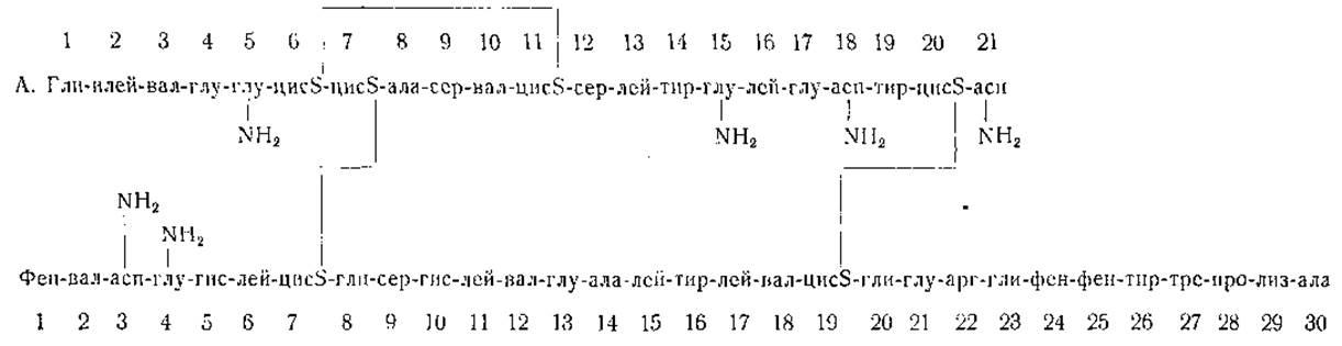
Fig. 2. Amino acid sequence in insulin (after Sanger).
|
Amino acid |
Abbreviation [573] |
Amino acid |
Abbreviation [573] |
|
Alanine |
ala |
Isoleucine |
ileu |
|
Arginine |
arg |
Leucine |
leu |
|
Asparagine |
asp-NH2 |
Lysine |
lys |
|
Aspartic acid* |
asp |
Methionine * |
met |
|
Cysteine * |
cysSH |
Phenylalanine |
phe |
|
Cystine |
(cysS-)2 |
Proline |
pro |
|
Glutamic acid |
glu |
Serine |
ser |
|
Glutamine |
glu-NH2 |
Threonine |
thr |
|
Glycine |
gly |
Tryptophan * |
try |
|
Histidine |
his |
Tyrosine |
tyr |
|
4-Hydroxyproline * |
opro |
Valine |
val |
* Absent in insulin.
Data obtained from studying the enzymatic degradation products of insulin [108] indicate that complete preservation of the insulin molecule may not be strictly required for biological activity. Such investigations are of considerable interest and may prove useful in elucidating the mechanism of insulin action.
Last update: 06/08/2026
Editorial and Educational Adaptation: This material has been compiled based on the primary/original source text. The project team performed an editorial review, corrected technical inaccuracies, structured sections, and adapted the content for an educational format.
What was processed:
- elimination of formatting defects (OCR errors, structural breaks, corrupted characters);
- editorial organization of content;
- standardization of terminology in accordance with academic sources;
- verification of factual statements against the original source text.
All mentions of the author, publication year, and origin of the primary text have been preserved in accordance with the source.