LEHNINGER PRINCIPLES OF BIOCHEMISTRY - VOL. 2. BIOENERGETICS AND METABOLISM - 2014
PART II. BIOENERGETICS AND METABOLISM
22. BIOSYNTHESIS OF AMINO ACIDS, NUCLEOTIDES, AND RELATED MOLECULES
22.3. Amino Acid Derivatives
Beyond serving as the Building Blocks of Proteins, Amino Acids function as precursors to a vast array of specialized Biomolecules, including Hormones, Coenzymes, NUCLEOTIDES, Alkaloids, Cell wall polymers, Porphyrins, Antibiotics, pigments, and Neurotransmitters. This section outlines the metabolic pathways leading to several of these key amino acid derivatives.
Glycine Is the Precursor to Porphyrins
The Biosynthesis of porphyrins, for which glycine serves as the primary precursor, provides our first example, as the porphyrin ring plays a fundamental role in forming the heme moiety of essential proteins such as Hemoglobin and Cytochromes. Porphyrins are constructed from four molecules of the monopyrrole derivative porphobilinogen, which itself is formed from two molecules of δ-aminolevulinate. There are two distinct pathways leading to δ-aminolevulinate. In higher eukaryotes (Fig. 22-23a), glycine condenses initially with succinyl-CoA to yield α-amino-β-ketoadipate, which is subsequently decarboxylated to form δ-aminolevulinate. In plants, Algae, and most Bacteria, δ-aminolevulinate is synthesized from glutamate (Fig. 22-23b). Glutamate is first converted to glutamyl-tRNAGlu (see Chapter 27, Vol. 3, on Transfer RNAs), which is then reduced by NADPH to glutamate-1-semialdehyde, followed by the Cleavage of the semialdehyde from the tRNA. The conversion of glutamate-1-semialdehyde into δ-aminolevulinate is catalyzed by an aminotransferase.
Class="center">Figure 22-23. Biosynthesis of δ-aminolevulinate. (a) In most animals, including mammals, δ-aminolevulinate is synthesized from glycine and succinyl-CoA. Atoms derived from glycine are highlighted in red. (b) In bacteria and plants, glutamate serves as the precursor for δ-aminolevulinate.

Figure 22-24. Biosynthesis of heme from δ-aminolevulinate. Ac = acetyl (—CH2COO−); Pr = propionyl (—CH2CH2COO−).

Box 22-2. MEDICINE. Biochemistry for Kings and Vampires
Porphyrias are a group of inherited Metabolic Disorders characterized by the accumulation of specific porphyrin precursors in erythrocytes, Body Fluids, and the Liver, resulting from enzymatic defects in the biosynthetic pathway from glycine to porphyrins. The most common form of this condition is acute intermittent porphyria. Most individuals with this disorder are heterozygous and typically remain asymptomatic because a single copy of the normal Gene provides sufficient enzymatic activity. However, specific dietary regimens or environmental triggers (not yet fully understood) can provoke an overproduction of δ-aminolevulinate and porphobilinogen, precipitating attacks of severe abdominal pain and neurological dysfunction. During the American Revolution, King George III of Great Britain experienced multiple episodes of apparent madness, although he was otherwise entirely rational in his behavior. His symptom profile strongly suggests that he suffered from acute intermittent porphyria.
One of the rarer forms of porphyria leads to the accumulation of uroporphyrinogen I, a precursor of protoporphyrin. This compound colors the urine red, causes Teeth to fluoresce intensely under ultraviolet light, and renders the Skin exquisitely sensitive to sunlight. Many afflicted individuals also suffer from anemia due to an inadequate synthesis of heme. This genetic disorder is widely considered to be the historical basis for folkloric myths about vampires.
The symptoms of many forms of porphyria can now be successfully managed through dietary modifications or the administration of heme and heme derivatives.
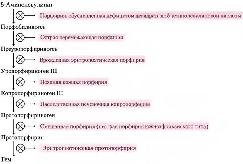
Across all organisms, two molecules of δ-aminolevulinate condense to form porphobilinogen, and through a series of complex enzymatic reactions, four porphobilinogen molecules are linked together to yield protoporphyrin (Fig. 22-24). Once the protoporphyrin macrocycle is assembled, an iron atom is inserted
in a reaction catalyzed by ferrochelatase. In higher eukaryotes, porphyrin biosynthesis is regulated by heme concentration, which acts as a feedback inhibitor of the initial steps in the pathway. Genetic Defects in porphyrin biosynthesis can result in the pathological accumulation of pathway intermediates, causing a group of human diseases collectively known as porphyrias (Box 22-2).
Heme Is the Source of Bile Pigments
Heme (the iron-containing porphyrin prosthetic group of hemoglobin), released upon the destruction of aged erythrocytes in the Spleen, is catabolized to yield free Fe3+ and ultimately bilirubin. The striking coloration of bilirubin draws particular attention to this degradative pathway.
The initial step of this two-stage pathway is catalyzed by heme oxygenase, which converts heme to biliverdin, a linear tetrapyrrole molecule containing four pyrrole rings joined in a row (Fig. 22-25). The other products of this reaction are a free ferric ion (Fe3+) and CO. The liberated Fe3+ is rapidly sequestered by ferritin. Carbon monoxide is toxic because it binds avidly to hemoglobin (see Box 5-1, Vol. 1); due to the ongoing activity of heme oxygenase in generating CO, about 1% of the body's hemoglobin has CO bound to it even under normal environmental conditions.
Figure 22-25. Bilirubin and its catabolic products. M = methyl; V = vinyl; Pr = propionyl; E = ethyl. For ease of comparison, all structures are depicted as linear chains of pyrrole rings rather than in their actual three-dimensional Conformations.

Biliverdin is converted to bilirubin In the second step, catalyzed by biliverdin reductase. This biochemical sequence is familiar to everyone: a severe bruise appears black and/or purple due to hemoglobin released from ruptured red Blood Cells; over time, it turns green as biliverdin accumulates, and eventually yellow as bilirubin is formed. Because bilirubin is extremely poorly soluble in Water, it is transported through the bloodstream bound to the plasma protein serum albumin. In the liver, bilirubin is converted into the bile pigment bilirubin diglucuronide. This derivative is sufficiently water-soluble to be secreted into the Small Intestine along with Other components of bile, where bacterial Enzymes transform it into various catabolites, principally urobilinogen. A fraction of the urobilinogen is reabsorbed into the bloodstream and carried to the Kidneys, where it is converted into urobilin, the compound responsible for the yellow color of urine (Fig. 22-25, left branch). The urobilinogen remaining in the intestinal tract is acted upon by microbial flora in a separate reaction to yield stercobilin (Fig. 22-25, right branch), which imparts the characteristic brown color to feces.
Liver dysfunction or an obstruction of bile flow causes bilirubin to "leak" from the liver into the bloodstream, manifesting clinically as a yellowing of the skin and the sclera of the eyes—a condition known as jaundice. Serum bilirubin assays are a standard diagnostic tool in evaluating liver disorders. In newborns, physiological jaundice frequently develops because their livers are temporarily deficient in bilirubin glucuronyltransferase. To clear the excess bilirubin, infants are typically treated with fluorescent phototherapy, which induces photochemical reactions that convert bilirubin into more soluble isomers that can be readily excreted by the body.
These heme degradation pathways play a vital role in protecting cells against oxidative damage and in regulating specific cellular Functions. Carbon monoxide, CO, produced during the heme oxygenase reaction, is toxic at high concentrations, but at the very low levels resulting from heme degradation, it exerts certain regulatory and/or signaling functions. It acts as a vasodilator (though less potent than nitric oxide), as discussed below. In small amounts, CO also acts as a regulator in neurotransmission. Bilirubin is a common antioxidant in mammalian Tissues and is the primary contributor to plasma antioxidant capacity. This protective function is particularly important during neonatal Brain development. The toxicity of bilirubin seen in jaundice may result from its excess relative to the plasma albumin that binds it.
Given these diverse roles of heme breakdown products, the degradation pathway is tightly regulated, primarily at the first step. At least three isoforms of heme oxygenase (HO) have been identified in humans. HO-1 is highly regulated: the expression of its gene is induced by a wide range of stress conditions (mechanical stress, angiogenesis — the uncontrolled proliferation of Blood Vessels, Hypoxia, hyperoxia, heat Shock, ultraviolet irradiation, hydrogen peroxide, and numerous other metabolic injuries). HO-2 is found predominantly in the brain and Testes, where it is constitutively expressed. The third isoform, HO-3, remains poorly characterized. ■
Amino acids as Precursors of Creatine and Glutathione
Phosphocreatine, derived from creatine, serves as an important energy buffer in Skeletal Muscle (see Fig. 13-15). Creatine is synthesized from glycine and Arginine (Fig. 22-26); Methionine, in the form of S-adenosylmethionine, acts as the methyl group donor.
Fig. 22-26. Biosynthesis of creatine and phosphocreatine. Creatine is synthesized from Three amino acids: glycine, arginine, and methionine. This pathway illustrates that Amino acids can also serve as precursors in the biosynthesis of other nitrogen-containing biomolecules.

Glutathione (GSH) is found in plants, animals, and some bacteria, often at high concentrations, and functions as a redox buffer. It is synthesized from glycine, glutamate, and Cysteine (Fig. 22-27). The y-carboxyl group of glutamate is activated by ATP to form an acyl-phosphate intermediate, which is then attacked by the α-amino group of cysteine. This is followed by a second Condensation reaction in which the α-carboxyl group of cysteine is activated by acyl phosphate, enabling it to react with glycine. The oxidized form of glutathione (GSSG), produced during its redox activity, consists of two glutathione molecules linked by a disulfide bridge.
Fig. 22-27. Glutathione METABOLISM. (a) Biosynthesis of glutathione. (b) Reduced form of glutathione.

Glutathione likely helps maintain protein sulfhydryl groups in the reduced state and heme iron in the ferrous state (Fе2+), and serves as a reductant for glutaredoxin in the synthesis of deoxyribonucleotides (see Fig. 22-39). Its redox properties are also utilized to scavenge toxic peroxides generated during normal aerobic metabolism:
2 GSН + R-О-О-Н —> GSSG + Н2O +R-ОН
This reaction is catalyzed by glutathione peroxidase, an enzyme notable for containing a selenium atom (Se) incorporated as selenocysteine (see Fig. 3-8a), which is essential for its catalytic activity.
D-Amino acids are Found Primarily in Bacteria
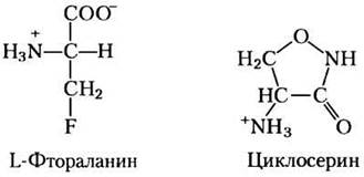
D-Amino acids are generally absent from proteins, yet they perform specialized functions in the Structure of Bacterial Cell Walls and are constituents of peptide antibiotics. Bacterial peptidoglycan (see Fig. 20-31) contains D-Alanine and D-glutamate. D-Amino acids are generated from their L-isomers by amino acid racemases, which employ Pyridoxal phosphate as a cofactor (see Fig. 18-6). Undoubtedly, Amino Acid Racemization is critically important for bacterial metabolism, making enzymes such as alanine racemase prime targets for pharmacological agents. One such agent, L-fluoroalanine, has been investigated as an antibacterial drug, while another, cycloserine, is used in the Treatment of tuberculosis. Unfortunately, these inhibitors also affect certain human PLP-dependent enzymes, which can lead to unwanted side effects. ■
Aromatic Amino Acids Are Precursors of Many Plant Substances
In plants, phenylalanine, Tyrosine, and Tryptophan are converted into a variety of essential compounds. Lignin, a tough, high-molecular-weight polymer derived from Phenylalanine and Tyrosine, is second only to Cellulose in Abundance among plant tissues. The polymeric structure of lignin is not yet fully understood. Tryptophan serves as the precursor for the plant Growth Hormone indole-3-acetate, or auxin (Fig. 22-28a), which regulates a vast array of biological processes in plants.
Fig. 22-28. Biosynthesis of two plant-specific substances from amino acids: (a) indole-3-acetate (auxin) and (b) cinnamate (the compound responsible for the flavor of cinnamon).

Phenylalanine and tyrosine also give rise to numerous commercially valuable natural products, including Tannins, which inhibit oxidation processes in wines; alkaloids such as morphine, which exert profound physiological effects on The Human Body; and alkaloids found in spices (cinnamon, nutmeg, cloves, vanilla, red pepper, and others).
Biogenic Amines Are Products of AMINO ACID DECARBOXYLATION
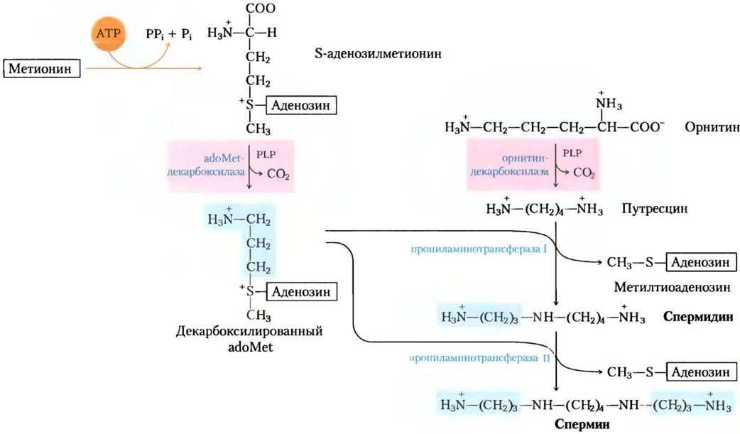
Many important neurotransmitters are primary or secondary amines derived from amino acids via straightforward pathways. In addition, certain Polyamines that form complexes with DNA are synthesized from Ornithine, an intermediate of The Urea Cycle. The decarboxylation reaction, common to several metabolic pathways, is another PLP-dependent process (see Fig. 18-6).
The synthesis of several neurotransmitters is illustrated in Fig. 22-29. Tyrosine is the precursor of the catecholamine family: dopamine, norepinephrine, and epinephrine. Among other functions, catecholamine levels correlate with Blood Pressure Regulation. Parkinson's disease, a neurological disorder, is caused by diminished dopamine production and is typically treated by administering L-dopa. Elevated dopamine release in the brain has also been implicated in psychological disorders such as Schizophrenia.
Fig. 22-29. Biosynthesis of selected neurotransmitters from amino acids. In each case, the key step is identical—a PLP-dependent decarboxylation (highlighted in pink).

The decarboxylation of glutamate yields γ-aminobutyric acid (GABA), a major inhibitory neurotransmitter. A deficiency in this substance triggers epileptic seizures. GABA analogues are widely used in the treatment of Epilepsy and Hypertension. Furthermore, GABA levels can be elevated by administering Inhibitors of the enzyme responsible for GABA degradation—GABA aminotransferase. Another vital neurotransmitter, serotonin, is synthesized from tryptophan in a two-stage process.
The decarboxylation of Histidine yields histamine, which exerts a potent vasodilatory effect in animal tissues. Histamine is released in large quantities during allergic reactions and also stimulates gastric acid secretion. Although a wide range of antihistamines is currently under active development, all of them ultimately affect either the synthesis or the storage of histamine. A well-known example of a histamine receptor antagonist is cimetidine (Tagamet), a structural analogue of histamine:
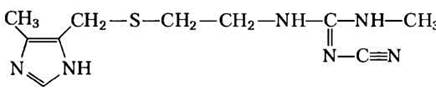
It is prescribed for duodenal ulcers to inhibit gastric acid secretion.
Box 22-3. MEDICINE. Treatment of African Sleeping Sickness with a Biochemical "Trojan Horse"
African sleeping sickness, or African trypanosomiasis, is caused by protozoan parasites (Unicellular Eukaryotes) known as trypanosomes (Fig. 1). This disease (along with related trypanosomal infections) poses a significant medical and economic burden in many developing nations. Until recently, the condition was virtually incurable. Vaccines are ineffective because these parasites employ an unusual mechanism to evade the host immune system.
Fig. 1. Trypanosoma brucei rhodesiense, one of the several known trypanosomes that cause African sleeping sickness.

The Cell wall of trypanosomes is coated with a single predominant protein—an antigen that triggers the host Immune Response. However, through genetic recombination (see Table 28-1, vol. 3), certain cells within the infecting trypanosome population periodically switch to synthesizing a new coat protein, thereby escaping immune recognition. This "coat-switching" process can occur hundreds of times. As a result, a chronic, relapsing infection develops: the infected individual suffers from bouts of fever that subside once The Immune System clears the initial wave of parasites, only for a newly coated variant to trigger another infection, causing the fever to return. This cycle may persist for weeks, ultimately leading to the death of the debilitated patient.
Some modern approaches to treating African sleeping sickness rely on a detailed understanding of parasite metabolism and enzyme kinetics. At least one therapeutic strategy employs Pharmaceuticals designed as mechanism-based enzyme inactivators (also known as suicide inactivators or irreversible inhibitors; see p. 298, vol. 1). A vulnerable metabolic pathway in trypanosomes is polyamine biosynthesis. Rapidly dividing cells require large amounts of the polyamines spermine and spermidine for DNA packaging. The initial step of their synthesis is catalyzed by the PLP-dependent enzyme ornithine decarboxylase (see Fig. 22-30). In mammalian cells, ornithine decarboxylase undergoes rapid turnover, meaning the enzyme is continuously synthesized and degraded. In certain trypanosomes, however, for reasons not yet fully understood, the enzyme is remarkably stable and is replaced very slowly through de novo synthesis. An ornithine decarboxylase inhibitor that binds tightly to the enzyme will have minimal impact on human cells—where the inactivated enzyme is rapidly replaced by newly synthesized molecules—while proving highly toxic to the parasite.
The initial steps of the ornithine decarboxylase reaction are illustrated in Fig. 2. Upon the removal of CO2, the electron flow reverses, yielding putrescine (see Fig. 22-30). Building upon this mechanism, several irreversible suicide inactivators have been developed, one of which is difluoromethylornithine (DFMO). DFMO is relatively inert in solution; however, upon binding to ornithine decarboxylase, the enzyme is rapidly inactivated (Fig. 3). The inhibitor acts by redirecting the electron flow via two strategically positioned fluorine atoms, which serve as excellent "leaving" groups. Instead of electrons being transferred to the cyclic structure (PLP), the reaction culminates in the displacement of a fluorine atom. The sulfur atom of an active-site Cys residue then forms a covalent complex with the highly reactive PLP-inhibitor adduct—an entirely irreversible reaction. Thus, the inhibitor hijacks the enzyme's own catalytic mechanism to halt the process.
Fig. 2. Mechanism of the ornithine decarboxylase reaction.

Fig. 3. Inhibition of ornithine decarboxylase by the irreversible suicide inactivator DFMO.

In clinical trials, DFMO demonstrated high efficacy against African sleeping sickness and is currently used to treat the disease caused by T. brucei gambiense. Therapeutic approaches to infections similar to the one shown here hold great promise for the treatment of a wide range of diseases. Drug development based on The Mechanism of the enzymatic reaction and The structure of the enzyme complex can Complement traditional trial-and-error Methods in modern pharmacology.
Polyamines, such as spermine and spermidine, are involved in DNA packaging. They are synthesized from methionine and ornithine via the pathway shown in Fig. 22-30. The first step is the decarboxylation of ornithine, a precursor of arginine (Fig. 22-10). Ornithine decarboxylase, a PLP-dependent enzyme, is inhibited by several potent therapeutic agents (Box 22-3). ■
Fig. 22-30. Biosynthesis of spermidine and spermine. PLP-dependent decarboxylation steps are highlighted in pink. In these reactions, S-adenosylmethionine (in its decarboxylated form) serves as the source of propylamine groups (highlighted in blue).
Arginine is the biological precursor of nitric oxide
A surprising discovery in the mid-1980s was the realization of the role played in living organisms by nitric oxide, NO, previously known mainly as a component of smog. This gaseous substance acts as a vital biological messenger. Nitric oxide readily diffuses across membranes, although due to its high reactivity, the distance it can travel is limited to ~ 1 mm from its site of synthesis. In humans, NO plays a crucial role in A number of physiological processes, including neurotransmission, blood clotting, and blood pressure regulation. The MECHANISM OF ACTION of NO is described in Chapter 12 (p. 628, Vol. 1).
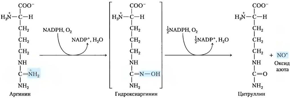
Nitric oxide is synthesized from arginine in an NADPH-dependent reaction catalyzed by nitric oxide synthase (Fig. 22-31), a dimeric molecule structurally similar to cytochrome P-450 reductase (see Box 21-1). The reaction is a five-electron oxidation. Each enzyme subunit binds one molecule of each of four distinct Cofactors: FMN, FAD, tetrahydrobiopterin, and heme iron Fe3+. Because NO is an unstable molecule, it cannot be stored. Its synthesis is stimulated by the interaction of NO synthase with Ca2+-calmodulin (see Fig. 12-21, Vol. 1).
Fig. 22-31. Biosynthesis of nitric oxide. Both steps are catalyzed by nitric oxide synthase. The nitrogen atom in the NO molecule originates from the guanidino group of arginine.
Summary of Section 22.3 Amino Acid Derivatives
■ Most important biomolecules are derived from amino acids. Glycine serves as a precursor for porphyrins. The degradation of the iron-containing porphyrin (heme) yields bilirubin, which is subsequently converted into bile pigments that perform a variety of physiological functions.
■ The energy buffers creatine and phosphocreatine are synthesized from glycine and arginine. Glutathione, formed from three different amino acids, acts as a crucial reducing agent within the cell.
■ Bacteria synthesize D-amino acids from L-amino acids via a racemization reaction that utilizes pyridoxal phosphate (PLP).
■ Aromatic amino acids serve as the source of numerous plant-specific substances. The PLP-dependent decarboxylation of Certain amino acids yields essential biological amines, including neurotransmitters.
■ Arginine acts as a precursor for nitric oxide, a key biological messenger.
Last update: 06/08/2026
Editorial and Educational Adaptation: This material has been compiled based on the primary/original source text. The project team performed an editorial review, corrected technical inaccuracies, structured sections, and adapted the content for an educational format.
What was processed:
- elimination of formatting defects (OCR errors, structural breaks, corrupted characters);
- editorial organization of content;
- standardization of terminology in accordance with academic sources;
- verification of factual statements against the original source text.
All mentions of the author, publication year, and origin of the primary text have been preserved in accordance with the source.