Human Biochemistry Volume 1 - Murray R. 1993
Protein and Amino Acid Metabolism
Porphyrins and Bile Pigments
Porphyrins
Porphyrins are cyclic compounds formed by four pyrrole rings linked together by methene bridges (Fig. 33.1). A characteristic property of porphyrins is their ability to form complexes with Metal Ions that bind to the nitrogen atoms of the pyrrole rings. Examples include iron porphyrins, notably heme, which is a component of Hemoglobin, and the magnesium-containing porphyrin chlorophyll, a plant pigment involved in Photosynthesis.
In nature, metalloporphyrins bind to Proteins, resulting in compounds that play a vital role in biological processes. These include:
A. Hemoglobin — iron porphyrins bound to the protein globin. Hemoglobins have The ability to reversibly bind oxygen, transporting this gas through the Circulatory system (see Ch. 6). The Structure of heme is shown in Fig. 6.2.
B. Erythrocruorins — iron porphyrinoproteins found in the Blood and tissue fluids of certain invertebrates; they perform the same function as hemoglobin.
C. Myoglobins — respiratory pigments found in the Muscle Cells of vertebrates and invertebrates. An example is Myoglobin from horse Heart muscle, crystallized by Theorell in 1934. The myoglobin molecule is structurally similar to a hemoglobin subunit.
Class="center">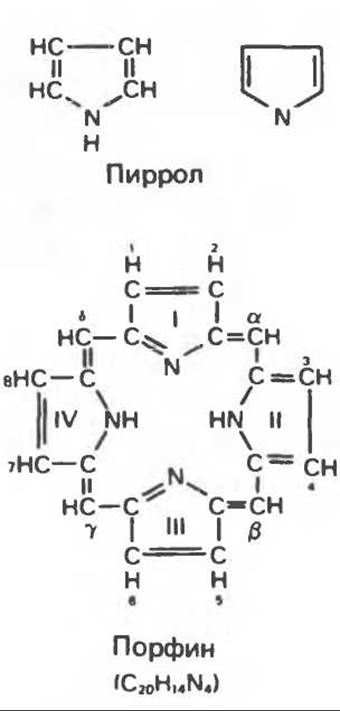
Fig. 33.1. Porphine molecule. The rings are designated by Roman numerals I, II, III, and IV. The attachment sites of substituent groups are indicated by numbers 1, 2, 3, 4, 5, 6, 7, 8. The methene bridges are designated by the letters a, ß, y, and δ.
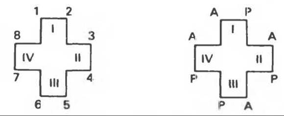
Fig. 33.2. Uroporphyrin III.
D. Cytochromes — compounds that function as electron carriers in oxidation-reduction reactions. An important example is cytochrome c, which has a Molecular Weight of approximately 13,000 and contains 1 gram-atom of iron per mole of protein.
E. Catalases — iron porphyrin Enzymes; several catalases have been obtained in crystalline form. In plants, catalase activity is negligible; a similar function is performed there by another iron porphyrin enzyme, peroxidase.
F. Tryptophan pyrrolase. This enzyme catalyzes The oxidation of tryptophan to formylkynurenine. It is also an iron porphyrin protein.
Structure of Porphyrins
Naturally occurring porphyrins are compounds in which 8 hydrogen atoms of the porphyrin core are replaced by various side chains, as shown in Fig. 33.1. A simplified method for depicting THE POSITION OF substituents was proposed by Fischer: the pyrrole rings (omitting the methene bridges) are represented as protrusions of a cross-like structure, whose numbered vertices serve as attachment sites for the substituents (Fig. 33.2). Various porphyrins are shown in Figs. 33.2, 33.3, and 33.4, using the following Abbreviations:
A (acetate) = —СН2СООН; Р (propionate) = —СН2СН2СООН; М (methyl) = —CH3; V (vinyl) = —СН = СН2.
The arrangement of substituent groups A and P in uroporphyrin is asymmetric (in ring IV, compared to the other rings, the order of attachment of acetate and propionate groups is reversed). A porphyrin with this type of asymmetric substitution is classified as a type III porphyrin. A porphyrin with a completely symmetric arrangement of substituent groups is classified as a type I porphyrin. Only type I and type III porphyrins are found in nature, with type type III being significantly more common (Fig. 33.3).
Both compounds shown in Fig. 33.4 belong to type III porphyrins (the methyl groups are arranged asymmetrically, as in coproporphyrin type III). However, they are sometimes classified as belonging to type IX because they occupied the ninth place in the series of isomers postulated by Hans Fischer, a pioneer in the chemistry of porphyrins.
Biosynthesis of Porphyrins
Chlorophyll, the plant pigment of the photosynthetic system, and heme, the iron protoporphyrin of animal hemoglobin, are synthesized in living cells via a common metabolic pathway. The starting Materials are "active succinate" — succinyl-CoA, formed in the Cell/35.html">Mitochondria during the reactions of The Citric Acid Cycle — and The amino acid Glycine. An "activation" of glycine by Pyridoxal phosphate is also required. Glycine presumably forms a Schiff base with pyridoxal; subsequently, the a-carbon of glycine attaches to the carbonyl carbon of succinate. The Condensation product of glycine and succinyl-CoA is a-amino-β-ketoadipic acid, which rapidly decarboxylates to form δ-aminolevulinate (ALA) (Fig. 33.5). This step is catalyzed by the enzyme ALA synthase. This enzyme is likely the rate-controlling step in porphyrin biosynthesis in mammalian Liver. The synthesis of aminolevulinic acid occurs in the mitochondria. In the Cytosol, the enzyme ALA dehydratase catalyzes the condensation of two molecules of ALA to form two molecules of Water and one molecule of porphobilinogen (Fig. 33.5). ALA dehydratase is a zinc-containing enzyme and is inhibited by lead ions.

Fig. 33.3. Uroporphyrins and coproporphyrins.
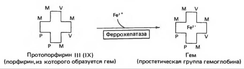
Fig. 33.4. The incorporation of iron into protoporphyrin leads to The formation of heme.
The Formation of the tetrapyrrole (i.e., porphyrin) proceeds via the condensation of four monopyrroles derived from porphobilinogen (Fig. 33.6). The amino-bearing carbon atom of the porphobilinogen molecule (the "former" a-carbon of glycine) becomes the carbon of the methylene group (a, ß, y, δ) linking adjacent pyrrole rings into the tetrapyrrole structure. Although The conversion of porphobilinogen into porphyrin can occur simply by heating in an acidic environment (e.g., in acidic urine), in Tissues this transformation is catalyzed by specific enzymes.
As noted above, only type I and type III porphyrins are found in nature; the fact that type III isomers are more widely distributed can be explained by the circumstance that biologically important porphyrins (heme and cytochromes) are type III isomers.
Currently, the details of uroporphyrinogen formation via porphobilinogen condensation remain unclear. The conversion of porphobilinogen into uroporphyrinogen III, an intermediate in heme biosynthesis, is catalyzed by a two-enzyme complex. Uroporphyrinogen I synthase, also known as porphobilinogen deaminase, catalyzes the in vitro condensation of porphobilinogen into uroporphyrinogen I (Fig. 33.6). However, in the presence of a second enzyme, uroporphyrinogen III cosynthase, the concerted action of both enzymes yields uroporphyrinogen III rather than its symmetrical isomer, uroporphyrinogen I (Fig. 33.6). Under normal conditions, almost exclusively the type III isomer is produced; however, in certain types of porphyria (discussed below), type I isomers are synthesized in significant quantities.

Fig. 33.5. Biosynthesis of porphobilinogen. ALA synthase is located in the mitochondria, whereas ALA dehydratase is located in the cytosol.
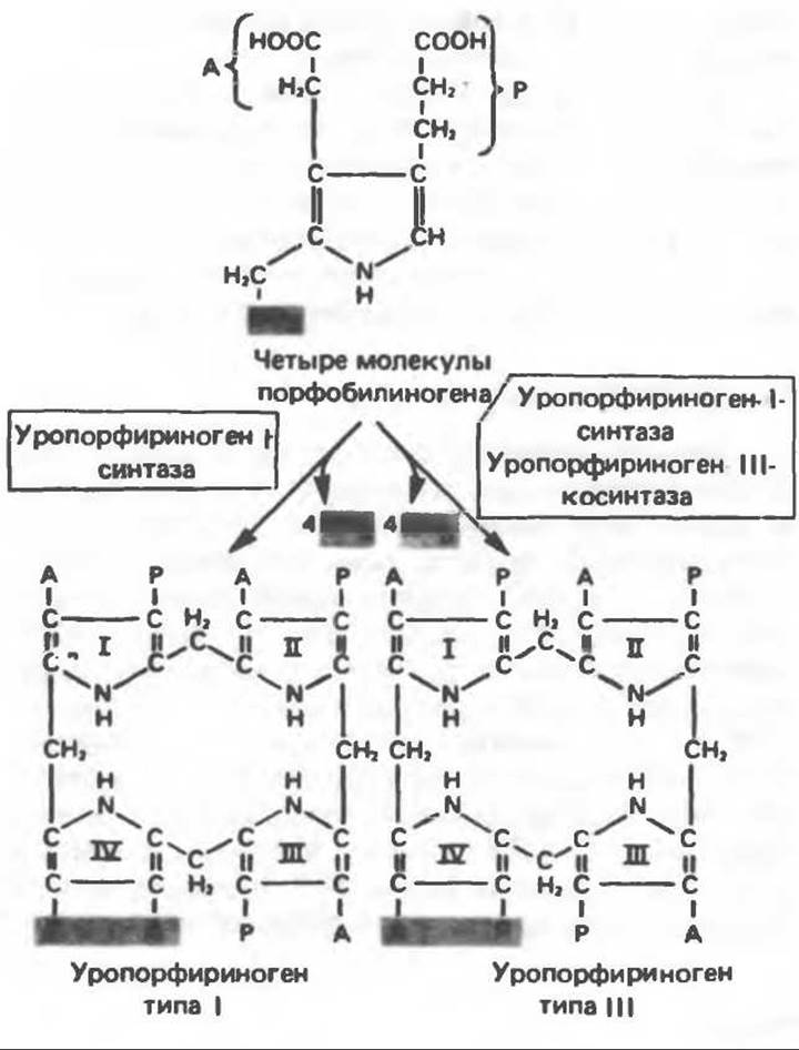
Fig. 33.6. Conversion of porphobilinogen into uroporphyrinogens.
Note that in these uroporphyrinogens, the pyrrole rings are linked by methylene bridges, meaning they lack a conjugated system. Consequently, these compounds (and all porphyrinogens in general) are colorless. However, porphyrinogens readily undergo auto-oxidation to the corresponding porphyrins. This oxidation is promoted by light and by porphyrins that have already been formed.
Uroporphyrinogen III is converted into coproporphyrinogen III by the decarboxylation of all acetate groups (A), which are replaced by methyl groups (M). This reaction is catalyzed by uroporphyrinogen decarboxylase, which can also catalyze the conversion of uroporphyrinogen I to coproporphyrinogen I (Fig. 33.7). Coproporphyrinogen III then enters the mitochondria, where it is converted into protoporphyrinogen III and subsequently into protoporphyrin III. This transformation likely involves multiple steps. The mitochondrial enzyme coproporphyrinogen oxidase catalyzes the decarboxylation and oxidation of two propionic side chains, leading to the formation of protoporphyrinogen. This enzyme acts exclusively on type III coproporphyrinogen, which apparently explains the absence of type I protoporphyrin in natural materials. The oxidation of protoporphyrinogen to protoporphyrin is catalyzed by another mitochondrial enzyme, protoporphyrinogen oxidase. In mammalian liver, the conversion of coproporphyrinogen to protoporphyrin requires molecular oxygen.

Fig. 33.7. Decarboxylation of uroporphyrinogens to form coproporphyrinogens (in the cytosol). A — acetate group, M — methyl group, P — propionyl group.
Heme Biosynthesis
The final step in heme synthesis is the incorporation of ferrous iron into protoporphyrin; this reaction is catalyzed by the mitochondrial enzyme heme synthase, or ferrochelatase (Fig. 33.4). The reaction proceeds readily even in the absence of the enzyme, but its rate is significantly enhanced upon The addition of tissue preparations due to the presence of enzymes that catalyze iron chelate formation.
A summary scheme of The biosynthesis of porphyrin derivatives from porphobilinogen is presented in Fig. 33.8. Heme biosynthesis occurs in most mammalian tissues, with the exception of mature erythrocytes, which lack mitochondria.
The porphyrinogens described above are colorless and contain 6 additional hydrogen atoms compared to the corresponding colored porphyrins. It is now well established that these reduced porphyrins (porphyrinogens), rather than the corresponding porphyrins, serve as intermediates in the biosynthesis of protoporphyrin and heme.
Regulation of Heme Biosynthesis
The rate-limiting step in heme synthesis is the condensation of succinyl-CoA and glycine to form ALA (Fig. 33.5); this reaction is catalyzed by $\delta$-aminolevulinic acid synthase (ALA synthase). In normal tissues capable of heme synthesis, the level of ALA synthase activity is significantly lower than that of Other Enzymes involved in the pathway. Nevertheless, ALA synthase is a regulatory enzyme. It is believed that heme acts as a negative regulator of ALA synthase synthesis by interacting with an aporepressor molecule. The Mechanism of repression is shown schematically in Fig. 33.9. Feedback inhibition by heme likely also occurs at this stage; however, the primary regulatory effect of heme is that the synthesis of ALA synthase is markedly accelerated in the absence of heme and slowed down in its presence. The turnover rate of ALA synthase in mammalian liver is normally high (with a half-life of about 1 h), which is expected for an enzyme catalyzing a rate-limiting step.
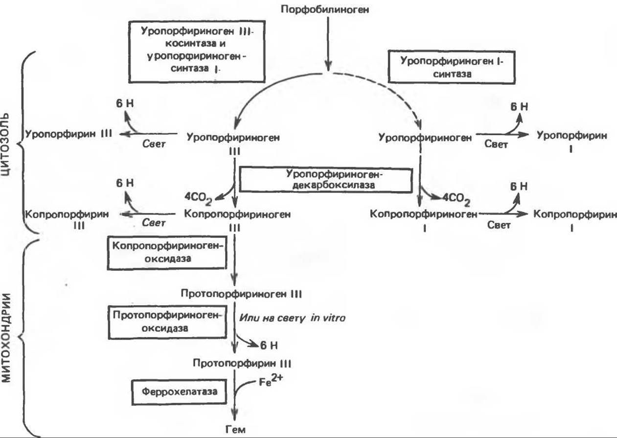
Fig. 33.8. Stages of the biosynthesis of porphyrin derivatives from porphobilinogen.
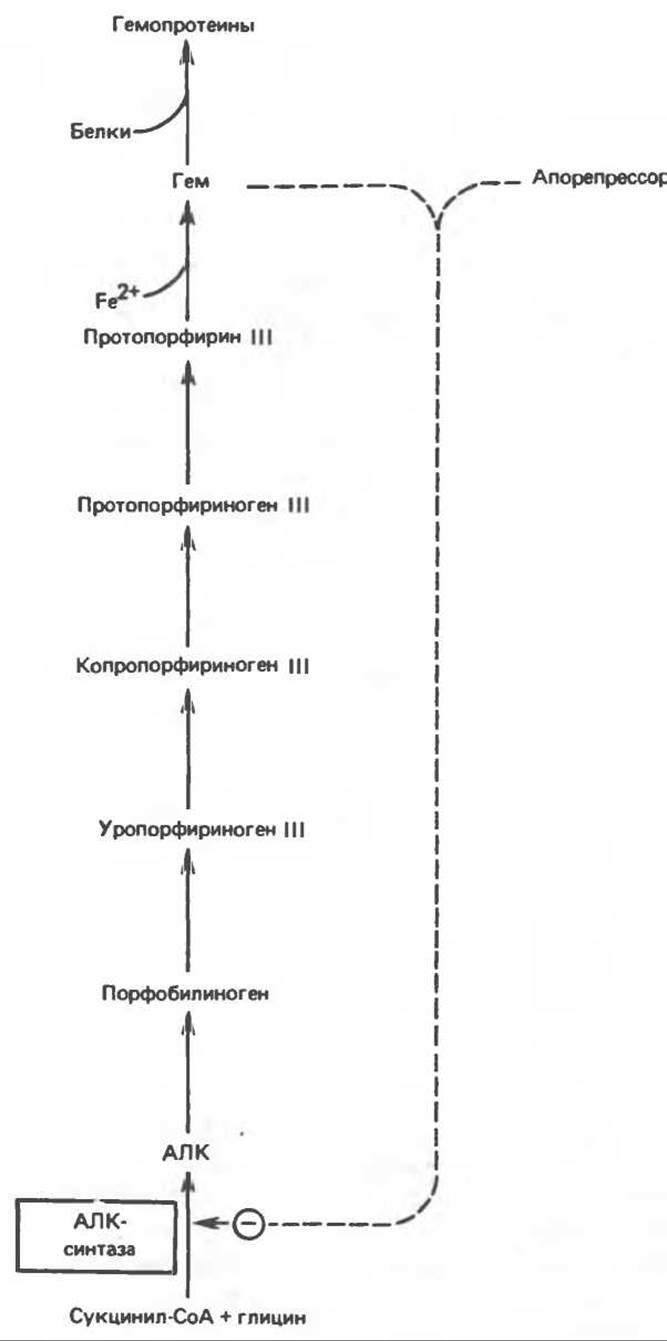
Fig. 33.9. Regulation of heme synthesis at the step catalyzed by ALA synthase, via a mechanism of repression and derepression involving heme and its hypothetical aporepressor. The dashed line indicates negative (⊝) regulation through the repression of ALA synthase synthesis.
Many structurally diverse compounds, including currently used insecticides, carcinogens, and Pharmaceuticals, can substantially increase hepatic levels of ALA synthase. Most drug compounds are metabolized in the liver by a system that includes the specific hemoprotein cytochrome P-450. During the METABOLISM of these agents, the consumption of heme by the cytochrome P-450 system increases significantly, leading to a drop in intracellular heme concentration. This, in turn, triggers the derepression of ALA synthase synthesis and, consequently, an increased rate of heme synthesis to meet cellular demands.
The induction of hepatic ALA synthase is also influenced by several other factors. Glucose can inhibit ALA synthase induction; chelated iron exerts a synergistic effect on the induction of liver ALA synthase; and Steroids promote the in vivo derepression of ALA synthase by drugs. Conversely, the administration of hematin can prevent ALA synthase derepression (a similar effect is characteristic of other Hemoproteins). In erythropoietic tissues, ALA synthase activity increases during Hypoxia, whereas hypoxia has no effect on ALA synthase activity in the liver.
The important role of these regulatory mechanisms will be discussed later in the context of diseases belonging to the porphyria group.
Chemistry of Porphyrins
Due to the presence of tertiary nitrogens in two of their pyrrole rings, porphyrins exhibit weak base properties. Porphyrins that contain carboxyl groups in one or more side chains also function as acids. Their isoelectric points typically range from 3.0 to 4.5; within this pH range, porphyrins readily precipitate.
Various porphyrinogens are colorless, whereas all porphyrins are colored. When studying porphyrins and their derivatives, their characteristic absorption spectra in both the visible and ultraviolet regions are of great importance. An example is the absorption curve of a porphyrin solution in 5% Hydrochloric acid (Fig. 33.10). Note the sharp absorption maximum near a wavelength of 400 nm. This is a hallmark of the porphyrin Nucleus, characteristic of all porphyrins regardless of The Nature of their side chains. This maximum is called the Soret band, named after its discoverer. In an acidic medium, hematoporphyrin exhibits, In addition to the Soret band, two weaker maxima at 550 and 592 nm.
When solutions of porphyrins in strong mineral acids or organic Solvents are irradiated with ultraviolet light, they emit an intense red fluorescence. This fluorescence is so characteristic that it is frequently used to detect small quantities of free porphyrins. The absorption and fluorescence of porphyrins are due to the presence of double bonds; as already mentioned, the reduction of methenyl (—НС =) bridges (by the addition of hydrogens) to methylene (—СН2—) groups leads to the formation of colorless porphyrinogens.
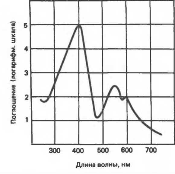
Fig. 33.10. Absorption spectrum of hematoporphyrin (0.01% hematoporphyrin solution in 5% HCl).
The incorporation of a metal into a porphyrin alters its visible absorption spectrum. An example is protoporphyrin, the iron-free precursor of heme. In alkaline solution, the porphyrin spectrum exhibits several narrow and intense absorption bands (at 645, 591, and 540 nm), whereas heme is characterized by a broad plateau-like absorption band extending from 540 to 580 nm.
Determination of Porphyrins
Coproporphyrins and uroporphyrins are of considerable clinical interest because their excretion is elevated in Porphyrias. Coproporphyrins I and III are soluble in mixtures of ether and glacial acetic acid, from which they can subsequently be extracted with hydrochloric acid. Uroporphyrins, by contrast, are insoluble in these mixtures, though partially soluble in ethyl acetate, and can be extracted with hydrochloric acid. Upon ultraviolet irradiation, the resulting hydrochloric acid solutions yield a characteristic red fluorescence. Characteristic absorption bands can be recorded using a spectrophotometer.
Table 33.1. Upper limits of normal excretion of porphyrins and their precursors, and their content in erythrocytes 1)
|
Urine (µg∙day-1) |
Feces (µg/g dry weight) |
Erythrocytes (µg/100 ml cell suspension) |
|
|
ALA |
4000 |
— |
— |
|
Porphobilinogen |
1500 |
— |
— |
|
Uroporphyrin |
50 |
5 |
Trace |
|
Coproporphyrin |
300 |
50 |
3 |
|
Protoporphyrin |
— |
120 |
80 |
1) Reproduced (with permission), with modifications, from the review by Meyer U. A., Schmid R. The porphyrias. In: The Metabolic Basis of Inherited Disease, 4th ed. Stanbury J. B., Wyngaarden J. B., Fredrickson D.S. (eds). McGraw-Hill, 1978.
Table 33.1 lists the upper limits of normal excretion for porphyrins and their precursors. In healthy individuals, urinary coproporphyrin excretion averages approximately 67 µg∙day-1, with type I isomer accounting for an average of 14 µg∙day-1 and type III isomer for 53 µg∙day-1. Deviations from this normal excretion ratio of type I and III coproporphyrins can serve as a diagnostic indicator in certain liver diseases.
The intermediates formed sequentially during heme biosynthesis from ALA become progressively more hydrophobic. The formation of coproporphyrinogen is accompanied by the removal of the acetyl groups of uroporphyrinogen; the conversion of coproporphyrinogen to protoporphyrinogen involves the decarboxylation of two propionyl groups. This increase in Hydrophobicity dictates the distribution of heme biosynthetic intermediates between urine and feces. The more polar uroporphyrinogen is excreted predominantly in the urine, whereas the more hydrophobic coproporphyrinogen and protoporphyrinogen are partitioned primarily into the Bile and eliminated via the feces.
Last update: 06/08/2026
Editorial and Educational Adaptation: This material has been compiled based on the primary/original source text. The project team performed an editorial review, corrected technical inaccuracies, structured sections, and adapted the content for an educational format.
What was processed:
- elimination of formatting defects (OCR errors, structural breaks, corrupted characters);
- editorial organization of content;
- standardization of terminology in accordance with academic sources;
- verification of factual statements against the original source text.
All mentions of the author, publication year, and origin of the primary text have been preserved in accordance with the source.