Fundamentals of Molecular Biology - V.I. Rezyapkin 2009
Replication
The mechanism of replication in E. coli
There are three DNA polymerases present in E. coli Cells. Let us characterize them.
DNA polymerase I
DNA polymerase I consists of a single polypeptide chain containing 2 active sites:
a) the 1st Active Site is responsible for polymerase and 3’→5’ exonuclease activities. The latter ensures the removal of incorrectly incorporated NUCLEOTIDES;
b) the 2nd active site is responsible for 5’→3’ exonuclease activity. This activity is required to remove the RNA primer during Replication.
DNA polymerase II
This enzyme exhibits polymerase and 3’→5’ exonuclease activities and preferentially acts on double-stranded DNA with gaps (Fig. 2.9). DNA polymerase II is involved in DNA Repair.
Class="center">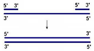
Fig. 2.9. DNA polymerase II preferentially acts on double-stranded
DNA with gaps
DNA polymerase III
This enzyme exhibits polymerase and 3’→5’ exonuclease activities and consists of ten types of subunits. Its primary function is METABOLISM/36.html">DNA replication. The synthesis rate is 500 nucleotides per second
The E. coli chromosome has a single origin of replication (oriC), with a size of 258 bp. The DnaA protein recognizes oriC and initiates replication. As a result, the ATP-dependent helicase begins unwinding the DNA duplex (Fig. 2.10). Topoisomerase, positioned ahead of the moving Replication fork, relieves the torsional strain generated by the unwinding of the DNA double helix. SSB Proteins bind to the resulting single-stranded DNA regions (Fig. 2.10). Primase synthesizes the primer (Fig. 2.10, 2.11), which is required for DNA polymerase activity. Then, DNA polymerase III comes into play, sequentially adding nucleotides to the 3’ end of the polynucleotide chain (Fig. 2.10). Since DNA Synthesis proceeds in the 5’→3’ direction, one strand (the leading strand) is synthesized continuously (Fig. 2.11), while the second strand is synthesized in fragments (the lagging strand) of 1000–2000 nucleotides (Okazaki fragments). Upon completion of an Okazaki fragment, DNA polymerase I uses its 5’→3’ exonuclease activity to remove the RNA primer and replace it with DNA. Following the action of this enzyme, a nick remains between the Okazaki fragments, which is then sealed by DNA ligase (Fig. 2.10, 2.11).

Fig. 2.10. Cooperative action of proteins at the replication fork

Fig. 11. Synthesis of DNA strands during replication
Termination of replication occurs after the duplication of the circular DNA molecule.
Last update: 12/08/2026
Editorial and Educational Adaptation: This material has been compiled based on the primary/original source text. The project team performed an editorial review, corrected technical inaccuracies, structured sections, and adapted the content for an educational format.
What was processed:
- elimination of formatting defects (OCR errors, structural breaks, corrupted characters);
- editorial organization of content;
- standardization of terminology in accordance with academic sources;
- verification of factual statements against the original source text.
All mentions of the author, publication year, and origin of the primary text have been preserved in accordance with the source.