Biochemistry - The Chemical Reactions of Living Cells, Volume 3 - D. Metzler 1980
Biochemical genetics and the synthesis of nucleic acids and proteins
DNA replication
Direction of replication
According to current understanding, METABOLISM/36.html">DNA Replication proceeds via the mechanism outlined in equation (15-3). As the DNA unwinds at the Replication fork, new DNA segments are synthesized along the parental strands. This raises an important question: does replication proceed in only a single direction, or do two forks originate at the Transcription initiation point and subsequently travel in opposite directions around the chromosome? This question was successfully answered by combining genetic techniques and Electron Cell/15.html">Microscopy.
One of the Methods used to determine the direction of replication in E. coli was as follows. Phage λ prophage was integrated into the bacterial chromosome at the att site (Fig. 15-1), while phage Mu-1 DNA was integrated at numerous other sites scattered along the chromosome [189]. Phage Mu-1 is particularly convenient for this purpose because its insertion can occur at many sites within well-mapped genes. Insertion within a Gene inactivates that gene (insertion mutation), making it possible to precisely map the Location OF THE Mu-1 prophage. A series of bacterial strains containing both λ prophages and the Mu-1 phage was successfully obtained, with the latter located at various positions along the chromosome. Furthermore, these Bacteria were auxotrophic for specific Amino Acids. This allowed replication to be halted by depriving the bacteria of the required amino acid (although a round of replication already underway was usually completed). When the missing amino acids were restored to the growth medium, replication resumed, starting from the origin. Simultaneously with the amino acids, bromouracil was added to the medium, which is incorporated into DNA in place of thymine. Consequently, the newly synthesized DNA strands had a higher density than the parental ones. At various time intervals following the initiation of replication1), the newly formed DNA strands were isolated by CsCl density gradient centrifugation (Chap. 2, Sec. 3, 1, d), after which their ability to hybridize with both λ and Mu-1 phage DNA was assessed.
1) Following The addition of amino acids, replication did not begin simultaneously in all Cells, which is why the newly synthesized DNA molecules varied in length.
Class="center">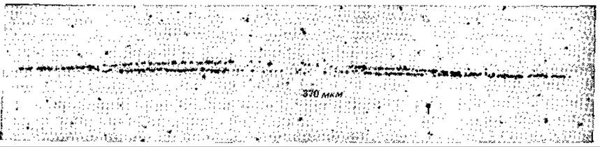
FIG. 15-28. Bidirectional Replication of the E. coli chromosome. Autoradiograph of a pair of replication forks obtained from a chromosome whose replication took place in the presence of 3H-thymine (5 Ci/mmol), with the cells subsequently incubated for 6 min at a specific stage of replication with 3H-thymidine of very high specific radioactivity (52 Ci/mmol). The total length of the grain track containing silver grains is 370 µm (Kuempel P. L. et al., in: DNA Synthesis in Vitro, R. Wells and R. Inman, eds., pp. 463–472, Copyright 1972 University Park Press, Baltimore).
Based on the ratios of Mu-1 and λ phage DNA across various strains, it was possible to map the spread of replication starting from the origin located near the ilv gene at approximately 74 min (Fig. 15-1). It was demonstrated that replication proceeds bidirectionally along the chromosome and terminates between the trp and his genes at roughly the 25th min.
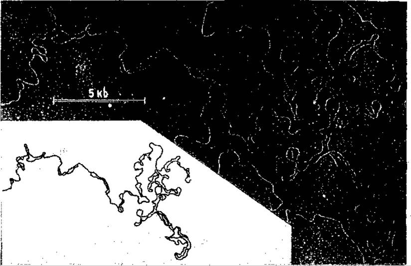
FIG. 15-29. A fragment of replicating chromosomal DNA from disrupted Drosophila melanogaster nuclei [191]. Spread in the presence of formamide, the DNA reveals several "eyes" formed at sites of RNA replication. See Fig. 2-23, B.
Autoradiographic studies confirmed the bidirectional nature of replication in E. coli using special amino acid auxotrophic strains characterized by low nucleoside triphosphate pools. The addition of amino acids following starvation induced replication initiation with a lag period of only 6 min. Cells were pulsed with 3H-thymidine, and once the replication forks had progressed a short distance from the origin, the cells were briefly pulse-labeled with 3H-thymidine of very high specific radioactivity. Under these conditions, bidirectionally moving replication forks were clearly visible on the autoradiographs [190] (Fig. 15-28). DNA replication in Drosophila Chromosomes was also investigated in rapidly dividing nuclei using electron microscopy [191]. The rate of replication in these nuclei was found to be approximately 300,000 Base Pairs per second; however, according to data from the same study, replication forks in animal chromosomes cannot travel faster than ~50 base pairs per second. Thus, one would expect there to be at least 6,000 forks in the chromosome, or one fork per 10,000 base pairs. Indeed, such a high number of forks was successfully detected [191]. The forks appear in pairs, and careful examination revealed that many short regions contain single-stranded DNA—as if one strand in the fork were replicating faster than the other. The Structure of the single-stranded regions between paired forks points to bidirectional replication (Fig. 15-29). Replication in Bacillus subtilis also proceeds bidirectionally, although the forks move in both directions at different rates [192]. DNA replication in phages λ and T7 is likewise bidirectional [193], whereas mouse Mitochondrial DNA replicates unidirectionally [194].
Last update: 06/08/2026
Editorial and Educational Adaptation: This material has been compiled based on the primary/original source text. The project team performed an editorial review, corrected technical inaccuracies, structured sections, and adapted the content for an educational format.
What was processed:
- elimination of formatting defects (OCR errors, structural breaks, corrupted characters);
- editorial organization of content;
- standardization of terminology in accordance with academic sources;
- verification of factual statements against the original source text.
All mentions of the author, publication year, and origin of the primary text have been preserved in accordance with the source.