Genetics - A. V. Sivolob 2008
The Nature of Genetic Material
Structural Organization of DNA in Cells
Chromosomes
In the somatic Cells of the vast majority of Multicellular Organisms, There is a double (paired) set of chromosomes: the homologous chromosomes of each pair contain nearly identical DNA molecules with almost the same set of genes, which are arranged along the DNA molecule in the same order for both chromosomes (occupying specific, defined positions known as chromosome loci). Thus, a Gene can be regarded as a specific chromosomal locus. During The formation of Germ Cells (Gametes), each receives one chromosome from each pair of homologous chromosomes (see the Meiosis process described below), meaning the germ Cell contains a single (haploid) set of chromosomes. Consequently, Fertilization brings together two haploid chromosome sets to form the diploid set of the offspring. Since the two homologous chromosomes originate from different individuals, they are not entirely identical: certain genes may be absent in one of the homologous chromosomes or may differ in their nucleotide sequences. The alternative variants of a single gene (locus) are referred to as the alleles of that gene.
It should be noted that the chromosome sets of many sexually reproducing organisms contain a single pair of sex chromosomes (all other chromosomes are termed autosomes), which are represented by two non-homologous types. As a rule, one sex is homogametic (containing two sex chromosomes of the same type) and the other is heterogametic (having different sex chromosomes), which forms the basis for determining the sex of the offspring upon fertilization (see Chapter 6). It is also worth noting that The Genome of a species is typically understood as the total collection of DNA sequences within its haploid set. It is precisely in this sense that data on genome sizes and chromosome numbers are presented in Table 1.1.
When a cell enters mitosis, following METABOLISM/36.html">DNA Replication (and consequently, the doubling of the diploid chromosome set), the Formation of the highly compact mitotic chromosome begins, although the details of its structural Organization remain insufficiently understood. Simultaneously with the compaction of the Chromatin fibril, a portion of the lamina "dissolves" along with the nuclear envelope, while another portion, together with the internal matrix, reorganizes to form the protein scaffold of the mitotic chromosome—the chromosomal scaffold—to which the bases of the chromatin loops remain anchored. The compact mitotic chromosome, which becomes visible under an optical Microscope after appropriate staining, consists of two arms (Fig. 1.14) separated by a centromeric constriction.
Class="center">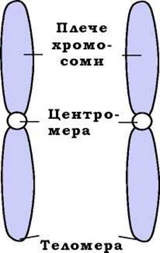
Fig. 1.14. Diagram of the Morphology of two homologous mitotic chromosomes
The centromere, which separates the two arms of a chromosome, is a chromosomal region where a complex multi-protein assembly—the kinetochore—is formed, which is necessary for the attachment of mitotic spindle microtubules. In the Cytology/cytology/16.html">Early stages of mitosis, As a result of prior DNA replication (see below), each chromosome is represented by two sister chromatids joined together at their centromeres through Protein-Structure/156.html">Protein Interactions. Later, the spindle apparatus ensures the segregation of the daughter chromosomes (see the Discussion of mitosis below).
The length of the centromeric DNA region varies widely (ranging from 125 Base Pairs in the Yeast Saccharomyces cerevisiae to 0.1–4 million base pairs in humans). In some species, such as the nematode Caenorhabditis elegans and the crayfish, individual chromosomes possess a diffuse centromere, where microtubules attach along the entire length of the chromosome (so-called holocentric chromosomes). Most frequently, the DNA sequence in the centromeric region is represented by tandem AT-rich repeats and lacks active genes. There is no Homology between the centromeric sequences of different organisms, nor are there consensus motifs exclusive to centromere DNA: the leading role in determining centromere structure is played by Proteins that interact with DNA in this region. Centromeric proteins (one of the principal ones being CENP-A, an analog of histone H3 incorporated into nucleosomes within the centromere zone) ensure the compaction of the chromosome's centromeric zone and recruit kinetochore proteins to this area.
THE POSITION OF the centromere on a given chromosome is a constant characteristic: during every Cell Division, the Location OF THE microscopic centromeric constriction remains unchanged. Depending on the position of the centromere, chromosomes are classified into three types (Fig. 1.15):
✵ metacentric chromosomes — the centromere divides the chromosome into two arms of approximately equal length;
✵ submetacentric chromosomes — the centromere divides the chromosome into two arms of unequal length; these arms are designated by the Latin letters p (short arm) and q (long arm);
✵ acrocentric chromosomes — the centromere divides the chromosome into two arms whose lengths differ so significantly that the short arms are barely noticeable under a microscope.
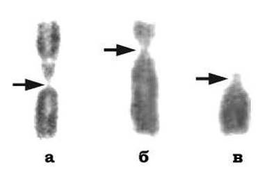
Fig. 1.15. Photographs of the Three types of human mitotic chromosomes (pairs of sister chromatids joined at their centromeres): metacentric (a), submetacentric (b), and acrocentric (c).
The arrow indicates the centromeric constriction
Each chromosome arm terminates in a telomere—a complex consisting of telomeric DNA at the chromosome end and specific telomeric proteins. The length of the telomeric region varies among organisms (for instance, over 30,000 base pairs in mice and 10,000–15,000 in humans). Furthermore, telomere length often depends on tissue type, cellular proliferative activity, and other factors.
Chromosome size, number, and relative arm lengths are all specific species characteristics. The set of morphological features used to characterize the Complement of mitotic chromosomes of a given Organism (species) is called the karyotype.
We should also consider a special case where individual chromosomes can be observed using an optical microscope outside of mitosis. In certain cells (various insect larval Tissues, mammalian trophoblast cells, plant embryo sac cells, etc.), chromosomes reach giant proportions and remain visible at all times. This is possible because the chromosome consists not of a single DNA molecule, but of hundreds or even thousands of identical molecules that accumulate as a result of repeated replication cycles without intervening mitotic divisions (endoreduplication). Such chromosomes are termed polytene chromosomes. In general, replication without subsequent cell division leads to the generation of multiple DNA molecule sets—polyploidization (see Chapter 4). However, unlike polyploid cells, in polyteny, identical copies of the DNA molecule remain tightly synapsed along their entire length, making it possible to visualize them as a single giant decondensed chromosome.
On cytological preparations, polytene chromosomes exhibit a characteristic transverse banding pattern: dark bands (disks, or chromomeres) alternate with light regions (interdisks), creating a pattern unique to each specific chromosome. It is believed that genes are predominantly localized within the disk regions, and their activation causes a characteristic Swelling of the band, known as a puff. The large size of polytene chromosomes and the relative ease of identifying specific genomic regions and chromosomal rearrangements make them exceptionally useful model objects in cytogenetics. The Biological Role of polyteny is likely related to the exceptionally high METABOLIC ACTIVITY OF certain specialized cells.
Last update: 11/08/2026
Editorial and Educational Adaptation: This material has been compiled based on the primary/original source text. The project team performed an editorial review, corrected technical inaccuracies, structured sections, and adapted the content for an educational format.
What was processed:
- elimination of formatting defects (OCR errors, structural breaks, corrupted characters);
- editorial organization of content;
- standardization of terminology in accordance with academic sources;
- verification of factual statements against the original source text.
All mentions of the author, publication year, and origin of the primary text have been preserved in accordance with the source.