LEHNINGER PRINCIPLES OF BIOCHEMISTRY - VOL 2. BIOENERGETICS AND METABOLISM - 2014
PART II. BIOENERGETICS AND METABOLISM
22. BIOSYNTHESIS OF AMINO ACIDS, NUCLEOTIDES, AND RELATED MOLECULES
22.2. Biosynthesis of Amino Acids
All Amino Acids are derived from intermediates of Glycolysis, The Citric Acid Cycle, or the Pentose Phosphate Pathway (Fig. 22-9). Nitrogen atoms enter these pathways via glutamate or glutamine. Some of these pathways are quite simple, while others are not. Ten amino acids are synthesized in just one or a few steps from common
precursor metabolites. The Biosynthesis of Other Amino Acids, such as the aromatics, is more complex.
Class="center">Figure 22-9. Overview of Amino acid biosynthesis. The precursors for the carbon skeletons of the molecules come from three sources: glycolysis (shown in pink), The Citric Acid cycle (blue), and The pentose phosphate pathway (purple).

Organisms vary widely in their ability to synthesize the 20 amino acids found in Proteins. Most Bacteria and plants can synthesize all of them, whereas mammals can synthesize only about half of the 20—primarily those amino acids formed by simple pathways. These are the so-called nonessential amino acids, as they do not necessarily need to be present in the diet (see Table 18-1). Essential Amino Acids must be obtained from dietary sources. The pathways for the Synthesis of the 20 amino acids by bacteria are presented below, unless otherwise noted.
In classifying these biosynthetic pathways, it is useful to group the amino acids into six families based on their common precursors (Table 22-1). In addition to the six precursors, There is a remarkable intermediate common to several Amino Acid and nucleotide pathways—5-phosphoribosyl-1-pyrophosphate (PRPP):

Table 22-1. Classification of amino Acids Based on Their Metabolic Precursors
α-Ketoglutarate |
|
Glutamic acid |
|
Glutamine |
Valine* |
Leucine* |
|
Isoleucine* |
|
3-Phosphoglycerate |
Phosphoenolpyruvate |
and erythrose 4-phosphate |
|
Phenylalanine* |
|
Oxaloacetate |
Tyrosine** |
Aspartic acid |
Ribose 5-phosphate |
Asparagine |
|
*Essential amino acids.
**Synthesized from phenylalanine in mammals.
The intermediate PRPP is formed from ribose 5-phosphate, which is synthesized in the pentose phosphate pathway (see Fig. 14-21) in a reaction catalyzed by ribose-phosphate pyrophosphokinase:
Ribose-5-phosphate + ATP —> 5-phosphoribosyl-1-phosphate + AMP
This enzyme is allosterically regulated by many of the molecules for which PRPP serves as a precursor.
α-Ketoglutarate is the precursor of glutamic acid, glutamine, proline, and arginine

We have already described the biosynthesis of glutamic acid (glutamate) and glutamine. Proline is a cyclic derivative of glutamic acid (Fig. 22-10). In the first step of proline synthesis, ATP reacts with the γ-carboxyl group of glutamate to form an acyl phosphate, which is reduced by NADPH or NADH to glutamate γ-semialdehyde. This intermediate undergoes rapid spontaneous cyclization followed by reduction to yield proline.
In animals, arginine is synthesized from glutamate via Ornithine in The Urea Cycle (Chap. 18). Ornithine can also be synthesized via Transamination of glutamate γ-semialdehyde, but because of the spontaneous cyclization of the latter to form proline, this reaction makes little contribution to ornithine synthesis. Bacteria possess a de novo pathway for ornithine (and arginine) synthesis, certain steps of which overlap with the reactions leading to proline, supplemented by two additional steps; here, Structure/149.html">The problem of the spontaneous cyclization of glutamate γ-semialdehyde is circumvented (Fig. 22-10). In the first step, the α-amino group of glutamate is protected by acy
tylation involving acetyl-CoA. Following the transamination step, the acetyl group is removed to yield ornithine.
Figure 22-10. Biosynthesis of Proline and Arginine from glutamic acid in bacteria. All five carbon atoms of proline originate from glutamic acid. In many organisms, Glutamate dehydrogenase utilizes NADH or NADPH as Cofactors. This is also typical of Other Enzymes. During proline synthesis, the γ-semialdehyde undergoes rapid reversible cyclization to Δ¹-pyrroline-5-carboxylate (P5C)—the equilibrium favors P5C formation. In ornithine/arginine synthesis, cyclization is prevented by Acetylation of the α-amino group of glutamate. Some bacteria lack arginase (and thus a complete urea cycle), but they can synthesize arginine from ornithine via reactions analogous to those in the mammalian urea cycle, involving the intermediates citrulline and argininosuccinate (see Fig. 18-10). Here and throughout, arrows indicate the direction toward the end product regardless of the reversibility of individual steps. For example, the second reaction of arginine synthesis, catalyzed by N-acetylglutamate dehydrogenase, is chemically similar to the glyceraldehyde 3-phosphate dehydrogenase reaction in glycolysis (see Fig. 14-7) and is readily reversible.

The pathways of proline and arginine synthesis in mammals share some similarities yet exhibit notable differences. Proline can be synthesized via the pathway shown in Fig. 22-10, but it can also be generated from arginine derived from dietary proteins or endogenous tissue proteins. Arginase, a urea cycle enzyme, converts arginine into ornithine and urea (see Figs. 18-10, 18-26). Ornithine is subsequently converted into glutamate-y-semialdehyde by the enzyme ornithine-δ-aminotransferase (Fig. 22-11). This semialdehyde cyclizes to form ∆1-pyrroline-5-carboxylate, which is then reduced to proline (Fig. 22-10). The pathway for arginine synthesis depicted in Fig. 22-10 is absent in mammals. When the supply of arginine from diet or protein turnover is insufficient, the reaction catalyzed by ornithine-δ-aminotransferase proceeds in the direction of ornithine formation. Ornithine is then converted into citrulline and subsequently into arginine via the urea cycle.
Fig. 22-11. The reaction catalyzed by ornithine-δ-aminotransferase, a key step in proline biosynthesis in mammals. This enzyme is localized in the mitochondrial matrix of most Tissues. Although the equilibrium favors P5C formation, the reverse reaction serves as the sole pathway for mammals to synthesize ornithine (and arginine) when dietary arginine is inadequate for Protein Synthesis.

Serine, glycine, and cysteine are derivatives of 3-phosphoglycerate

The primary pathway for serine biosynthesis is identical across all organisms (Fig. 22-12). In the first step, the hydroxyl group of 3-phosphoglycerate is oxidized by a dehydrogenase (utilizing NAD+) to yield 3-phosphohydroxypyruvate. Subsequent transamination (transfer of an amino group from glutamate) produces 3-phosphoserine, which is then hydrolyzed to free serine by phosphoserine phosphatase.
Serine (a three-carbon amino acid) serves as the precursor for glycine (a two-carbon amino acid), which is formed upon the removal of a single carbon atom by serine hydroxymethyltransferase (Fig. 22-12).
Fig. 22-12. Biosynthesis of serine from 3-phosphoglycerate and of glycine from serine in all organisms. Glycine is also synthesized from CO2 and NH4+ by the action of glycine synthase, utilizing N5, N10-methylenetetrahydrofolate as a methyl group donor (see text).

Tetrahydrofolate accepts the β-carbon (C-3), which forms a methylene bridge between N-5 and N-10, resulting in N5, N10-methylenetetrahydrofolate (see Fig. 18-17). This overall reaction is reversible and requires Pyridoxal phosphate. In vertebrate Liver, glycine can also be produced via an alternative pathway that is the reverse of the one shown in Fig. 18-20c, catalyzed by the glycine-Cleavage enzyme glycine synthase:
CO2 + NH4+ + N5, N10-methylenetetrahydrofolate + NADH + H+ —> glycine + tetrahydrofolate + NAD+
Plants and bacteria are capable of utilizing environmental sulfates to obtain the reduced sulfur required for the synthesis of cysteine (and methionine, whose synthesis is discussed below); this pathway is illustrated in Fig. 22-13. Sulfate is activated in a two-step process to form 3-phosphoadenosine-5'-phosphosulfate (PAPS), which is subsequently reduced to sulfide by accepting eight electrons. The resulting sulfide is then incorporated into serine to form cysteine through a two-step pathway. Mammals synthesize cysteine using Two amino acids: methionine provides the sulfur atom, while serine provides the carbon Skeleton. Methionine is first converted into S-adenosylmethionine (see Fig. 18-18), which can transfer its methyl group to various acceptors, yielding S-adenosylhomocysteine (adoHcy). This demethylated product is hydrolyzed to free homocysteine, which condenses with serine in a reaction catalyzed by cystathionine-β-synthase to form cystathionine (Fig. 22-14). Finally, cystathionine-y-lyase, a PLP-dependent enzyme, catalyzes the removal of an ammonium group and the cleavage of cystathionine to yield cysteine.
Fig. 22-13. Biosynthesis of cysteine from serine in bacteria and plants. The Water/144.html">Origin of the reduced sulfur is indicated on the right.
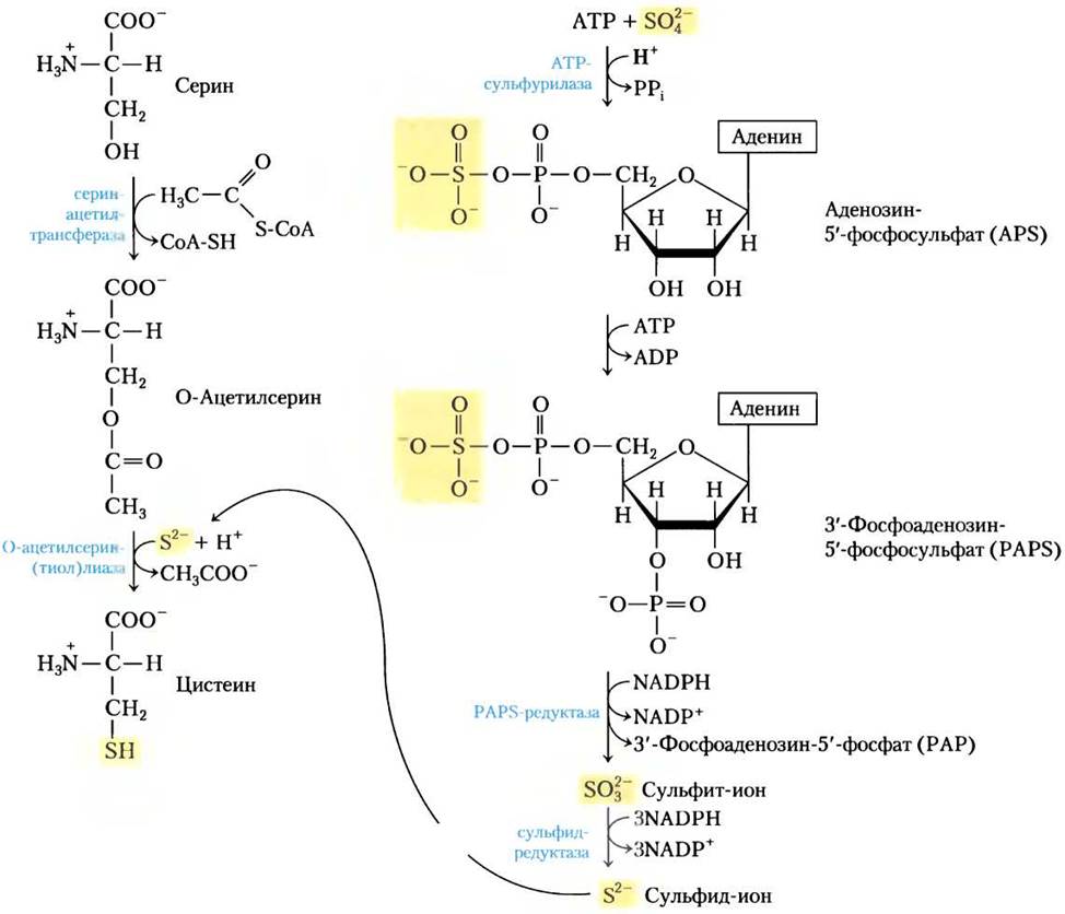
Fig. 22-14. Biosynthesis of cysteine from homocysteine and serine in mammals. Homocysteine is derived from methionine as described in the text.

Three non-essential and six essential amino acids are synthesized from oxaloacetate and pyruvate

Alanine and aspartic acid are synthesized from pyruvate and oxaloacetate (respectively) via transamination, wherein an amino group is transferred from glutamate. Asparagine is formed through the amidation of aspartate, with the NH+4 group provided by glutamine. All of these are non-essential amino acids; their biosynthesis proceeds via straightforward pathways and is ubiquitous across living organisms.
For reasons not yet fully understood, tumor lymphocytes found in children with acute lymphoblastic leukemia (ALL) heavily consume asparagine during tumor proliferation. During Chemotherapy for ALL, patients are administered exogenous bacterial L-asparaginase to deplete plasma asparagine levels. This combination therapy achieves complete remission rates of up to 95% in children (compared to 40–60% with L-asparaginase alone). However, The Use of asparaginase is associated with certain adverse side effects, and approximately 10% of patients experience disease relapse with drug-resistant tumors following initial remission. Currently, specific human asparagine synthetase inhibitors are under development for the Treatment of pediatric ALL. ■
Methionine, threonine, lysine, isoleucine, valine, and leucine are essential amino acids. Their biosynthetic pathways are complex and intricately interconnected (Fig. 22-15). In bacteria, Fungi, and plants, these pathways can differ significantly; the pathways specific to bacteria are outlined in Fig. 22-15.
Fig. 22-15. Bacterial biosynthesis of six essential amino acids—methionine, threonine, lysine, isoleucine, valine, and leucine—from oxaloacetate and pyruvate. Here, as in other multi-step biosynthetic pathways, enzymes (and their corresponding reactions) are designated by numbers (see boxed list). Note that L, L-α, ε-diaminopimelate, the product of reaction (14), is symmetrical. Beyond this step, carbon atoms originating from the pyruvate molecule are no longer specifically tracked, as subsequent reactions may position them at either end of the lysine molecule.


Aspartic acid serves as the precursor for methionine, threonine, and lysine. The key branch points in these pathways are aspartate-β-semialdehyde, a common intermediate for all Three amino acids, and homoserine, the direct precursor to threonine and methionine. Threonine, in turn, serves as one of the precursors for isoleucine. The biosynthetic pathways for valine and isoleucine share three common enzymes (Fig. 22-15, reactions (18) through (21)). Pyruvate acts as the precursor for both valine and isoleucine; the pathway initiates with the Condensation of a pyruvate molecule (in the form of hydroxyethylthiamine pyrophosphate; see Fig. 14-14) with either another pyruvate molecule (in valine synthesis) or an α-ketobutyrate molecule (in isoleucine synthesis). α-Ketobutyrate is generated from threonine in a pyridoxal phosphate-dependent reaction (Fig. 22-15, reaction (17)). The four-step pathway for leucine synthesis (reactions (22)–(25)) originates from α-ketoisovalerate, an intermediate in the valine biosynthetic pathway.
Chorismate is a key intermediate in the biosynthesis of tryptophan, phenylalanine, and tyrosine.

Aromatic rings are not readily available from the environment, even though the benzene ring is exceptionally stable. The primary pathway for aromatic ring formation in bacteria, fungi, and plants is the biosynthesis of tryptophan, phenylalanine, and tyrosine. This pathway begins with the cyclization of an aliphatic precursor, followed by the stepwise Introduction of double bonds. The first four reactions yield shikimate, a seven-carbon molecule derived from erythrose 4-phosphate and phosphoenolpyruvate (Fig. 22-16). Shikimate is then converted into chorismate through three additional reactions that incorporate three more carbons from another molecule of phosphoenolpyruvate. Chorismate serves as the first major branch point in the pathway, with one branch leading to tryptophan and the other to Phenylalanine and Tyrosine.
Fig. 22-16. Biosynthesis of chorismate, an intermediate in the biosynthesis of aromatic amino acids in bacteria and plants. All carbon atoms originate from erythrose 4-phosphate (light purple) or phosphoenolpyruvate (pink). Note that NAD+, required as a cofactor in reaction (2), remains unchanged overall; it is temporarily reduced to NADH during the reaction and subsequently regenerated as the oxidized intermediate is formed. Reaction (6) is competitively inhibited by glyphosate (-COO-CH2-NH-CH2-PO32-), the active ingredient in the widely used herbicide Roundup. This herbicide is relatively safe for mammals, which lack this biosynthetic pathway. The names quinate and shikimate are derived from the plants (cinchona tree and star anise [shikimi], respectively) in which the accumulation of these substances was first discovered.

In the synthesis of tryptophan (Fig. 22-17), chorismate is converted to anthranilate in a reaction where glutamine serves as the nitrogen donor for the nitrogen atom that eventually becomes incorporated into the indole ring. Anthranilate then condenses with 5-phosphoribosyl-1-pyrophosphate (PRPP). The indole ring of tryptophan is assembled from the ring carbons and amino group of anthranilate, along with carbons contributed by PRPP. The final reaction is catalyzed by tryptophan synthase. This enzyme has an α2β2 subunit structure and can dissociate into two α subunits and a β2 dimer, each catalyzing distinct steps of the overall reaction:

Fig. 22-17. Biosynthesis of tryptophan from chorismate in bacteria and plants. In E. coli, the enzymes catalyzing reactions (1) and (2) are subunits of a single complex.


The Second Stage utilizes pyridoxal phosphate (PLP) (Fig. 22-18). The indole produced in The First stage is not released by the enzyme; instead, it travels through an internal tunnel from the Active Site of the α subunit to the active site of the β subunit, where it condenses with a Schiff base intermediate formed between serine and PLP. This substrate channeling is a remarkable feature of the entire pathway from chorismate to tryptophan. In some fungi and bacteria, the active sites catalyzing successive (or even non-sequential) steps of tryptophan biosynthesis reside on separate Polypeptides, whereas in other species these steps are catalyzed by distinct proteins. Furthermore, the expression of certain catalytic activities requires non-covalent association with other Enzymes of the pathway. It is becoming increasingly clear that in both PROKARYOTES AND EUKARYOTES, all enzymes of this pathway are organized into large multienzyme complexes. Such complexes often do not survive traditional biochemical fractionation Procedures, but evidence for their existence in this and other metabolic pathways continues to accumulate (p. 187).
Fig. 22-18. Reaction mechanism of tryptophan synthase. This enzymatic process involves multiple steps with various chemical rearrangements. These PLP-assisted transformations occur at the β-carbon (C-3) of The amino acid, distinguishing them from Reactions Involving the α-carbon (discussed in Fig. 18-6). The β-carbon of serine becomes linked to the indole ring. Mechanism of tryptophan synthase.

In plants and bacteria, phenylalanine and tyrosine are synthesized from chorismate via a more complex sequence of reactions than that of tryptophan. Their common intermediate is prephenate (Fig. 22-19). In both cases, the final step is a transamination reaction with glutamate.
Fig. 22-19. Biosynthesis of phenylalanine and tyrosine from chorismate in bacteria and plants. The conversion of chorismate to prephenate represents a rare biological example of a Claisen rearrangement.

Animals can synthesize tyrosine directly from phenylalanine by hydroxylating the C-4 position of the phenyl ring using phenylalanine hydroxylase; this enzyme is also involved in phenylalanine degradation (see Figs. 18-23, 18-24). Tyrosine is considered a conditionally essential amino acid because it can be produced from the essential amino acid phenylalanine.
Histidine biosynthesis utilizes precursors of Purine Biosynthesis

In all plants and bacteria, the pathway for histidine synthesis differs significantly from the biosynthetic routes of other amino acids. Histidine is derived from three precursors (Fig. 22-20): five carbons come from PRPP, nitrogen and carbon atoms are contributed by the purine ring of ATP, and a second ring nitrogen is provided by glutamine. The key steps include: the condensation of ATP and PRPP, in which N-1 of the purine ring attaches to the activated C-1 carbon of the ribose moiety in PRPP (step (1) in Fig. 22-20); the opening of the purine ring, which allows The formation of a bond between N-1 of adenine and C-2 of ribose (step (3)); and the Formation of the imidazole ring in a reaction where glutamine supplies the nitrogen atom (step (5)). The use of ATP as a metabolic precursor rather than a high-energy cofactor is quite unusual, but it is not wasteful because it is coordinated with purine biosynthesis. The remainder of the ATP molecule, modified at the N-1 and C-2 positions, is 5-aminoimidazole-4-carboxamide ribonucleotide (AICAR), an intermediate in purine biosynthesis (see Fig. 22-33) that is rapidly recycled back into ATP.
Fig. 22-20. Biosynthesis of histidine in bacteria and plants. Atoms derived from PRPP and ATP are highlighted in red and light blue, respectively. The two nitrogen atoms of the imidazole ring originate from glutamine and glutamate (green). Note that after step (5), an ATP derivative (AICAR) remains, which serves as an intermediate in purine biosynthesis (see Fig. 22-33, step (9)), allowing ATP to be rapidly regenerated.

Amino acid biosynthesis is regulated allosterically
As discussed in detail in Chapter 15, metabolic flux is frequently governed by the coordinated activity of multiple enzymes functioning along a pathway. In Amino acid synthesis, Metabolic Regulation is often achieved through feedback inhibition, where the end product of the pathway inhibits the first committed step. This initial step is typically irreversible and is catalyzed by an allosteric enzyme that plays a crucial role in controlling the overall flux through the pathway. For instance, Figure 22-21 illustrates the Allosteric Regulation of isoleucine synthesis from threonine (see Fig. 22-15). The end product, isoleucine, acts as an allosteric inhibitor of the first enzyme in this reaction sequence. In bacteria, such allosteric modulation of amino acid synthesis ensures immediate adaptation to the changing metabolic needs of The Cell.
Fig. 22-21. Allosteric regulation of isoleucine biosynthesis. The first reaction in the pathway from threonine to isoleucine is inhibited by the end product of the pathway, isoleucine. This was one of the earliest discovered Examples of allosteric feedback inhibition. The steps from α-ketobutyrate to isoleucine correspond to reactions (18) through (21) in Figure 22-15 (comprising five steps, as reaction (19) proceeds in two stages).

Allosteric regulation can be considerably more complex. As an example, let us consider the remarkable array of Allosteric regulators of E. coli Glutamine Synthetase (Fig. 22-6). Six products derived from glutamine provide negative feedback inhibition of this enzyme, and the combined effect of these and other modulators is not merely additive. Such regulation is termed cooperative inhibition.
Other mechanisms also contribute to the Regulation of Amino acid biosynthesis. For protein synthesis, all 20 amino acids must be produced in the cell in the correct proportions; consequently, Cells possess mechanisms to control not only The rate of synthesis of each individual amino acid, but also the coordination of their accumulation. The coordination of relative amounts is particularly evident in rapidly growing bacterial cells. Figure 22-22 illustrates how E. coli cells control the synthesis of lysine, methionine, threonine, and isoleucine—amino acids derived from aspartate. Several important inhibitory connections are apparent. The formation of aspartyl-β-phosphate from aspartate is catalyzed by three isozymes, each of which is independently controlled by different modulators. Enzyme multiplicity prevents key pathway steps from being shut down by a single end product of biosynthesis, as other products of the pathway may still be required. In the reaction sequences from aspartate-β-semialdehyde to homoserine, and from threonine to α-ketobutyrate (detailed in Fig. 22-15), the steps are catalyzed by two independently controlled isozymes. One isozyme responsible for converting aspartate to aspartyl-β-phosphate is allosterically inhibited by two different modulators, lysine and isoleucine, whose combined effect is greater than additive—yet another example of cooperative inhibition. The pathway from aspartate to isoleucine is subject to multiple Cross-Regulation via negative feedback; for instance, isoleucine inhibits the conversion of threonine to α-ketobutyrate (as described above), whereas threonine inhibits its own formation at three points: from homoserine, from aspartate-β-semialdehyde, and from aspartate (steps (4), (3), and (1) in Fig. 22-15). Such a comprehensive regulatory mechanism is known as sequential feedback inhibition.
Fig. 22-22. Interlocking inhibitory regulatory mechanisms in the Biosynthesis of certain aspartate-derived amino acids in E. coli METABOLISM. Three enzymes (A, B, C) have two or three isozymes each, denoted by subscripts. In each case, one isozyme (A2, B1, or C2) lacks allosteric regulation; these isozymes are regulated by the level of their synthesis (ch. 28, vol. 3). The synthesis of isozymes A2 and B1 is repressed by high levels of methionine, and the synthesis of isozyme C2 is repressed by high levels of C2. Enzymes: A — aspartokinase; B — homoserine dehydrogenase; C — Threonine dehydratase.

Similar motifs are found in the pathways leading to the formation of aromatic amino acids. Early in the pathway leading to the common intermediate chorismate, the first step is catalyzed by the enzyme 3-deoxy-D-arabino-heptulosonate-7-phosphate synthase (DAHP synthase, derived from 2-keto-3-deoxy-D-arabino-heptulosonate 7-phosphate) (step (1) in Fig. 22-16). Most microorganisms and plants possess three DAHP synthase isozymes. One of these isozymes is allosterically inhibited (via negative feedback) by phenylalanine, another by tyrosine, and the third by tryptophan. This arrangement helps the entire pathway respond to the cell's demand for one or more aromatic amino acids. Additional regulation occurs after the pathways diverge from chorismate. For example, the enzymes catalyzing the first two steps in tryptophan synthesis are allosterically inhibited by tryptophan.
Summary of Section 22.2 BIOSYNTHESIS OF AMINO Acids
■ Plants and bacteria synthesize all 20 Proteinogenic Amino Acids. Mammals can synthesize only about half of these; the remaining amino acids must be obtained from the diet (essential amino acids).
■ Among the nonessential amino acids, glutamate is formed by the reductive amination of α-ketoglutarate and serves as a precursor for glutamine, proline, and arginine. Alanine and aspartate (along with asparagine) are produced via transamination reactions from pyruvate and oxaloacetate, respectively. The carbon skeleton of serine originates from 3-phosphoglycerate. Serine is a precursor for glycine; the β-carbon atom of serine is transferred to tetrahydrofolate. In microorganisms, Cysteine is synthesized from serine and sulfide generated by the reduction of environmental sulfate. Mammals synthesize cysteine from methionine and serine through a sequence of reactions requiring S-adenosylmethionine and cystathionine.
■ Among the essential amino acids, the aromatic amino acids (phenylalanine, tyrosine, and tryptophan) are synthesized via pathways in which chorismate serves as a key branch point. Phosphoribosyl pyrophosphate acts as a precursor for both tryptophan and histidine. The histidine biosynthetic pathway intersects with purine biosynthesis. Tyrosine can also be produced by the hydroxylation of phenylalanine (making it a conditionally essential amino acid). The biosynthetic pathways for other essential amino acids are more complex.
■ Amino acid biosynthetic pathways are allosterically regulated by their end products, with the regulatory enzyme typically located at THE START OF the pathway. The regulation of various synthetic pathways proceeds in a coordinated manner.
Last update: 06/08/2026
Editorial and Educational Adaptation: This material has been compiled based on the primary/original source text. The project team performed an editorial review, corrected technical inaccuracies, structured sections, and adapted the content for an educational format.
What was processed:
- elimination of formatting defects (OCR errors, structural breaks, corrupted characters);
- editorial organization of content;
- standardization of terminology in accordance with academic sources;
- verification of factual statements against the original source text.
All mentions of the author, publication year, and origin of the primary text have been preserved in accordance with the source.