LEHNINGER PRINCIPLES OF BIOCHEMISTRY - VOL. 2. BIOENERGETICS AND METABOLISM - 2014
PART II. BIOENERGETICS AND METABOLISM
Class="center">Time passes quickly when you have nothing to be sad about. To see a person who was at death's door, under changed conditions, get up and clearly do well, gives a joyous lift of the spirit... that cannot be expressed in words. And the Nobel Prize is just the icing on the cake.
Gertrude Elion, Science, 2002
22. BIOSYNTHESIS OF AMINO ACIDS, NUCLEOTIDES, AND RELATED MOLECULES
In the living world, nitrogen ranks fourth by mass after carbon, hydrogen, and oxygen. Most of nitrogen is found in bound forms, incorporated into Amino Acids and NUCLEOTIDES. This chapter examines all aspects of Nitrogen METABOLISM, with the exception of Amino Acid Catabolism, which was covered in Chapter 18.
The biosynthetic pathways for amino acids and nucleotides are usually considered together, not only because both classes of compounds contain nitrogen (derived from the same biological source), but also because their two pathways intersect at many points, sharing several Key Intermediates. Certain amino acids or parts of their molecules are incorporated into Purines and Pyrimidines, and a single amino acid—Histidine—contains a purine ring. These two synthetic pathways also share much in common from a purely chemical perspective, being dominated by reactions that involve The transfer of nitrogen and one-carbon groups.
To the student just beginning The Study of biochemistry, the pathways described in this chapter may seem complex. However, this complexity is not so much due to the chemistry itself—the reaction mechanisms are generally straightforward—as it is to the daunting number of steps and the wide variety of intermediates. The best way to study these metabolic pathways is by keeping in mind the Principles of Metabolism, Key intermediates and precursors, and common reaction types already discussed. A mere glance at these pathways reveals that biological systems are the site of some of the most unusual chemical transformations, such as rare Reactions Involving the transition metals molybdenum, selenium, and vanadium. Your efforts will be well rewarded, especially if you are a medical or veterinary student, as many inherited Human and Animal diseases stem from the absence of one or more Enzymes of Amino Acid and/or Nucleotide Metabolism, and many drugs used to combat infections are inhibitors of enzymes in these pathways, as is a broad array of antitumor chemotherapeutic agents.
The regulation of nitrogen compound Biosynthesis is vital. However, because nucleotides and Amino acids are required in relatively small amounts, the throughput of most of these reactions is not nearly as high as that of lipid and carbohydrate synthesis in animal Tissues. At the same time, PROTEIN AND NUCLEIC acid synthesis requires various amino acids and nucleotides in the correct proportions and at specific times, making precise and Coordinated regulation of all their biosynthetic processes essential. Furthermore, because both Proteins AND AMINO acids are typically ionized—that is, they can carry a charge—maintaining electrochemical balance is also required when regulating their intracellular levels. As discussed in previous chapters, Metabolic control can be exerted either through changes in enzyme activity or through Changes in the amounts of specific enzymes. The metabolic pathways discussed in this chapter provide some of the clearest, most easily understood Examples of the Introduction/15.html">Regulation of enzyme Activity. The quantitative control of various enzymes within The Cell (i.e., the balance between the rates of Synthesis and degradation) is described in Chapter 28 (Vol. 3).
22.1. Overview of Nitrogen Metabolism
Nitrogen is essential for the synthesis of both amino acids and nucleotides. In the environment, soluble compounds containing biologically available nitrogen are present in very limited quantities, forcing most organisms to operate under a regime of strict conservation of ammonia, amino acids, and nucleotides. Indeed, as we will see later, free amino acids, purines, and pyrimidines derived from the Breakdown of Proteins and Nucleic Acids are often reused over and over again; in other words, Cells function by taking up and releasing essential substances in a cyclic manner. First, we will examine the pathways by which environmental nitrogen is incorporated into biological systems.
The Nitrogen Cycle Maintains a Pool of Biologically Available Nitrogen
Air, our most abundant reservoir of nitrogen, consists of about 4/5 molecular nitrogen (N2). Nonetheless, very few biological species are capable of converting atmospheric nitrogen into forms accessible to living organisms. Within the biosphere, the metabolic processes of various species are interconnected, enabling nitrogen to be captured and reused in the natural nitrogen cycle (Figure 22-1). The First stage of this cycle is fixation (reduction) of atmospheric nitrogen by nitrogen-fixing Bacteria to yield ammonia or ammonium ions (NH3 or NH+4). Although ammonia can be utilized by many living organisms, soil bacteria that derive energy by oxidizing ammonia to nitrite (NO2-) and subsequently to nitrate (NO3-) are so widespread and active that almost all ammonia entering the soil is oxidized to nitrate. This process is called nitrification. Plants and many bacteria can take up and rapidly reduce nitrites and nitrates via the action of nitrite and nitrate reductases. The ammonia thus produced is incorporated into amino acids in plants. Animals then consume plants as a source of both essential and nonessential amino acids for the synthesis of their own proteins. When an Organism dies, its proteins are broken down into ammonia by soil microorganisms, where nitrifying bacteria convert it back into nitrites and nitrates. The balance between fixed and atmospheric nitrogen is maintained by bacteria that convert nitrate into N2 under anaerobic conditions; this process is called denitrification (Figure 22-1). Soil bacteria use NO3- instead of O2 as a terminal electron acceptor in a series of reactions (such as Oxidative Phosphorylation) that establish a transmembrane proton gradient used to synthesize ATP.
Figure 22-1. The nitrogen cycle. The total amount of nitrogen fixed annually in the biosphere exceeds 1011 kg. Reactions indicated by red arrows occur almost exclusively under anaerobic conditions. The oxidation states of nitrogen compounds are shown on the scale at the bottom of the figure.

Bacteria capable of carrying out ammonia oxidation under anaerobic conditions (Figure 22-1) have recently been discovered; this process is termed anammox (anaerobic ammonium oxidation) and involves The conversion of AMMONIA AND NITRITE into N2. In the biosphere, 50% to 70% of the conversion of NH3 to N2 occurs via this metabolic pathway, which was discovered only in the 1980s. Obligate anaerobes that carry out anammox are of great intrinsic interest and also offer solutions to certain wastewater Treatment problems (Box 22-1).
Now let us examine the first stage of the nitrogen cycle—the Fixation of Atmospheric nitrogen.
Box 22-1. The Unusual Lifestyle of Numerous Yet Enigmatic Creatures
Because we breathe air, we easily overlook bacteria and archaea inhabiting anaerobic environments. These organisms are rarely discussed in introductory biochemistry textbooks, yet they comprise a substantial fraction of our planet's biomass and make a vital contribution to the global carbon and nitrogen balances upon which all other life forms depend.
As discussed in detail in previous chapters, the energy required for living organisms to survive is coupled to the generation of a hydrogen ion gradient across membranes. Electrons from a reduced substrate are delivered to membrane-bound electron carriers and, following a series of transfers, reach a terminal electron acceptor. As a result, protons accumulate on one side of the membrane, creating a proton gradient. This gradient is used to synthesize ATP or drive other energy-requiring cellular processes. In all eukaryotes, the reduced substrate is typically a carbohydrate (glucose or Pyruvate), and oxygen serves as the electron acceptor.
The metabolism of bacteria and archaea is far more versatile. Under anaerobic conditions, such as those found in marine and freshwater sediments, the variety of life strategies is remarkable. Virtually any redox pair can serve as an energy source for a specialized organism or group of organisms. Many lithotrophic bacteria (chemotrophs that use inorganic substances as an energy source; see Figure 1-5, Vol. 1) possess an enzyme called Hydrogenase, which uses molecular hydrogen to reduce NAD:

NADH serves as an electron source for numerous membrane-bound electron acceptors; the transfer of electrons generates the proton gradient necessary for ATP synthesis. Other lithotrophs are capable of oxidizing sulfur compounds (H2S, elemental sulfur, or thiosulfates) or ferrous iron compounds. A widely distributed group of archaea known as methanogens consists of strict anaerobes that obtain energy by reducing CO2 to methane. These are merely a few examples of how anaerobic organisms adapt to their environments. Their metabolic pathways involve a multitude of fascinating reactions operating via specialized Cofactors not found in the world of obligate aerobes. Harnessing certain anaerobic bacteria can yield tangible practical benefits. Furthermore, studying such organisms helps provide Answers regarding THE ORIGIN OF life on Earth under conditions when molecular oxygen was absent.
The nitrogen cycle is driven by the activities of a diverse array of specialized bacteria. Nitrifying bacteria are divided into two groups: those that oxidize ammonia to nitrite, and those that oxidize the resulting nitrite to nitrate (Figure 22-1). Nitrate is the second most common biological electron acceptor after oxygen; many bacteria and archaea can catalyze the denitrification of nitrate to dinitrogen gas, which is subsequently converted back into ammonia by nitrogen-fixing bacteria. Ammonia is a major pollutant in municipal wastewater and agricultural animal waste; it is also a byproduct of fertilizer manufacturing and petroleum refining. Wastewater treatment plants employ communities of nitrifying and denitrifying bacteria to convert wastewater ammonia into atmospheric nitrogen. This process consumes both oxygen and Organic compounds.
During the 1960s and 1970s, several scientific publications emerged proposing that ammonium could be oxidized to nitrogen gas under anaerobic conditions using nitrite as an electron acceptor; this process was termed anammox. These reports attracted little attention until bacteria capable of mediating this process were discovered in the mid-1980s at a wastewater treatment plant in Delft, Netherlands. A group of Dutch microbiologists led by Gijs Kuenen and Mike Jetten set out to investigate these bacteria, which were soon identified as belonging to an unusual group of planctomycete bacteria. Further surprises awaited the researchers.
The biochemical mechanisms underlying the anammox process gradually became clear (Fig. 1). Unexpectedly, it turned out that the reaction intermediate is hydrazine (N2H4)—a reactive chemical compound used even as a component of rocket propellant. The molecules of this substance are small, highly toxic, and easily cross phospholipid membranes. Anammox bacteria solve this problem by enclosing hydrazine within specialized Organelles called anammoxosomes. The membranes of these organelles are composed of unique Lipids known as ladderanes (Fig. 2), which had not been previously discovered in biological systems. The interconnected cyclobutane rings that form the ladderane Structure are tightly packed against one another, creating a very dense barrier that significantly slows down the diffusion of hydrazine. Because cyclobutane rings possess a strained geometry, their synthesis is quite challenging. To this day, the exact mechanisms by which bacteria synthesize these lipids remain a mystery.
Fig. 1. Reactions of the anammox process. Ammonia and ammonium hydroxide are converted into hydrazine and H2O by hydrazine hydrolase; hydrazine is then oxidized by hydrazine oxidoreductase to yield N2 and protons, generating a proton gradient for ATP synthesis. On the outer surface of the anammoxosome, these protons are consumed by nitrite reductase, which produces nitric oxide and completes the cycle. All enzymes participating in this process are enclosed within the anammoxosome membrane.

Fig. 2. (a) Ladderanes in the anammoxosome membrane. The synthesis mechanism for this unstable structure consisting of fused cyclobutane rings remains unknown. (b) Ladderanes pack into a remarkably dense, impermeable hydrophobic Membrane Structure, which restricts the diffusion of hydrazine produced during anammox.

The discovery of anammoxosomes came as a surprise. Typically, bacterial cells lack distinct intracellular compartments, and the presence of a membrane-bound Nucleus is frequently cited as a primary distinction between eukaryotes and bacteria. The mere discovery of such organelles was of great scientific interest; however, planctomycetes also possess a nucleus-like structure, with their chromosomal DNA enclosed within a membrane envelope (Fig. 3). The finding of such subcellular Organization stimulated further research into the origin of planctomycetes and the evolution of The eukaryotic nucleus. Planctomycetes represent an ancient bacterial Lineage comprising numerous genera, with bacteria from three of these genera capable of carrying out anammox. A more detailed study of these bacteria may bring us closer to solving a fundamental question in evolutionary biology: identifying the organism that serves as the last universal common ancestor (LUCA) of all life on our planet.
Fig. 3. Transmission electron micrograph (TEM) of a cross-section of a Gemmata obscuriglobus cell, revealing DNA within a nucleus (N) enclosed by a nuclear envelope (NE). Bacteria of the genus Gemmata (planctomycetes) are incapable of performing anammox.

Currently, anammox bacteria are actively employed in wastewater treatment. This approach cuts ammonia removal costs by up to 90%—entirely bypassing the conventional denitrification stage while reducing aeration and nitrification expenses—and minimizes the yield of by-products. Clearly, gaining a detailed understanding of the bacteria that inhabit the biosphere is immensely valuable for humanity in the 21st century.
Nitrogen is fixed by the enzyme complex Nitrogenase
Only certain bacteria and archaea are capable of fixing atmospheric nitrogen. These include soil, freshwater, and marine cyanobacteria, other free-living soil bacteria such as Azotobacter species, and nitrogen-fixing symbiotic bacteria of legumes that reside in ROOT nodules. The primary useful product of Nitrogen Fixation is ammonia, which can be utilized by all organisms either directly or after conversion into other soluble compounds such as nitrites, nitrates, or amino acids. The reduction of nitrogen to ammonia is exergonic:
N2+3 H2 —> 2 NH3 ∆G'° = -33.5 kJ/mol
However, the N≡N triple bond is exceptionally strong, with a bond dissociation energy of 930 kJ/mol. Nitrogen fixation is therefore a process requiring a very high activation energy, making atmospheric nitrogen an inert substance under standard conditions. In industrial settings, Ammonia is produced via the Haber process (named after its inventor, Fritz Haber) from a mixture of nitrogen and hydrogen at temperatures of 400–500 °C and high pressures reaching tens of thousands of kilopascals (several hundred atmospheres) to provide the necessary activation energy. Biological Nitrogen Fixation, conversely, must proceed at ambient biological temperatures and an N2 pressure of 0.8 atm. Here, the high activation barrier is overcome through alternative means—at least in part, via the binding and Hydrolysis of ATP. In general terms, the reaction can be written as follows:
N2+ 10 H+ + 8 e- + 16 ATP —> 2 NH4+ + 16 ADP + 16 Pi + H2
Biological nitrogen fixation is catalyzed by a highly conserved protein complex known as the nitrogenase complex (or simply nitrogenase) (Fig. 22-2), whose most critical components are dinitrogenase reductase and dinitrogenase (Fig. 22-3). Dinitrogenase reductase (Mr = 60,000) is a homodimer consisting of two identical subunits. It contains a single 4Fe-4S redox center (see Fig. 19-5) situated at the interface between the subunits, which can be reversibly oxidized or reduced by a single electron. The complex also possesses two ATP/ADP-binding sites (one per subunit). Dinitrogenase (Mr = 240,000) is a tetramer composed of two pairs of Different types of subunits and contains both iron and molybdenum.
Fig. 22-2. Nitrogen fixation by the nitrogenase complex. Electrons from pyruvate are transferred to dinitrogenase via ferredoxin (or flavodoxin) and dinitrogenase reductase. Dinitrogenase reductase reduces dinitrogenase by transferring one electron at a time, whereas the fixation of a single N2 molecule requires a minimum of six electrons. Additionally, two electrons are consumed to reduce two H+ protons to H2 in a process that invariably accompanies nitrogen fixation in anaerobes. Thus, a total of eight electrons are expended for the fixation of one N2 molecule. The subunit architecture and metal-containing cofactors of dinitrogenase reductase and dinitrogenase proteins are described in the text and in Figure 22-3.

Fig. 22-3. Enzymes and cofactors of the nitrogenase complex (PDB ID 1N2C). (a) Dinitrogenase subunits are shown in gray and pink, and dinitrogenase reductase subunits in blue and green. A bound ADP molecule is highlighted in red. Note the 4Fe-4S clusters (Fe atoms in orange, S in yellow) and the iron-molybdenum cofactor (Mo in black, homocitrate in light gray). P-clusters (closely spaced pairs of 4Fe-4S complexes) are also visible. (b) Protein-free cofactors of the dinitrogenase complex (color-coded as in a). (c) The iron-molybdenum cofactor contains one Mo atom (black), seven Fe atoms (orange), nine S atoms (yellow), and a homocitrate molecule (gray).
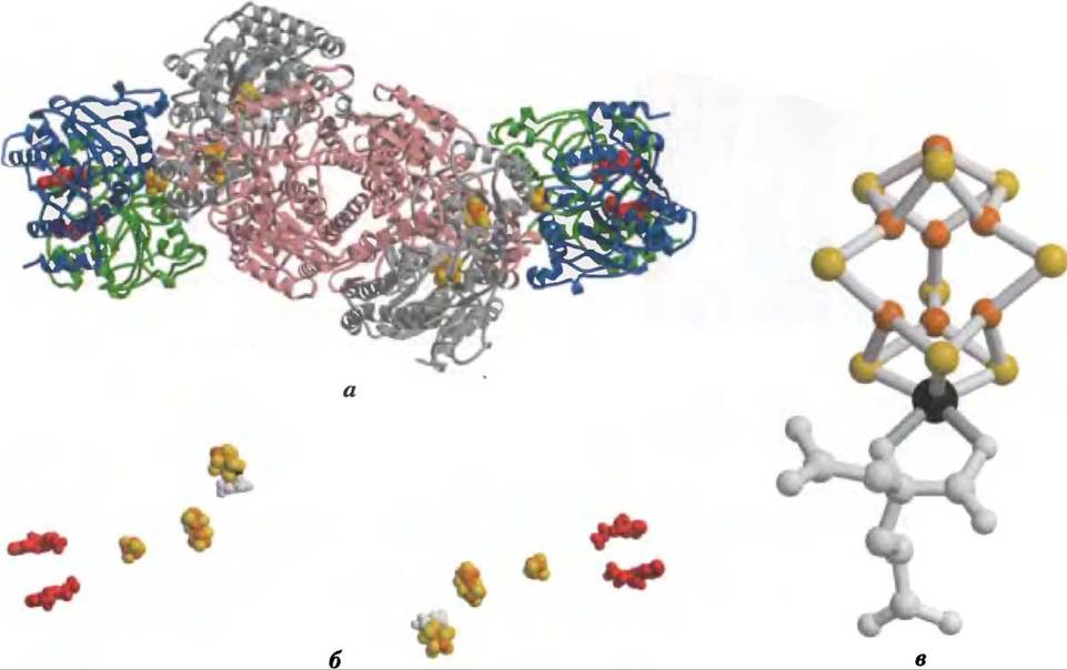
The active center of the tetramer contains a total of 2 Mo atoms, 32 Fe atoms, and 30 S atoms. Roughly half of the iron and sulfur atoms reside within P-clusters, each containing a pair of closely interacting 4Fe-4S centers, while the remaining Mo, Fe, and S atoms comprise the unusual iron-molybdenum cofactor. Furthermore, a variant form of nitrogenase containing vanadium instead of molybdenum has been discovered, and certain bacterial species can utilize both types of nitrogenases. Under specific conditions, vanadium-containing forms can serve as the primary nitrogen-fixing systems, though they are not yet as thoroughly characterized as their molybdenum-containing counterparts.
Nitrogen fixation is performed by the fully reduced form of dinitrogenase and requires eight electrons: six for the reduction of N2 and two for the generation of a molecule of H2, an obligate byproduct of this reaction mechanism. Dinitrogenase reduction occurs via Electron transfer from dinitrogenase reductase (Fig. 22-2). The dinitrogenase tetramer features two binding sites for the reductase. The electrons required for the reaction are transferred from the reductase to dinitrogenase one by one: a reduced reductase molecule binds to dinitrogenase, transfers a single electron, and then the oxidized reductase dissociates from dinitrogenase, allowing the cycle to repeat. Each cycle entails the hydrolysis of two ATP molecules per reductase dimer. The electron donor used to regenerate reduced dinitrogenase can vary and may include reduced ferredoxin (p. 371, see also Fig. 19-5), reduced flavodoxin, or potentially other Donors. In at least one species, pyruvate serves as the ultimate source of electrons for ferredoxin reduction (Fig. 22-2).
The Role of ATP in this process is somewhat unconventional. As will be discussed further, ATP can supply chemical energy not only through the hydrolysis of one or more phosphodiester bonds, but also by utilizing binding energy (pp. 227, 425, vol. 1) derived from noncovalent interactions that lower the activation energy. In the reaction catalyzed by dinitrogenase reductase, both ATP binding and hydrolysis induce conformational changes that help overcome the high activation barrier of nitrogen fixation. The binding of two ATP molecules by the reductase shifts its reduction potential ($E^{\circ\prime}$) from -300 to -420 mV, thereby enhancing its reducing power sufficiently to drive electron transfer to dinitrogenase. ATP hydrolysis then takes place immediately prior to the transfer of each electron to dinitrogenase.
Another critical characteristic of the nitrogenase complex is its extreme sensitivity to oxygen. The reductase is rapidly inactivated in air, with a half-life of 30 seconds, whereas the half-life of dinitrogenase in air is 10 minutes. Free-living nitrogen-fixing bacteria cope with this challenge through various strategies. Some inhabit strictly anaerobic niches or suppress nitrogenase synthesis in the presence of oxygen. Certain aerobes, such as Azotobacter vinelandii, partially uncouple electron transport from ATP synthesis so that oxygen is immediately "scavenged" as soon as it enters the cell (see Box 19-1). To protect themselves against oxygen, cultures of such bacteria indeed exhibit a Temperature rise during active nitrogen fixation.
The Symbiosis between legumes and rhizobial nitrogen-fixing bacteria (Fig. 22-4) addresses both the high energy demand and the oxygen vulnerability of the nitrogenase complex. The Energetic Cost of nitrogen fixation was likely a driving force in the evolution of this plant-bacterial association. The bacteria inhabiting root nodules gain access to an abundant energy reservoir in the form of copious CARBOHYDRATES and Tricarboxylic Acid Cycle intermediates supplied by the host plant. This allows the bacteria to fix hundreds of times more nitrogen than their free-living soil-dwelling relatives can sustain. To mitigate oxygen toxicity, the bacteroids are "bathed" in a solution of an oxygen-binding, heme-containing protein called leghemoglobin, synthesized by the plant (though in some instances the heme component may be of bacterial origin). Leghemoglobin binds virtually all free oxygen, preventing it from interfering with nitrogen fixation while efficiently delivering O2 to the bacterial Electron Transport Chain. The benefit to the plant is a steady supply of reduced, readily assimilable nitrogen. The agricultural value of this plant-bacterial symbiosis is manifested in soil nitrogen enrichment by legumes, which forms The basis of crop rotation practices: fields previously planted with non-leguminous crops (such as corn) that deplete soil nitrogen are sown every few years with legumes like alfalfa, peas, or clover.
Fig. 22-4. Nitrogen-fixing root nodules. (a) Root nodules of bird's-FOOT trefoil (Lotus corniculatus), a member of the Fabaceae family. The flowers of this plant are shown in the inset. (b) False-color transmission electron micrograph of a thin section of a pea root nodule. Symbiotic nitrogen-fixing bacteria, or bacteroids (colored red), reside within nodule cells, enclosed by a peribacteroid membrane (blue). The bacteroids synthesize the nitrogenase complex, which converts atmospheric nitrogen (N2) into ammonium (NH4+); in the absence of bacteroids, the plant cannot fix N2. The infected root cells exhibit key specializations required for nitrogen fixation, notably the synthesis of leghemoglobin; this heme protein displays a high affinity for oxygen, which would otherwise inhibit nitrogenase. (Plant cell nuclei are depicted in yellow/green; other typical plant cell organelles are not visible.)

Due to its immense practical importance, nitrogen fixation has been studied extensively. The industrial production of ammonia (ammonium) for use as a fertilizer is a very energy-intensive and therefore expensive process. Currently, considerable efforts are directed toward developing recombinant or transgenic organisms capable of fixing nitrogen. Recombinant DNA technologies (see Vol. 1, Ch. 9) are used to transfer genes encoding nitrogen-fixing enzymes into non-nitrogen-fixing bacteria and plants. Success in this endeavor depends on overcoming The problem of oxygen toxicity for any cell producing nitrogenase.
Ammonium is incorporated into Biomolecules via glutamic acid (glutamate) and glutamine
Reduced nitrogen in the form of NH+4 is incorporated first into amino acids and subsequently into other nitrogen-containing biomolecules. This key branch point of nitrogen assimilation is mediated by Two amino acids: Glutamic Acid and Glutamine. Recall that these two amino acids play a central role in the catabolism of ammonia and amino groups during amino acid oxidation (see Ch. 18). Glutamic acid serves as an amino group donor for most Other Amino Acids via Transamination reactions (for the reverse reaction, see Fig. 18-4). The amide nitrogen of glutamic acid acts as an amino group source in A wide variety of biosynthetic processes. In higher organisms, the intracellular and extracellular concentrations of one or both of these amino acids are frequently higher than those of any other amino acids, sometimes by an order of magnitude or more. Escherichia coli cells require such large amounts of glutamic acid that it is one of the major solutes in the Cytosol. Its concentration is regulated not only to meet the cell's demand for nitrogen, but also to maintain osmotic balance between the cytosol and the external environment.
The biosynthetic pathways for glutamic acid and glutamine are straightforward, with all or at least some of their stages found in all organisms. The most important pathway for ammonia incorporation into glutamic acid requires a two-step reaction. First, glutタミン synthetase catalyzes the reaction between glutamate and NH4+ to yield glutamine. This reaction proceeds in two steps via an enzyme-bound y-glutamyl phosphate intermediate (see Fig. 18-8):
(1) Glutamate + ATP —> y-glutamyl phosphate + ADP
(2) y-Glutamyl phosphate + NH4+ —> glutamine + Pi + H+
Overall equation:
glutamate + NH4+ + ATP —> glutamine + Pi + H+ (22-1)
Glutamine Synthetase is found in all organisms. In addition to its crucial role in NH4+ assimilation in bacteria, this enzyme plays a central role in mammalian Amino acid metabolism by converting toxic ammonia into glutamine, which enters the bloodstream (Ch. 18).
In bacteria and plants, glutamate is synthesized from glutamine in a reaction catalyzed by glutamate synthase. α-Ketoglutarate, an intermediate of The Citric Acid Cycle, undergoes reductive amination using glutamine as the nitrogen donor:
α-Ketoglutarate + glutamine + NADPH + H+ —> 2 glutamate + NADP+ (22-2)
The overall reaction carried out by glutamine synthetase and glutamate synthase (Equations 22-1 and 22-2):
α-Ketoglutarate + NH4+ + NADPH + ATP —> L-glutamate + NADP+ + ADP + Pi
Glutamate synthase is absent in animals, which maintain high levels of glutamate through processes such as the transamination of α-ketoglutarate during amino acid catabolism.
Glutamate can also be produced via an alternative, less frequently used pathway involving a single-step reaction between α-ketoglutarate and NH4+. This reaction is catalyzed by L-Glutamate dehydrogenase, an enzyme present in all organisms. The reductive power is provided by NADPH:
α-Ketoglutarate + NH4+ + NADPH —> L-glutamate + NADP+ + H2O
We discussed this reaction in the section on amino acid catabolism (see Fig. 18-7). In Eukaryotic cells, L-glutamate dehydrogenase is localized in the mitochondrial matrix. The reaction equilibrium strongly favors the substrates, and the Michaelis constant $K_m$ for NH+4 is so high (~1 mM) that this pathway likely makes only a minor contribution to the incorporation of NH+4 into amino acids and other metabolites. (Recall that, conversely, the glutamate dehydrogenase reaction supplies ammonium to The Urea Cycle; see Fig. 18-10.) Concentrations of NH+4 high enough for glutamate dehydrogenase to contribute significantly to the glutamate pool are typically observed only when NH3 is added to soil or when organisms are grown in the laboratory in the presence of high ammonia concentrations. In general, soil bacteria and plants rely predominantly on the two-enzyme pathway discussed above (Equations 22-1 and 22-2).
Glutamine synthetase is the primary regulatory point in nitrogen metabolism
The activity of glutamine synthetase is regulated in virtually all organisms—hardly surprising given its pivotal metabolic role in catalyzing the first step of nitrogen assimilation into biological molecules. In enteric bacteria such as E. coli, this regulation is exceptionally complex. The enzyme is composed of 12 identical
subunits with $M_r = 50,000$ (Fig. 22-5) and is regulated both allosterically and by covalent modification. Alanine, Glycine, and at least six other End products of glutamine metabolism act as allosteric Inhibitors of the enzyme (Fig. 22-6). Each inhibitor alone causes only partial inhibition, but multiple inhibitors exert a cumulative inhibitory effect, and all eight combined shut down the synthetase almost completely. This control mechanism allows for fine-tuning of glutamine levels to match the cell's current metabolic demands.
Fig. 22-5. Subunit structure of glutamine synthetase based on X-ray crystallographic data (PDB ID GLS). (a) Side view; all 12 subunits are identical, shown in different colors to illustrate their packing and arrangement; (b) top view, with active sites highlighted in green.

Fig. 22-6. Allosteric Regulation of glutamine synthetase. The enzyme is cumulatively regulated by six end products of glutamine metabolism. Alanine and glycine likely serve as indicators of the cell's overall amino acid metabolic status.

Allosteric regulation is further reinforced by adenylylation—the attachment of an AMP moiety to Tyr397, located adjacent to the enzyme's Active Site (Fig. 22-7). This covalent modification increases the enzyme's sensitivity to allosteric inhibitors, thereby reducing its catalytic activity in proportion to the number of adenylylated subunits. Both adenylylation and deadenylylation are catalyzed by adenylyl transferase (AT in Fig. 22-7), a component of the complex enzymatic cascade responsible for monitoring the intracellular levels of glutamine, α-ketoglutarate, ATP, and Pi. The activity of adenylyl transferase is modulated by binding to the regulatory protein PII, whose own activity is regulated via covalent modification (uridylylation), also at a Tyrosine residue. The complex of adenylyl transferase with uridylylated PII (PII-UMP) stimulates deadenylylation, whereas the complex with deuridylylated PII stimulates the adenylylation of glutamine synthetase. Both the uridylylation and deuridylylation of PII are carried out by a single enzyme, uridylyl transferase. Uridylylation is inhibited by the binding of glutamine and Pi to uridylyl transferase, and is stimulated by the association of α-ketoglutarate and ATP with PII.
Fig. 22-7. The second level of glutamine synthetase regulation: covalent modifications. (a) Adenylylated tyrosine residue, (b) the cascade leading to the adenylylation (inactivation) of glutamine synthetase. AT, adenylyl transferase; UT, uridylyl transferase. Details of the cascade are discussed in the text.

However, regulation does not end here. Uridylylated PII also mediates the transcriptional activation of the glutamine synthetase Gene, thereby increasing the Enzyme Concentration. Deuridylylated PII leads to a downregulation of Transcription for the same gene. This mechanism involves the interaction of PII with additional proteins implicated in Transcriptional Regulation, as detailed in Chapter 28. The net result of this intricate control system is a decrease in glutamine synthetase activity when glutamine levels are high, and an increase in its activity when α-ketoglutarate and ATP (the enzyme's substrates) are readily available. This multi-tiered regulatory network ensures a highly sensitive response, tailoring glutamine synthesis to the immediate metabolic needs of the cell.
Several Types of Reactions Essential to Amino Acid and Nucleotide Biosynthesis
The pathways described in this chapter feature a series of fascinating group-transfer reactions. Several of these reactions recur frequently and therefore warrant special consideration before we examine the specific pathways themselves. These reactions are: (1) transamination and other rearrangements mediated by Pyridoxal phosphate-dependent enzymes; (2) the transfer of one-carbon groups facilitated by cofactors such as tetrahydrofolate (typically transferring groups at oxidation states equivalent to -CHO and -CH2OH) or S-adenosylmethionine (-CH3, the most reduced state); and (3) the transfer of amino groups derived from the amide nitrogen of glutamine. Pyridoxal phosphate (PLP), tetrahydrofolate (H4-folate), and S-adenosylmethionine (adoMet) are discussed in detail in Chapter 18 (see Figs. 18-6, 18-17, and 18-18). Here, we will focus specifically on amino group transfers involving the amide nitrogen of glutamine.
In more than 10 known biosynthetic reactions, glutamine serves as the primary physiological source of amino groups, with the majority of these reactions occurring within the pathways outlined in this chapter. The enzymes catalyzing these reactions are termed glutamine amidotransferases. Each of these enzymes features a two-domain structure: one domain binds glutamine, while the other binds a second substrate that acts as the amino group acceptor (Fig. 22-8). A conserved Cysteine residue within the glutamine-binding domain is believed to function as a nucleophile, attacking the amide bond of glutamine to form a covalent glutamyl-enzyme intermediate. The resulting NH3 is not released into solution, but is instead channeled via an internal "ammonia tunnel" directly to the second active site, where it reacts with the second substrate to yield the animated product.
Fig. 22-8. Reaction mechanism. Proposed catalytic mechanism for glutamine amidotransferases. Each enzyme contains two domains. The glutamine-binding domain contains Structural motifs that are conserved across these enzymes, including a cysteine residue essential for catalytic activity. The domains that accept NH3 (the second reaction substrate) vary. Two Types of amine acceptors are shown. X represents an activated group, typically a phosphoryl group derived from ATP, which facilitates the displacement of a hydroxyl group in R-OH by NH3.

Summary of Section 22.1 Overview of Nitrogen Metabolism
■ Molecular nitrogen, which accounts for over 80% of Earth's atmosphere, remains inaccessible to the vast majority of living organisms until it is reduced. Atmospheric nitrogen fixation is carried out by certain species of free-living soil bacteria and symbiotic root-nodule bacteria associated with legumes.
■ The global nitrogen cycle is sustained by several key processes: the synthesis of ammonia via bacterial nitrogen fixation, the nitrification of ammonium to nitrates by soil microorganisms, the reduction of nitrates back to ammonia by higher plants, the synthesis of amino acids from ammonium across All living organisms, and the conversion of nitrates back to N2 by denitrifying soil bacteria. Bacterial anammox anaerobically oxidizes ammonium to molecular nitrogen, utilizing nitrite as the electron acceptor.
■ The fixation of nitrogen into NH3 is catalyzed by nitrogenase in an ATP-dependent reaction. The nitrogenase complex is extremely oxygen-sensitive.
■ In living systems, reduced nitrogen is first incorporated into amino acids and subsequently into a vast array of other biomolecules, including nucleotides. Glutamic acid serves as the pivotal hub in this metabolic network. Glutamic acid and glutamine act as nitrogen donors in a wide variety of biosynthetic reactions. Glutamine synthetase, which catalyzes the Synthesis of Glutamine from glutamic acid, is the master regulatory enzyme of nitrogen metabolism.
■ The biosynthetic PATHWAYS OF AMINO acids and nucleotides repeatedly employ key biological cofactors: pyridoxal phosphate, tetrahydrofolate, and S-adenosylmethionine. Pyridoxal phosphate is required for glutamate-involved transaminations and various other amino acid transformations. Tetrahydrofolate and S-adenosylmethionine are essential for the transfer of one-carbon units. Glutamine amidotransferases catalyze reactions that incorporate nitrogen atoms derived from the amide group of glutamine into target molecules.
Last update: 06/08/2026
Editorial and Educational Adaptation: This material has been compiled based on the primary/original source text. The project team performed an editorial review, corrected technical inaccuracies, structured sections, and adapted the content for an educational format.
What was processed:
- elimination of formatting defects (OCR errors, structural breaks, corrupted characters);
- editorial organization of content;
- standardization of terminology in accordance with academic sources;
- verification of factual statements against the original source text.
All mentions of the author, publication year, and origin of the primary text have been preserved in accordance with the source.