Review of Medical Physiology - William F. Ganong 2002
Functions of the Digestive System
Digestion and Absorption
Proteins and Amino Acids
Protein digestion begins in The Stomach, where pepsins cleave some of the peptide bonds. Like many Other Enzymes involved in digestion, pepsins are secreted as inactive precursors (proenzymes) and activated within the gastrointestinal tract. These precursors are called pepsinogens, and they are activated by Hydrochloric acid produced by the stomach. The human gastric mucosa contains A number of related pepsinogens that can be divided into two distinct groups based on histochemical characteristics: pepsinogen I and pepsinogen II. Pepsinogen I is found exclusively in the acid-secreting zones, whereas pepsinogen II is also present in the pyloric region. Maximum acid secretion correlates accordingly with the level of pepsinogen I.
Class="center">Table 25-2. Normal Transport of substances and sites of maximum absorption or secretion1
Absorption |
Colon |
|||
proximal2 |
middle |
distal |
||
Sugars (glucose, galactose, and others) |
++ |
+++ |
++ |
0 |
++ |
+++ |
++ |
0 |
|
Water- and Fat-soluble Vitamins, except for B12 |
+++ |
++ |
0 |
0 |
Betaine, dimethylglycine, Sarcosine |
+ |
++ |
++ |
? |
Antibodies in newborns |
+ |
++ |
+++ |
? |
Pyrimidines (thymine and uracil) |
+ |
+ |
? |
? |
Absorption of long-chain Fatty acids and |
+++ |
++ |
+ |
0 |
conversion into triglycerides |
||||
Bile salts |
+ |
+ |
+++ |
|
Vitamin B12 |
0 |
+ |
+++ |
0 |
Na+ |
+++ |
++ |
+++ |
+++ |
К+ |
+ |
+ |
+ |
Sec |
Са2+ |
+++ |
++ |
+ |
? |
Fe2+ |
+++ |
++ |
+ |
? |
Cl- |
+++ |
++ |
+ |
+ |
SO42- |
++ |
+ |
0 |
? |
1 Absorption levels are rated from + to +++. Sec – secreted if luminal K+ is low.
2 The proximal PARTS OF THE small intestine include the jejunum and duodenum (with the noted exception that the duodenum secretes HCO3 - and exhibits minor net absorption or secretion of NaCl).
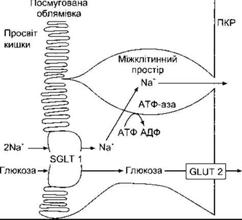
Fig. 25-3. Mechanism of glucose Transport Across the intestinal epithelium. Glucose Transport into enterocytes is coupled with Na+ transport via the SGLT 1 cotransporter. Subsequently, Na+ is actively pumped out of The Cell, and glucose enters the interstitial space via Facilitated Diffusion through GLUT 2, from which it ultimately diffuses into the bloodstream.
Pepsins hydrolyize bonds between aromatic amino acids, such as phenylalanine or Tyrosine, and another amino acid; thus, the products of peptic digestion are Polypeptides of various sizes. The stomach also contains gelatinase, which liquefies gelatin. Chymosin, a gastric milk-clotting enzyme also known as rennin, is present in the stomach of young animals and is sometimes absent in humans.
Since pepsins are optimally active at pH 1.6–3.2, their action ceases when the gastric contents mix with the alkaline pancreatic juice in the duodenum and jejunum. The pH of the intestinal contents in the duodenal bulb ranges from 2.0 to 4.0, whereas in other segments it is approximately 6.5.
In the small intestine, polypeptides formed during gastric proteolysis are digested by highly active Proteolytic Enzymes from the Pancreas and the intestinal mucosa. Trypsin, Chymotrypsin, and Elastase hydrolyze internal peptide bonds within peptide molecules, which is why they are classified as Endopeptidases. The generation of active endopeptidases from their inactive precursors is described in Chapter 26. Pancreatic Carboxypeptidases are exopeptidases that hydrolyze amino acids at the carboxyl and amino terminals of polypeptides. Some free Amino acids are released into the intestinal lumen, whereas others are liberated at the cell surface following prior Cleavage by aminopeptidases, carboxypeptidases, endopeptidases, and dipeptidases of the mucosal cell brush border. Certain di- and tripeptides are actively transported into intestinal Cells, where intracellular peptidases hydrolyze them into amino acids that subsequently enter the bloodstream. Consequently, the final digestion of Proteins into amino acids occurs at three sites: the intestinal lumen, the brush border, and the Cytoplasm of mucosal cells.
Absorption
Enterocytes possess seven distinct transport systems for moving amino acids. Five of these require Na+ ions, and amino acid–Na+ cotransport Functions similarly to Na+–glucose cotransport (see Fig. 25-3). Two transport systems also require Cl-. In two systems, transport is Na+-dependent.
Di- and tripeptides enter enterocytes via a system that requires H+ rather than Na+. This represents the primary absorption pathway for large Peptides. Within enterocytes, amino acids are released from peptides via intracellular Hydrolysis. Additionally, amino acids absorbed from the intestinal lumen and brush border are transported out of enterocytes across their basolateral membrane via five transport systems, from which they enter the hepatic portal Venous system. Two of these transport systems are Na+-dependent, while the rest are not. A significant quantity of small peptides also enters the portal system.
Amino acid absorption is rapid in the duodenum and jejunum, and slower in the ileum. Dietary intake accounts for about 50% of all digested proteins, digestive juice proteins contribute 25%, and desquamated mucosal cells provide 25%. Only 2–5% of proteins in the small intestine escape DIGESTION AND ABSORPTION. A fraction of unabsorbed proteins reaches the colon, where they are eventually digested by Bacteria. The presence of protein in fecal waste is not of dietary origin, but rather the result of bacterial and cellular breakdown. This is evidenced by cases where brush border and mucosal Cell Cytoplasm peptidase activity increases following resection of part of the ileum or during independent damage associated with starvation. Thus, these enzymes are subject to homeostatic regulation. In humans, an inherited defect in the mechanism transporting neutral amino acids in the intestine and renal tubules causes Hartnup disease. An inherited defect in basic Amino Acid Transport results in cystinuria.
In newborns, moderate amounts of undigested Proteins can also be absorbed. Protein antibodies in maternal colostrum are large secretory IMMUNOGLOBULINS (IgA) that cross the mammary epithelium via transcytosis; their production in the mammary gland increases toward the end of gestation. When breastfed, IgA is absorbed from the gut into the infant's Circulatory system, providing passive Immunity against infections. Absorption occurs via endocytosis followed by exocytosis.
Protein absorption declines with age, although it can still be detected in small amounts in adults. Foreign proteins entering the bloodstream trigger antibody formation and antigen–antibody reactions; upon repeated exposure to larger amounts of the same protein, these reactions cause allergic symptoms following the consumption of specific foods. The prevalence of food allergies in children exceeds 8%. Certain foods are much stronger allergens than others. Crustaceans, Mollusks, and fish are well-known allergens, although allergic responses to cow's milk, eggs, and various delicacies are also quite common.
The absorption of protein Antigens, particularly bacterial and viral proteins, takes place in large M cells (microfold cells)—specialized intestinal epithelial cells located in Aggregated lymphoid follicles (Peyer's patches). These cells deliver antigens to lymphoid Tissues, thereby initiating lymphoblast activation. Activated lymphoblasts enter the circulatory system and later return to the intestinal mucosa and other epithelial cells, where they secrete IgA in response to the reappearance of the same antigen. Such secretory immunity is a crucial defense mechanism and is described in detail in Chapter 27.
In the intestine, nucleic acids are broken down into NUCLEOTIDES by pancreatic Nucleases, which are subsequently split into nucleosides and phosphoric acid by enzymes located on the luminal membrane of mucosal cells. Nucleosides are further degraded into their constituent sugars and purine and pyrimidine bases, which are absorbed via Active Transport.
Last update: 10/08/2026
Editorial and Educational Adaptation: This material has been compiled based on the primary/original source text. The project team performed an editorial review, corrected technical inaccuracies, structured sections, and adapted the content for an educational format.
What was processed:
- elimination of formatting defects (OCR errors, structural breaks, corrupted characters);
- editorial organization of content;
- standardization of terminology in accordance with academic sources;
- verification of factual statements against the original source text.
All mentions of the author, publication year, and origin of the primary text have been preserved in accordance with the source.