Biological Chemistry - Berezov T. T., Korovkin B. F. 1998
Metabolism of Simple Proteins
Protein Digestion
Endopeptidases
Pepsin. One of the well-studied and principal Proteolytic Enzymes of the digestive tract is pepsin. Its presence in The Stomach was discovered as early as 1783 by L. Spallanzani, although it was isolated in crystalline form only in 1930 (see Chapter 1). Pepsin is produced in the chief Cells of the gastric mucosa in an inactive form as pepsinogen. The conversion of pepsinogen into active pepsin occurs within the gastric contents, yet the exact Molecular Mechanism of this transformation remains to be fully elucidated. The most widely accepted hypothesis suggests that this process is sequential, taking place in several stages in the presence of Hydrochloric acid via an autocatalytic mechanism driven by pepsin itself. The molecular mass of pepsinogen is approximately 40400, whereas that of pepsin is 32700; consequently, the Conversion of the former into the latter involves the Cleavage of peptide fragments. Both Enzymes can be relatively easily obtained in crystalline form. It is noteworthy that, unlike other proteinases, pepsin exhibits high stability in strongly acidic environments and is characterized by a low isoelectric point (pI < 1). Such conditions are typically established in the gastric contents, where hydrochloric acid secreted by the mucosal parietal cells is delivered*; the pH of pure gastric juice ranges from 1.0 to 2.0. This environment is optimal for the catalytic activity of pepsin. Evidence indicates that in the human stomach, pepsinogen likely yields not only active pepsin but also several structurally similar pepsins, including the pepsin-like enzyme gastrixin, which possesses a distinct pH optimum of 3.0.
Rennin. The enzyme rennin has been isolated in crystalline form from the juice of the fourth stomach compartment (abomasum) of calves. It is also present in the gastric juice of infants. In terms of its mechanism and catalytic Specificity, rennin differs significantly from pepsin, yet it is structurally close to it, consisting of a single polypeptide chain with a molecular mass of 40000. The isoelectric point of rennin is 4.5.
Three other major endopeptidases—Trypsin, Chymotrypsin, and Elastase—along with one exopeptidase, carboxypeptidase, which participate in the subsequent Digestion of Proteins following the action of pepsin, are synthesized in the Pancreas. All of them are produced in an inactive form as proenzymes, and their conversion into active enzymes takes place in the Small Intestine, where they are delivered via pancreatic juice.
Trypsin. Trypsinogen and trypsin have been obtained in crystalline form; their Primary Structure has been fully elucidated, and the molecular mechanism of the proenzyme-to-enzyme conversion is well understood. In vitro experiments demonstrate that the conversion of trypsinogen into trypsin is catalyzed not only by enteropeptidase and trypsin itself, but also by other proteinases and Ca2+ ions.
Chemical Activation of Trypsinogen involves the cleavage of 6 amino acid residues (Val—Asp—Asp—Asp—Asp—Lys) from the N-terminus of the polypeptide chain, resulting in a corresponding shortening of the chain (Fig. 12.1).
It should be emphasized that this seemingly minor chemical event—the cleavage of a hexapeptide from the precursor—carries profound biological significance, as it drives The formation of the Active Site and the ESTABLISHMENT OF THE three-dimensional structure of trypsin. As is well known (see Chapters 1 and 4), proteins are biologically active only in their native three-dimensional conformation. The fact that trypsin, like other proteinases, is synthesized in The Pancreas in an inactive form also serves a clear physiological purpose; otherwise, trypsin would exert destructive proteolytic effects not only on the cells of the gland itself, but also on Other Enzymes synthesized within it (amylase, lipase, etc.). At the same time, the pancreas protects itself through another mechanism: the synthesis of a specific pancreatic trypsin inhibitor. This inhibitor is a low-molecular-weight peptide (molecular mass 6000) that tightly binds to the active sites of trypsin and chymotrypsin, causing their reversible inhibition. The pancreas also synthesizes a1-antiproteinase (molecular mass 50000), which predominantly inhibits elastase.
* The entry of dietary protein into the stomach stimulates the secretion of the hormone gastrin, which in turn stimulates the secretion of HCl and pepsinogen by the mucosal cells.
Class="center">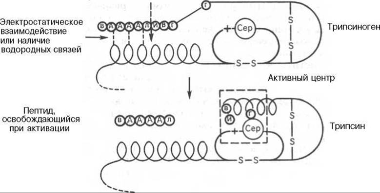
Fig. 12.1. Mechanism of bovine trypsinogen activation (diagram).
In acute pancreatitis, when trypsin and other enzymes are "washed out" from the damaged pancreas into the bloodstream, their Blood levels correlate with the size of the necrotic area. Under these conditions, the determination of serum trypsin activity serves as a reliable enzymatic test for diagnosing acute pancreatitis. It is worth noting that the substrate specificity of trypsin is restricted to the cleavage of peptide bonds formed exclusively by the carboxyl groups of Lysine and Arginine.
Chymotrypsin. The pancreas synthesizes a series of chymotrypsins (a-, ß-, and л-chymotrypsins) from two precursors: chymotrypsinogen A and chymotrypsinogen B. These proenzymes are activated in the intestine by the action of active trypsin and chymotrypsin. The Amino Acid Sequence of chymotrypsinogen A has been fully mapped and shares extensive Homology with that of trypsin. Its molecular mass is approximately 25000, and it consists of a single polypeptide chain containing 246 amino acid residues. Activation of the proenzyme does not involve the cleavage of a large segment of the molecule (see Fig. 4.3). Evidence has shown that the cleavage of a single peptide bond between arginine and isoleucine in chymotrypsinogen A by trypsin leads to the formation of л-chymotrypsin, which possesses the highest enzymatic activity. Subsequent Cleavage of the Ser—Arg dipeptide yields δ-chymotrypsin. The autocatalytic activation process driven by chymotrypsin first generates an inactive intermediate, neochymotrypsin, which is converted into a-chymotrypsin by active trypsin; this same product is also formed from δ-chymotrypsin through the action of active chymotrypsin.
Thus, through the combined cross-action of Chymotrypsin and trypsin, chymotrypsinogen is converted into various chymotrypsins that differ both in enzymatic activity and in certain physicochemical properties, notably electrophoretic mobility.
It should be noted that chymotrypsin exhibits a broader substrate specificity than trypsin. It catalyzes the Hydrolysis not only of Peptides, but also of esters, hydroxamates, amides, and other acyl derivatives, although its highest activity is directed toward peptide bonds involving the carboxyl groups of aromatic Amino Acids: phenylalanine, Tyrosine, and Tryptophan*.
Elastase. The pancreas synthesizes yet another endopeptidase, elastase, in the form of proelastase. The conversion of the proenzyme into elastase in the small intestine is catalyzed by trypsin. The enzyme derives its name from its substrate, Elastin, which it hydrolyzes. Elastin is a component of Connective Tissue characterized by a high content of Glycine and Serine residues. Elastase displays broad substrate specificity, yet preferentially hydrolyzes peptide bonds formed by amino acids with small hydrophobic side chains, specifically glycine, Alanine, and serine. Interestingly, neither trypsin nor chymotrypsin hydrolyzes the peptide bonds of the elastin molecule, even though all three enzymes, including elastase, share similar amino acid sequence regions, identical positions of disulfide bridges, and the same key active-site serine residue (see Table 4.2). This is supported by inhibition experiments using diisopropyl fluorophosphate, which chemically binds to the OH group of serine. It has been hypothesized that all three pancreatic endopeptidases—trypsin, chymotrypsin, and elastase—may share a common evolutionary precursor, and that the specificity of each active enzyme is primarily determined by Conformational Changes in the proenzyme during activation.
Exopeptidases. A family of exopeptidases plays an active role in Protein Digestion within the small intestine. Some of these, the Carboxypeptidases, are synthesized in the pancreas as procarboxypeptidases and activated by trypsin in the intestine; others, the aminopeptidases, are secreted by the intestinal mucosal cells and are likewise activated by trypsin.
Carboxypeptidases. Two carboxypeptidases, A and B, have been studied in detail; they belong to the metalloprotein class and catalyze the removal of C-terminal amino acids from Polypeptides. Carboxypeptidase A preferentially cleaves peptide bonds formed by C-terminal aromatic amino acids, whereas carboxypeptidase B cleaves bonds involving C-terminal lysine and arginine. Purified carboxypeptidase A exhibits bifunctional activity—both peptidase and esterase—and contains a Zn2+ ion (one atom per mole of enzyme). Replacing Zn2+ ions with Ca2+ ions completely abolishes peptidase activity while enhancing the intrinsic esterase activity, although no significant alterations in the Tertiary Structure of the enzyme are observed.
* Chymotrypsin is one of the most thoroughly investigated enzymes, for which the Mechanism of Enzymatic catalysis has been elucidated in detail, including the formation of an acyl-enzyme intermediate. The critical role of the serine hydroxyl group and an unprotonated Histidine residue in the active site has been definitively proven.
Aminopeptidases. Two enzymes have been discovered in intestinal juice: alanine aminopeptidase, which predominantly catalyzes the hydrolysis of peptide bonds involving an N-terminal alanine, and leucine aminopeptidase, which lacks strict substrate specificity and hydrolyzes peptide bonds formed by any N-terminal amino acid. Both enzymes carry out the stepwise cleavage of amino acids from the N-terminus of the polypeptide chain.
Dipeptidases. The digestive breakdown of peptides into free amino acids in the small intestine is completed by dipeptidases. Among the dipeptidases of intestinal juice, glycylglycine dipeptidase has been well studied; it hydrolyzes the corresponding dipeptide into two molecules of glycine. Two other dipeptidases are also known: prolyl dipeptidase (prolinase), which catalyzes the hydrolysis of peptide bonds involving the COOH group of Proline, and prolidase (proline dipeptidase), which hydrolyzes dipeptidases where the nitrogen of proline is bound via an acid-amide linkage.
Until relatively recently, proteinases were traditionally associated solely with digestive processes. Today, a growing body of evidence points to a much broader biological role for proteolytic enzymes of Organs and Tissues in regulating various extracellular and intracellular processes. Certain proteinases fulfill protective Functions (blood clotting, The Complement System, Cell lysis), while others generate Hormones, toxins, and vasoactive agents (angiotensin, kinins). A number of proteinases regulate the activation of digestive enzymes, cell-to-cell and cell-surface interactions, as well as Fertilization (Chitin synthetase) and differentiation. In most cases, this regulation involves the conversion of an inactive precursor into an active protein through the cleavage of a limited number of peptide bonds. This process, first described by K. Linderstrøm-Lang back in the 1950s, has recently come to be known as Limited proteolysis. Its significance is crucial for understanding The Nature of biological synthesis of inactive pre- and proproteins within cells. Furthermore, this process has found widespread Practical Application in both laboratories and industry. The regulation of proteolytic enzyme activity is also mediated by proteinaceous proteinase inhibitors discovered not only in the pancreas, but also in Blood Plasma, chicken egg white, and elsewhere.
The secretion of Pancreatic and Intestinal juices is regulated by neurohormonal factors, which are covered in detail in courses on physiology. Evidence Supports The Role of hydrochloric acid as a trigger for The production of specialized hormones in the intestine. Specifically, upon entering the duodenum, hydrochloric acid stimulates the secretion of secretin (see Chapter 8); the latter stimulates the secretion and flow of alkaline pancreatic juice while facilitating Bile outflow. Studies have shown that secretin rapidly disappears from the bloodstream and is not replenished because hydrochloric acid is neutralized by the alkaline pancreatic juice. Thus, through this negative feedback mechanism, the secretion and flow of pancreatic juice are meticulously regulated. Pancreatic juice obtained under METABOLISM/18.html">The Influence of secretin contains negligible amounts of enzymes, yet is rich in bicarbonates, which establish a mildly alkaline environment (pH 7.5–8.5) optimal for the action of digestive enzymes in the intestine. The second hormone, also synthesized in the duodenum and involved in regulating pancreatic secretion, is cholecystokinin (pancreozymin); it stimulates the output of a juice rich in enzymes and poor in bicarbonates.
Last update: 06/08/2026
Editorial and Educational Adaptation: This material has been compiled based on the primary/original source text. The project team performed an editorial review, corrected technical inaccuracies, structured sections, and adapted the content for an educational format.
What was processed:
- elimination of formatting defects (OCR errors, structural breaks, corrupted characters);
- editorial organization of content;
- standardization of terminology in accordance with academic sources;
- verification of factual statements against the original source text.
All mentions of the author, publication year, and origin of the primary text have been preserved in accordance with the source.