Biochemistry - The Chemical Reactions of Living Cells, Volume 2 - D. Metzler 1980
Types of enzyme-catalyzed reactions
Substitution reactions at carbonyl groups
Chymotrypsin and trypsin
The most thoroughly studied proteinase, Chymotrypsin, exists in several slightly different forms generated by the Cleavage of specific peptide bonds in the chymotrypsinogen molecule. The latter is a single polypeptide chain composed of 245 Amino Acids; the amino acids in the active enzyme are conventionally numbered according to their positions in the precursor zymogen. Data obtained from studies on acetylcholinesterase played a crucial role in elucidating the Mechanism of Chymotrypsin action. It was demonstrated that this key enzyme of The Nervous system is irreversibly inactivated by a group of potent phosphorus-containing poisons used as insecticides and nerve gases.
a. Active-Site Serine
In 1949, it was shown that diisopropyl fluorophosphate (DFP), a nerve agent, inactivates chymotrypsin.
Class="center">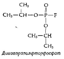
When chymotrypsin was treated with diisopropyl fluorophosphate labeled with 32P, the 32P became firmly bound to the enzyme via a covalent bond. Further experiments revealed that Denaturation of the labeled enzyme followed by acid Hydrolysis did not result in the release of 32P. The resulting radioactive fragment was identified as O-phosphoserine. From a chemical standpoint, the reaction between diisopropyl fluorophosphate and chymotrypsin proceeds simply and consists of the following.
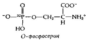
The hydroxyl group of the serine side chain attacks the diisopropyl fluorophosphate molecule at the phosphorus atom, displacing a fluorine atom. Notably, this reaction is a nucleophilic substitution at a phosphorus atom (Type 1.B reaction, Table 7-1), yet it occurs on an enzyme that typically catalyzes substitution reactions at the carbonyl carbon atom. The diisopropyl fluorophosphate molecule can be regarded as a pseudosubstrate (quasi-substrate) that reacts with the enzyme similarly to a true substrate, but fails to support the normal completion of the reaction.
Based on The Study of Peptides formed during the partial hydrolysis of 32P-labeled chymotrypsin, the Amino Acid Sequence surrounding the reactive serine residue was determined, and it was ultimately established that this serine residue occupies position 195 in the overall chain sequence. The preceding positions (193 and 194) are occupied by Glycine and aspartic acid, while position 196 contains another glycine molecule.
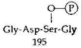
This same amino acid sequence surrounding the reactive serine residues was soon discovered in Trypsin, Thrombin, Elastase, and the trypsin-like enzyme coconase, which is used by the silkworm to emerge from its cocoon [27]. A structurally similar sequence containing Glu-Ser-Ala was found in acetylcholinesterase. Thus, there exists a family of serine peptidases and esterases characterized by a common amino acid sequence around the reactive serine and The ability to be inhibited by diisopropyl fluorophosphate [28].
b. The Acyl-Enzyme Intermediate. Another pseudosubstrate, p-nitrophenyl acetate,
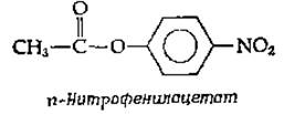
reacts with chymotrypsin at pH 4 (well below the pH optimum for the hydrolysis reaction rate) with the rapid release of p-nitrophenol and The formation of an acetyl derivative of the enzyme. The resulting acetyl enzyme undergoes very slow hydrolysis at pH 4, but much more rapidly at higher pH values. These experiments suggest that chymotrypsin, much like sucrose phosphorylase, acts via a double-displacement mechanism:
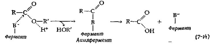
What is The Nature of the —B- group in this equation? Experiments with diisopropyl fluorophosphate indicate that the —O- group belongs to Ser-195. However, this hypothesis is contradicted by the extremely weak acidity of the —CH2OH group. Furthermore, other evidence suggests that The Role of the —B- group is played by the imidazole group of Histidine. For instance, it is known that the catalytic activity of chymotrypsin in ester hydrolysis varies with changes in pH. While the Michaelis constant changes very little with pH, the dependence of Vmax on pH is graphically represented by a typical bell-shaped curve (Fig. 6-13) with an optimum between pH 8 and 9. From the curve profiles for several different esterases, two pKa values have been determined: one between 6.1 and 6.8, and the second between 9.0 and 9.6. The pKa value lying between 6.1 and 6.8 is most logically attributed to the imidazole group of histidine, which is in the unprotonated state in the catalytically active form of the enzyme. There are also other experiments pointing to the involvement of a histidine residue in the catalytic process. For example, chymotrypsin reacts with 2,4-dinitrofluorobenzene, and the attack by this reagent on one of the two histidine residues in the enzyme molecule leads to its inactivation.
c. Nonenzymatic Models
It has been shown that imidazole-containing compounds catalyze the nonenzymatic hydrolysis of p-nitrophenyl acetate, yielding unstable acetylimidazoles as intermediates:
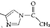
It was highly tempting to assume that the acyl-enzyme intermediates represented in equation (7-14) might have a similar Structure. Thus, there were two candidates for the role of the —B group: serine and histidine. Although there was no doubt that the stable End products of the reaction with pseudosubstrates are unquestionably serine derivatives, the possibility remained that these were side products and that histidine participates in the formation of intermediates that are rapidly formed and rapidly broken down.
d. Three-Dimensional Structure
The structures of chymotrypsin and trypsin have now been elucidated using X-Ray Diffraction Analysis [29–32], which confirmed the hypotheses put forth on The basis of chemical studies. Both Ser-195 and His-57 are located in the Active Site of the Enzymes (Fig. 7-2). It should be borne in mind that X-ray crystallography of the enzyme crystal does not allow the positions of hydrogen atoms within the enzyme molecule to be resolved, and they are positioned in the figure according to chemical logic. Thus, the short distance (0.30 nm) between the nitrogen of the His-57 residue and the oxygen of the Ser-195 residue indicates the presence of a Hydrogen bond. Similar reasoning led to the Conclusion that other Hydrogen Bonds shown in the figure are present. If histidine is in the unprotonated form and the serine hydroxyl group is protonated, we can see that histidine can act as a proton acceptor from the —CH2OH group of serine (i.e., as a general base catalyst), thereby increasing the nucleophilicity of the hydroxyl oxygen.
X-ray diffraction studies have shown that His-57 also forms a hydrogen bond with the carboxyl group of the Asp-102 residue, which in turn forms hydrogen bonds with two other groups. The side-chain carboxyl group of the Asp-102 residue is one of the few carboxyl groups buried deep within the protein molecule. Blow [29] suggests that such a structure must possess a charge-Relay system through which protons can be synchronously transferred from Ser-195 to imidazole and from imidazole to Asp-102. The Significance of this system for Serine proteinases is not yet fully established, but this question should be considered in light of METABOLISM/2.html">THE CONCEPT OF "tautomeric catalysis" (Chap. 6, Sec. D,5, e). It is also possible that the charge transfer is driven by electron-density shifts along the peptide chain. The fact that an identical charge-relay system evolved in another, otherwise completely distinct serine proteinase, subtilisin (from Bacillus subtilis), leads to the conclusion that this system may indeed be essential for the Mechanism of Enzymatic catalysis [33, 34].
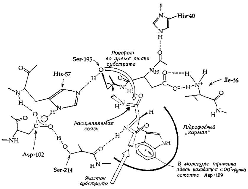
FIG. 7-2. Active site of chymotrypsin with a bound substrate fragment (after Blow [29] and Henderson and Young [13]).
Direct NMR observations of the proton involved in the hydrogen bond between His-57 and Asp-102 support the charge-relay concept [35]. However, attempts to construct an efficient nonenzymatic model incorporating the charge-relay system have proved unsuccessful [36].
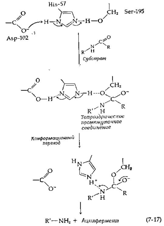
Increasing the nucleophilicity of the serine hydroxyl group is not the only possible role of the His-57 residue. The R'—NH- group displaced from the substrate is a poor leaving group unless it is converted into a protonated form (general acid catalysis):
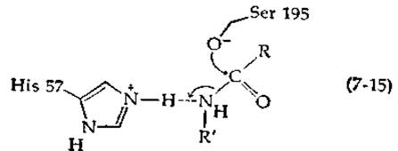
As Jencks [37] pointed out, it is often difficult to distinguish between general base and general acid catalysis. His-57 likely performs both Functions simultaneously, first abstracting a proton from the serine hydroxyl group and then donating a proton to the leaving group [13, 38].
Another probable Mechanism of enzyme participation in carbonyl substitution reactions involves the protonation of the carbonyl oxygen by an acidic group of the enzyme:
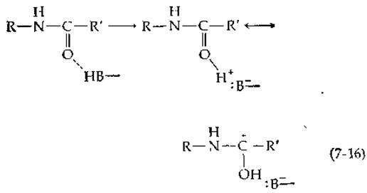
This should lead to a significant increase in the positive charge of the carbon atom, which in turn should facilitate nucleophilic attack. Such an interaction should also stabilize the tetrahedral intermediate [Equation (7-13)]. Carbonyl oxygen exhibits very weak basicity, but it can be protonated by an appropriately oriented acidic group of the enzyme [HB in Equation (7-16)]. In serine proteases, this function is apparently performed by two NH groups of amide bonds, one of which in chymotrypsin belongs to the Ser-195 residue (Fig. 7-2). Apparently, the fit of the substrate into the oxyanion hole between the two NH groups is optimal only for the tetrahedral intermediate [33].
It has been suggested [38] that the Formation of the tetrahedral oxyanion (with charge transferred from the masked carboxylate of the Asp-102 residue to the substrate) triggers a conformational transition, whereby during The breakdown of the tetrahedral intermediate, the imidazole group of the His-57 residue is able to take back the proton from Asp-102 and protonate the leaving group [Equation (7-17)].
Why does a conformational change occur? The enzyme molecule contains a complex network of hydrogen bonds. The appearance of a negative charge on the oxygen atom of the tetrahedral intermediate inevitably affects the electron density distribution within certain hydrogen bonds located far from the active center. If a protein molecule can exist in several energetically equivalent Conformations, a situation may arise where A change in charge distribution causes a sharp transition from one conformational state to another, and such a conformational transition can be an essential part of the catalytic process.
At high pH values, The activity of chymotrypsin decreases, and the Nature of the pH dependence indicates the presence of a group with a pKa of ~ 8 to 9 in the active center. This pKa value may correspond to the N-terminal amino group of Ile-16. The amino group of Ile-16 is involved in the formation of one of the bonds cleaved during The conversion of the zymogen into the active enzyme. This amino group forms an ionic bond (ion pair) with the Asp-194 residue (Fig. 7-2), which is located near the active-site serine. Possibly, this ionic bond helps maintain the enzyme in the conformation required for the reaction. Deprotonation at pH above 8–9 leads to inactivation [39].
Another possibility is that the group with the high pKa value belongs to a second histidine of the enzyme, His-40. However, this histidine is absent in bacterial serine protease, which exhibits the same type of pH dependence. His-40 is located close to the Asp-194 residue and is hydrogen-bonded to a peptide carbonyl. It is believed to play a certain role in the zymogen–active enzyme interconversion.
d. Substrate Specificity
Like most enzymes, trypsin and chymotrypsin exhibit pronounced specificity toward certain substrates. Rapid cleavage by chymotrypsin is observed when the C=O group of the cleaved peptide bond belongs to one of the aromatic amino acids. Thus, the substrate-binding site of the enzyme must contain a region that preferentially binds large, flat aromatic groups. Analysis of the crystal STRUCTURE OF THE enzyme shows that this center (Fig. 7-2) consists of hydrophobic amino acid side chains. In trypsin, however, the region of the active center responsible for specific substrate binding contains a fixed negative charge due to the carboxyl group of the Asp-189 side chain. This explains why trypsin cleaves only those peptide bonds flanked by Arginine or Lysine residues, which carry a positive charge at neutral pH [32].
Last update: 06/08/2026
Editorial and Educational Adaptation: This material has been compiled based on the primary/original source text. The project team performed an editorial review, corrected technical inaccuracies, structured sections, and adapted the content for an educational format.
What was processed:
- elimination of formatting defects (OCR errors, structural breaks, corrupted characters);
- editorial organization of content;
- standardization of terminology in accordance with academic sources;
- verification of factual statements against the original source text.
All mentions of the author, publication year, and origin of the primary text have been preserved in accordance with the source.