Molecular Biology of the Cell - Volume 3 - Alberts B., Bray D., Lewis J., Raff M., Roberts K., Watson J. 1994
From Cells to Multicellular Organisms
The Nervous System
Cells of the Nervous System: Structure and Function
The Nervous System provides rapid communication between distant PARTS OF THE body. Acting as a communication network, it governs reactions to external stimuli, processes information, and generates complex signal patterns to regulate sophisticated behaviors. Furthermore, the nervous system is capable of self-learning: as sensory information about the outside world is processed and memorized, the nervous system undergoes appropriate "fine-tuning," which alters The Nature of subsequent actions.
The primary pathways for neural signal transmission were mapped out more than a century ago, even before The Role of individual Nerve Cells was fully understood. Figure 19-1 illustrates the general layout of neural connections. Much like a large computer, the vertebrate nervous system consists of a main processor—the Central Nervous System, comprising the Brain AND SPINAL cord—which is linked via "wires," or nerves, to numerous peripheral structures: Sensory Organs that supply input, and Muscles (along with, to a lesser extent, glands) that execute output commands. There are also connections to groups of peripheral nerve cells called ganglia, which in some cases simply maintain communication between the periphery and the center, and in others serve as auxiliary mini-computers. Invertebrates share a similar Organization, but their central nervous system is less developed, whereas ganglia play a much larger and more autonomous role.
Class="center">
Fig. 19-1. A highly simplified diagram of the vertebrate nervous system organization. It shows how sensory information is transmitted from the periphery to the central nervous system (CNS), and motor commands from the CNS to the periphery. The transmission of sensory and motor signals is carried out by nerve cells, whose Cell bodies (indicated by large black dots) are in many cases grouped into ganglia (colored circles) outside the CNS, while their axons are bundled together to form nerves (black lines). Nerves, ganglia, and Sense Organs collectively make up the Peripheral Nervous System. Some ganglia function simply as "Relay stations," whereas others (particularly the enteric ganglia of the gut, which regulate peristalsis) are complex networks of interconnected Neurons capable of functioning entirely independently of the CNS. Interneurons, which are part of the central nervous system, are not shown.
While the detailed Structure of neural connections varies widely across different animal species within the framework of this general plan, the Properties of Individual neurons remain remarkably similar, whether in Mollusks, insects, amphibians, or mammals.
19.1.1. The function of a nerve cell is determined by the length of its processes [2]
The fundamental task of a neuron is to receive and transmit signals. Its unusual size and shape enable a neuron to perform these Functions: in humans, the length of a nerve cell connecting the Spinal Cord to a FOOT Muscle, for instance, can reach up to one meter. Each neuron consists of a cell body (containing The Nucleus) and long, slender processes extending from it. Typically, these include one long axon, which conducts signals away from The Cell body to distant targets, and several shorter, branching dendrites that act like antennae, receiving signals from the axons of other nerve cells (Fig. 19-2). The neuron's cell body also receives signals. The distant end of the axon usually branches, allowing it to transmit a signal simultaneously to multiple target cells. The degree of dendritic branching can also be extensive—in some cases, a single neuron can receive up to 100,000 signals. The branching patterns of axons and dendrites in functionally distinct types of neurons can be strikingly diverse (Fig. 19-3).
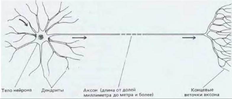
Fig. 19-2. Schematic representation of a typical vertebrate neuron. Arrows indicate the direction of signal transmission. The largest human neurons reach lengths of about one meter and have axons up to 15 µm in diameter.
19.1.2. Nerve cells transmit electrical signals [3]
The Significance of the signals transmitted by a nerve cell depends on the cell's specific role within the nervous system as a whole. In motor neurons, signals serve as commands for the contraction of specific muscles. In sensory neurons, they convey information about particular stimuli acting on a region of the body, such as light, mechanical force, or chemicals. In interneurons, which link one neuron to another, signals facilitate complex interactions and the integration of information from multiple sources, participating in The regulation of complex behavior.
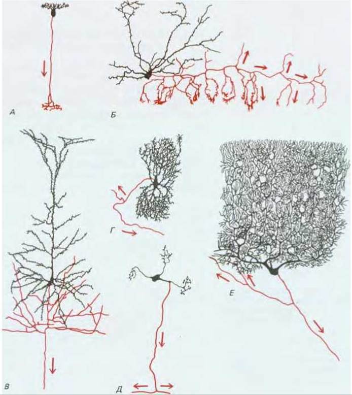
Fig. 19-3. Some of the numerous types of vertebrate nerve cells, as visualized by Golgi staining. This technique, which involves immersing tissue in a solution of metal salts, completely stains a small fraction of the cells in a preparation black, making all the branches of their processes visible. Numerous dendrites, which receive incoming signals from other cells, extend from the neuron cell body, along with a single thin, branching axon that transmits outgoing signals in the direction indicated by the arrows. Axons are shown in red, while the cell body and dendrites are black. Cells A and B have short axons, which are shown in their entirety. Cells C through E have very long axons, and only their proximal segments are illustrated. A—bipolar cell from the retina of a lizard; B—basket cell from the mouse Cerebellum; C—pyramidal cell from the Cerebral Cortex of a rabbit; D—neuron from the human Brainstem; E—one of the granule cells from the cat cerebellum; F—Purkinje cell from the human cerebellum. This latter cell, featuring an extensively branched dendritic tree, receives signals from more than 100,000 other neurons; it represents an element of the brain circuitry that regulates complex movements. The drawings are not to scale: the length of the bipolar cell (A) is about 100 µm, whereas the depicted portion of the Purkinje cell (F) is about 400 µm wide (its axon can reach several centimeters in length).
Despite the varying significance of these signals, their underlying nature is identical in all cases, involving Changes in the electrical potential across the neuron's Plasma Membrane. Communication occurs because an electrical disturbance generated in one region of the cell spreads to others. These disturbances decay with distance from their source unless they receive additional Amplification along the way. Over short distances, this attenuation is negligible, and many small neurons conduct signals passively without amplification. However, such passive propagation is insufficient for long-distance communication. Therefore, neurons with longer processes employ an active signaling mechanism, which constitutes one of the most remarkable and Characteristic Properties of a neuron. An electrical stimulus exceeding a certain threshold triggers a "spike" in electrical activity that propagates rapidly along the neuron's plasma membrane, sustained by automatic amplification throughout its journey. This traveling wave of electrical excitation, known as an Action Potential or Nerve Impulse, is capable of transmitting information without attenuation from one end of the neuron to the other at speeds of 100 m/s or more.
19.1.3. Communication between neurons occurs at synapses via chemical signals [4]
Signals conducted by neurons are passed from one cell to another specialized contact sites called synapses. Typically, this transmission takes place—rather surprisingly at first glance—via an indirect route. The cells are electrically isolated from one another: the presynaptic cell is separated from the postsynaptic cell by a gap known as the synaptic cleft. A change in the electrical potential of the presynaptic cell triggers the release of a substance called a neurotransmitter, which is stored in membrane-bound synaptic vesicles and released via exocytosis. The neurotransmitter diffuses across the synaptic cleft and alters the electrophysiological state of the postsynaptic cell (Fig. 19-4). As we will see later, The Mechanism of signal transmission through such chemical synapses is far more flexible and adaptable than direct electrical coupling via Gap Junctions (see Section 14.1.7), which is also utilized, though much less frequently.

Fig. 19-4. Diagram of a typical synapse. An electrical signal arriving at the axon terminal of cell A prompts the release of a chemical messenger (neurotransmitter) into the synaptic cleft, which induces an electrical change in the membrane of the dendrite of cell B. The broad arrow indicates the direction of signal transmission.
A chemical synapse is a site of intense biochemical activity, involving the breakdown, turnover, and secretion of Proteins and other molecules. However, the biochemical control center of the neuron is its cell body, where the primary instructions for Protein Synthesis reside. Consequently, the neuron requires an efficient Intracellular Transport system to move molecules from the cell body to the most distant Regions of the axon and dendrites. How is this transport system organized, and what molecules are actually transported?
19.1.4. Newly synthesized Materials are transported from the nerve cell body to axons and dendrites via Slow and fast transport mechanisms [5]
Electron Microscopy has revealed that the cell body of a typical large neuron contains a vast number of Ribosomes, some located in the Cytosol and others attached to the membranes of the rough Endoplasmic reticulum (Fig. 19-5, A). Although dendrites often contain a small number of ribosomes, axons lack them entirely, meaning axonal proteins must be synthesized on ribosomes in the cell body (Fig. 19-5, B). The demands placed on the axon are substantial: for instance, a large human motor neuron can have a diameter of up to 15 µm and a length of 1 m, corresponding to a volume of roughly 0.2 mm3, which is nearly 10,000 times the volume of a Liver cell. Because such a neuron contains only a single nucleus, its Cytoplasm-to-DNA ratio is much higher than that of any other cell type in The Human Body.

Fig. 19-5. ULTRASTRUCTURE OF THE cytoplasm of a typical large neuron (a spinal motor neuron). A. Low-power schematic view of the cell body, showing that ribosome-rich cytoplasmic regions are interspersed between bundles of neurofilaments and other cytoskeletal proteins. B. Electron micrograph of one such ribosome-rich region; some ribosomes are attached to the rough endoplasmic reticulum, while others are free. C. Electron micrograph of a cross-sectional portion of an axon, displaying a high density of microfilaments and microtubules, with a complete absence of ribosomes. Membrane-bound vesicles presumably move along nearby microtubules via fast axonal transport mechanisms. (Courtesy of Jennifer La Vail (B) and John Hopkins (C).)
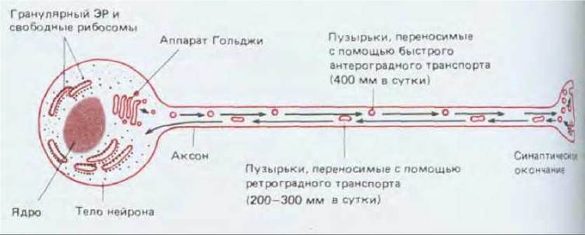
Fig. 19-6. The neuron as a secretory cell, where the site of secretion (the axon terminal) is located far away from the site of macromolecule synthesis (the cell body). This arrangement necessitates a mechanism for rapid axonal transport. One should not conclude from this diagram that all synaptic vesicles are transported all the way from the neuron cell body: in most neurons, they are primarily generated through local membrane recycling at the axon terminal.
Microtubule-forming proteins, neurofilaments (a class of Intermediate filaments), and Actin filaments are found in the highest Abundance within the axon (Fig. 19-5, B). These cytoskeletal proteins originate in the cell body and travel down the axon at rates ranging from 1 to 5 mm per day. This process is known as slow axonal transport (a similar transport mechanism also operates in dendrites, which contain a somewhat distinct set of microtubule-associated proteins—see Section 11.4.7). Other cytosolic proteins, including numerous Enzymes, are likewise carried via slow axonal transport, though its precise mechanism remains elusive.
Non-cytosolic materials essential for synaptic function, such as secretory proteins and membrane-bound molecules, are dispatched from the cell body through a significantly faster mode of axonal transport. These proteins and Lipids are ferried from their sites of synthesis in The endoplasmic reticulum to the Golgi apparatus, which is situated near the nucleus (frequently at the axon hillock). From there, these molecules—packaged into membrane vesicles—are dispatched via fast axonal transport at speeds up to 400 mm per day along tracks formed by microtubules within the axon and dendrites (Section 11.4.8). Mitochondria are transported in an identical manner. Because different classes of proteins are routed to axons and dendrites via this pathway, it is believed that the transported molecules are sorted into distinct types of transport vesicles within the cell body (Section 8.9.4).
Proteins destined for release at the synapse also belong to the category of rapidly transported axonal proteins. Examples include Neuropeptides, which are secreted by numerous neurons to act as Neurotransmitters, often in conjunction with non-protein mediators. From the perspective of internal organization, neurons can be conceptualized as secretory cells in which the site of secretion lies at an immense distance from the site where proteins and membranes are synthesized (Fig. 19-6).
19.1.5. Retrograde transport maintains a feedback chemical link between nerve terminals and the cell body [5, 6]
Fast axonal transport is essential during cellular development for the growth of axons and dendrites, which elongate through The addition of new membrane material at their tips. This rapid transport system persists in mature, fully grown neurons where The amount of membrane material at the tips of the processes remains constant. Under these conditions, the rapid anterograde transport of membranes away from the cell body must be precisely balanced by a rapid retrograde transport of membranes in the reverse direction—from the tips of the cellular processes back toward the soma. Although the mechanisms governing these two opposing directions of fast transport are similar, they are not identical. Fast retrograde transport, which proceeds at roughly half the velocity of fast anterograde transport, relies on distinct motor proteins (Section 10.4.9) and is utilized to carry slightly larger vesicles. The structures returning to the cell body consist partly of Aging cytoplasmic Organelles, such as mitochondria, and partly of vesicles generated by intensive endocytosis, which is required to reclaim the membrane at the axon terminal following neurotransmitter release (see Fig. 19-20). Extracellular molecules surrounding the axon terminal can be internalized by these endocytic vesicles and carried alongside them to the cell body. Consequently, the biosynthetic machinery housed within the soma is able to "sense" environmental changes occurring at the axon terminal and respond accordingly, as we will explore later (Section 19.7.10).
Retrograde transport enables neuroanatomists to easily trace Neural Pathways using the straightforward methodology illustrated in Fig. 19-7.

Fig. 19-7. Using fast axonal transport to identify and localize distant neurons whose axons terminate within a region of interest. Horseradish peroxidase (HRP) is the most widely employed marker enzyme because its molecules can be detected in extremely minute quantities via the colored products of the enzymatic reaction it catalyzes.
19.1.6. Neurons are surrounded by various types of glial cells [7]
All Nervous Tissue, both peripheral and central, is composed of two primary classes of cells. Neurons play the starring role, but supporting glial cells vastly outnumber them, accounting for roughly 10 times more cells than neurons in the mammalian brain. Glial cells envelop neurons (both their cell bodies and processes) and occupy the intercellular spaces between them. The most thoroughly investigated are Schwann cells in vertebrate peripheral nerves and oligodendrocytes in the vertebrate central nervous system. These cells wrap around axons to construct an insulating myelin sheath (Section 19.2.4). Three additional types of glial cells populate the central nervous system: microglia, ependymal cells, and astrocytes (Fig. 19-8). Microglia constitute a somewhat distinct class; these cells are functionally related to macrophages (Section 17.5.1) and, similarly, originate from hematopoietic tissue. All other glial cells share a common embryonic origin with the neurons they are associated with, yet unlike most neurons, Glia generally lack electrical excitability. Furthermore, whereas neurons lose the capacity to divide after differentiation, the majority of glial cells retain this ability throughout life.
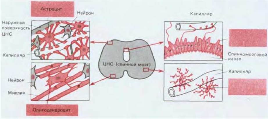
Fig. 19-8. Three Major Classes of glial cells from the vertebrate central nervous system. Glial cells are highlighted in color. Astrocytes, the most numerous glial cells, feature numerous radially branching processes. Some of these processes terminate on neuronal surfaces, whereas others terminate in expanded end-feet that form the outer limiting surface of the CNS, encircling Blood Vessels and collaborating with capillary endothelial cells to construct the blood-brain barrier. Ependymal cells form a ciliated epithelium that lines the internal cavities of the CNS; like astrocytic processes, their projections frequently terminate on blood vessels. Oligodendrocytes generate the insulating myelin sheath surrounding CNS axons. Microglial cells resemble macrophages in both function and origin; they participate in tissue responses to injury and infection and are typically located in close proximity to blood vessels.
Ependymal cells line the internal cavities of the brain and spinal cord (Fig. 19-8), and their epithelial organization serves as a reminder that the central nervous system originates from the embryonic epithelial tube (Section 19.7.1).
Astrocytes (Fig. 19-8) are the most numerous and morphologically diverse glial cells, yet they remain among the most enigmatic. Although their exact functions are still largely undetermined, there is little doubt that they play a crucial role in The Development of the nervous system (Section 19.7.2) and regulate the chemical and ionic COMPOSITION OF THE microenvironment surrounding neurons. For instance, one subclass of astrocytes possesses processes with expanded end-feet linked by junctional complexes akin to those found in epithelia (Section 14.1), thereby creating an insulating barrier on the outer surface of the central nervous system. Other processes from these same astrocytes form similar end-feet upon blood vessels (capillaries and venules), where endothelial cells are joined by exceptionally well-developed tight junctions, forming the blood-brain barrier. This barrier prevents Water-soluble molecules in the blood from penetrating brain tissue unless they are ferried by specialized transport proteins embedded in The Plasma Membrane of the endothelial cells. As a result, neurons are maintained within a tightly controlled and protected environment, which is vital for the molecular mechanisms underlying electrical signaling.
Summary
Nerve cells, or neurons, are specialized cells featuring exceptionally long processes designed to transmit electrical signals in the form of action potentials—propagating waves of electrical excitation. Typically, a nerve cell body gives rise to several branching dendrites and a single long axon. Signals are generally received by the dendrites and the cell body, subsequently propagating along the axon to be relayed to other cells at chemical synapses. Upon reaching the presynaptic axon terminal, the arriving electrical signal triggers the secretion of a neurotransmitter, which in turn induces an electrical shift in the postsynaptic cell.
A neuron can be viewed as a secretory cell that discharges its product—a neurotransmitter—at a vast distance from the cell body where macromolecules are synthesized. Newly synthesized secretory proteins and membrane-building materials are transported along axons and dendrites via fast axonal transport, wherein small membrane vesicles move along tracks formed by microtubules. Microtubules and other non-membrane-bound cytoplasmic components migrate away from the cell body through an entirely distinct mechanism known as slow axonal transport. Fast axonal transport also operates in the reverse, retrograde direction, carrying membrane vesicles back from the axon terminals to the cell body.
Neurons are surrounded by glial cells, which contribute in diverse ways to regulating the chemical and electrical Properties of the neuronal microenvironment.
Last update: 12/08/2026
Editorial and Educational Adaptation: This material has been compiled based on the primary/original source text. The project team performed an editorial review, corrected technical inaccuracies, structured sections, and adapted the content for an educational format.
What was processed:
- elimination of formatting defects (OCR errors, structural breaks, corrupted characters);
- editorial organization of content;
- standardization of terminology in accordance with academic sources;
- verification of factual statements against the original source text.
All mentions of the author, publication year, and origin of the primary text have been preserved in accordance with the source.