Peptide Self-Regulation of Living Systems (Facts and Hypotheses) - Shataeva L. K. 2003
Peptides in Aqueous Solutions
Tissue-Specific Regulatory Peptides
Neuropeptides
The vertebrate Nervous system performs several key Functions: monitoring physiological parameters within the Organism as well as environmental conditions, transmitting the acquired information to the Central Nervous System, integrating this data and comparing it with information stored in memory, sending command signals to effector Organs across all body systems, and monitoring the execution of these commands. In other words, The Nervous System exerts direct regulatory control driven by continuous Information Flow regarding the internal and external environment.
The human nervous system consists of Nerve Cells—Neurons—interconnected by a network of synapses. The human Brain contains approximately 1011 neurons, each typically synapsing with hundreds or thousands of others. In addition to neurons, the human brain contains Connective Tissue comprising various types of glial cells, which outnumber neurons 5-to 10-fold; neuroglia accounts for roughly 40% of the volume of the human BRAIN AND SPINAL cord. The integrating and commanding information networks of the brain are complemented by several Endocrine glands that synthesize Peptide Hormones.
The classical nomenclature of endocrine glands includes the pituitary, Pineal Gland, Hypothalamus, thyroid and Parathyroid glands, Pancreas, Adrenal Glands, and Gonads (Pankov, 1996). Historically, the regulators isolated from these glands were termed hormones, encompassing both high-molecular-weight Proteins and non-protein compounds. Later, a large group of Peptides isolated from neuroendocrine glands came to be known as neuropeptides (NPs). Today, these represent the most extensively studied regulatory peptides.
The Classification of peptides proposed by I. P. Ashmarin (Ashmarin, Kamenskaya, 1988) encompasses exclusively peptides of neuronal origin. Adhering to this principle and taking into account the specific Tissues from which the peptides were isolated, Appendix Table I presents the simplest regulatory NPs (Regulatory peptides, 1989; Ashmarin, Karazeeva, 1999).
Many NPs and peptide hormones are generated from high-molecular-weight precursors (such as protachykinin, proopiomelanocortin, chromogranins, etc.), the synthesis of which is encoded by respective genes. In particular, chromogranin A—a precursor to several statins—is found in all neuroendocrine tissues and is secreted from pancreatic and thyroid C-cells alongside Other Hormones. Its Structure contains blocks of two amino acid residues with basic side groups (R—R, R—K, K—R, and K—K) that are specifically cleaved by intracellular Endopeptidases. This process yields the functional regulatory NPs: parastatin, pancreastatin, and vasostatin, which inhibit catecholamine release, with vasostatin additionally reducing Blood pressure. Other RPs, produced via the specific Hydrolysis of protachykinin and proopiomelanocortin, are stored packaged in neurosecretory granules within the presynaptic terminals of neurons until a specific signal arrives. Their release appears to occur via standard exocytosis triggered by an electrosecretory or chemical signal received at the outer membrane, specifically via a membrane receptor.
The NP structures presented in Appendix Table 1 vary in molecular size, with the number of amino acid residues per molecule ranging from 3 (thyroliberin) to 44 (somatoliberin). Work by I. P. Ashmarin and M. A. Kamenskaya (1988) provides data on the half-life of NPs in plasma and CEREBROSPINAL FLUID, demonstrating that short peptides of 3–8 amino acid residues have a half-life of 1–2 minutes, although thyroliberin remains active in various tissues for up to 1 hour. Longer peptides maintain their efficacy for several hours.
A peptide's stability in the cellular environment is primarily governed by The Nature of its initial, N-terminal amino acid. Methionine, Alanine, Serine, Threonine, valine, Glycine, Cysteine, and Proline protect peptides from hydrolysis. Conversely, the remaining 12 Amino Acids at the N-terminus trigger proteolytic attack (Alberts et al., 1994). Consequently, NPs exhibit a more prolonged action compared to conventional Neurotransmitters (acetylcholine, histamine, serotonin, and GABA), which remain active for only 10-2–1 s. In some cases, peptides released into the extracellular humoral environment (blood, Lymph, tissue fluid) undergo hydrolysis mediated by peptidases. A classic example is the cascade hydrolysis (Fig. 6, A) of the bradykinin precursor by Serine proteinases of the kallikrein family (Margolis, Buse, 1983).
A comparison of the structures of endorphin and enkephalins suggests that a similar stepwise hydrolysis occurs within the opioid group as well.
The structural relationship among various NPs derived from a single precursor can be illustrated using Pituitary Hormones as an example. Several of these share an identical heptapeptide core, which is boxed within the Amino Acid Sequence of a-melanotropin (melanocyte-stimulating hormone) (Fig. 6, B).
Class="center">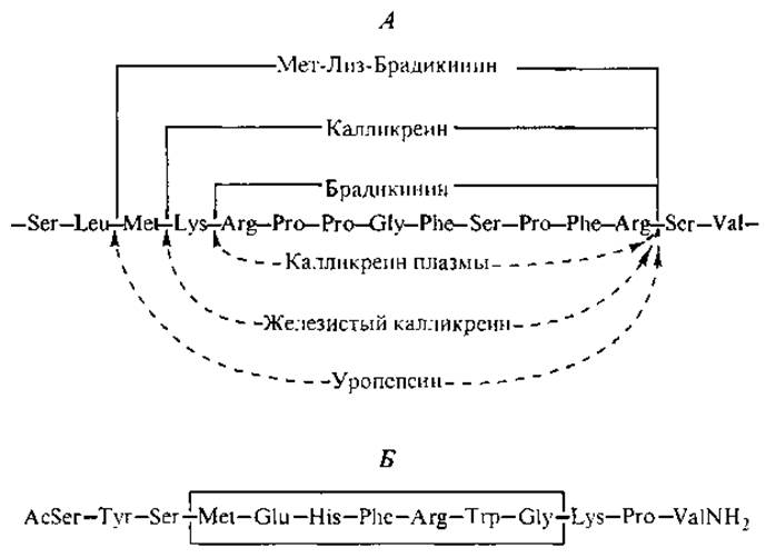
Fig. 6. Sequential proteolysis of a plasma globulin kininogen chain segment by specific Hydrolases (A) and The amino acid sequence of a-melanotropin (B).
Corticotropin (ACTH) contains not only this designated heptapeptide but the entire amino acid sequence of a-melanotropin as well (compare the final two rows in Appendix Table I). An identical core is found in pituitary ß- and y-lipotropins (containing 91 and 56 amino acid residues, respectively), which stimulate lipid hydrolysis reactions in adipose tissues. Like ACTH, these hormones belong to the family of pituitary hormones derived from proopiomelanocortin, a prohormone comprising 241 amino acid residues. In fact, the lipotropic activity of lipotropins per se is no higher than that of ACTH and melanotropin, and it remains unclear whether they possess any distinct specific physiological activity of their own, though both lipotropins are present in human plasma.
Data on the multifunctional nature of corticotropin are complemented by findings that peptide segments 4–7 and 4–10 of its chain stimulate attention and learning (Ponomareva-Stepnaya et al., 1984). Furthermore, the synthetic peptide Semax—designed based on the ACTH (4–10) sequence by substituting the last Three amino acids to protect against Enzymatic hydrolysis—acts as a long-acting learning enhancer and is recommended for clinical use (Gomazkov, 1997):
Met—Glu—His—Phe—Pro—Gly—Pro.
On the one hand, the presence of a common heptapeptide core in The structure of pituitary hormones points to the evolutionary relatedness of this group. On the other hand, the Structural Organization of these hormones and the sequential alteration of their functions as a specific number of amino acid residues are hydrolytically cleaved demonstrate the remarkable economy of these peptide regulators. Within corticotropin itself, a segment of the amino acid chain (residues 7–38) is capable of inhibiting corticosteroid synthesis—one of the primary processes the entire corticotropin molecule is designed to stimulate. This peptide was also isolated from the Pituitary Gland and designated corticotropin-inhibiting peptide:

A similar relationship is observed in the structure of ß-endorphin. A segment of its peptide chain (residues 6–31) was synthesized and shown to exert an inhibitory effect on ß-endorphin-induced analgesia (Choh Hao Li, 1984). Accordingly, this peptide was named ß-endorphin-inhibiting peptide, and its structure is illustrated in the diagram below:

However, fragments remaining after the Cleavage of a parent NP molecule do not always inhibit the original peptide's action. The galanin 1–15 fragment potentiates The activity of the full-length peptide by increasing the sensitivity of its selective receptors in the hippocampus.
Common architectural principles among peptide regulators are evident not only through literal structural Homology but also via shared "Structural motifs." This is particularly crucial when analyzing the Regulatory Functions of peptides from different groups, such as neuropeptides and Gastrointestinal Hormones. Below is a structural comparison of melanostatin (MS)—a neuropeptide found in the brain, adrenal glands, and Heart—along with the intestinal peptide YY (which inhibits exocrine pancreatic secretion) and pancreatic peptide (PP) (which regulates Digestion and stimulates pancreatic functions) (Oxford Dictionary..., 1967):
MS: YPSKPDN PGEDAPAEDMARYYSALRHYIN LITRQRY-N H2
YY: YPIKPEAPGEDASPEELNRYYASLRHYLNLVTRQRY—NH2
PP: APLFPVYPGDNATPEQMAQYAADLRRYINMLTRPPY-NH2
Clearly, these structures are homologous yet exhibit distinct activities. Not only the removal of a polypeptide chain segment, but even the substitution of a single amino acid residue within a given sequence can reverse the biological meaning of the signal: for instance, the inhibitory signal of YY becomes stimulatory upon the substitution of just a few amino acids. Concurrently, a slight C-terminal elongation can significantly prolong a peptide's action while preserving its specific activity, as demonstrated with ACTH4-10 and tuftsin (Ponomareva-Stepnaya et al., 1984; Potaman et al., 1992). This gives the impression that the N-terminus of an NP carries greater semantic significance than its C-terminal regions.
Last update: 06/08/2026
Editorial and Educational Adaptation: This material has been compiled based on the primary/original source text. The project team performed an editorial review, corrected technical inaccuracies, structured sections, and adapted the content for an educational format.
What was processed:
- elimination of formatting defects (OCR errors, structural breaks, corrupted characters);
- editorial organization of content;
- standardization of terminology in accordance with academic sources;
- verification of factual statements against the original source text.
All mentions of the author, publication year, and origin of the primary text have been preserved in accordance with the source.