Human Anatomy - Kotsan I. Y. 2009
Endocrine glands
Pituitary gland
The Pituitary Gland (hypophysis) is a small, spherical or oval-shaped gland located in the hypophyseal fossa of the Sphenoid bone (Fig. 227). Superiorly, the pituitary gland is covered by a fold of the dura mater known as the diaphragma sellae, which separates it from the cranial cavity. Through an opening in this Diaphragm, the gland connects with the infundibulum of the Hypothalamus in the Diencephalon.
The mass of the pituitary gland is approximately 0.5 g in men and 0.6 g in women. Its dimensions are quite small: length is 8–10 mm, width is 12–15 mm, and height is 5–6 mm. The pituitary gland reaches its maximum size by the age of 20, after which it remains unchanged. In women, the pituitary gland enlarges significantly during Pregnancy and remains in this state postpartum throughout life.
Although anatomically a single Structure, the pituitary gland is divided into two lobes that differ in origin, structure, and function: the anterior lobe (lobus anterior) and the posterior lobe (lobus posterior). The anterior lobe, or adenohypophysis (adenohypophysis), develops from the epithelium of the oral bay through a finger-like protrusion of the pharyngeal (hypophyseal) pouch. It initially arises as an exocrine gland, but its duct soon regresses, transforming it into an endocrine gland. Remnants of the former duct may occasionally persist as a channel (canalis craniopharyngeus) extending from the floor of the sella turcica to the Pharynx. The posterior lobe, or neurohypophysis (neurohypophysis), forms later than the anterior lobe and represents an outgrowth of the diencephalon (via a protrusion of the floor of the Third ventricle). The upper part of this protrusion remains hollow, forming the tuber cinereum and infundibulum, while the lower part forms the posterior lobe of the pituitary gland and the pituitary stalk.
Class="center">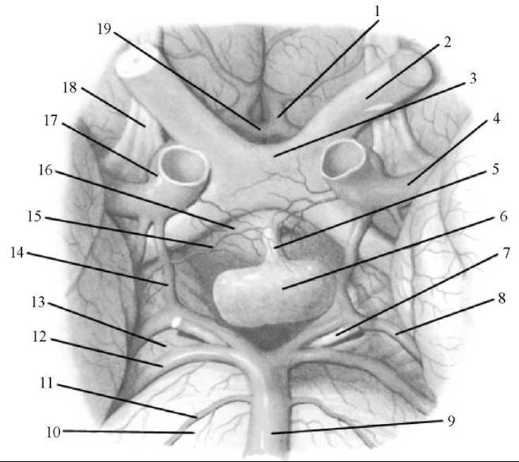
Fig. 227. The pituitary gland (after R. D. Sinelnikov)
1 — anterior cerebral artery; 2 — Optic nerve; 3 — optic chiasm; 4 — middle cerebral artery; 5 — infundibulum; 6 — pituitary gland; 7 — Oculomotor nerve; 8 — posterior cerebral artery; 9 — Basilar artery; 10 — Pons; 11 — labyrinthine artery; 12 — superior cerebellar artery; 13 — cerebral peduncle; 14 — posterior communicating artery; 15 — hypophyseal artery; 16 — tuber cinereum; 17 — Internal Carotid Artery; 18 — olfactory tract; 19 — anterior communicating artery.
The anterior lobe of the pituitary gland is significantly larger than the posterior lobe, accounting for 70–80% of its total mass. The anterior lobe comprises the pars distalis, which is the largest part occupying the anterior region of the hypophyseal fossa; the pars intermedia, located at the border with the posterior lobe; and the pars tuberalis, which extends upward to connect with the hypothalamic infundibulum. The parenchyma of the anterior lobe consists of several types of glandular Cells, between the cords of which lie sinusoidal Blood capillaries. Due to the abundant vascular supply, the anterior lobe has a pale pinkish-red hue.
The posterior lobe consists of the neural lobe (lobus nervosus), located in the posterior part of the hypophyseal fossa, and the infundibulum, situated posterior to the pars tuberalis of the adenohypophysis. The posterior lobe is formed by neuroglial cells (pituicytes), nerve fibers extending from the neurosecretory nuclei of the hypothalamus into the neurohypophysis, and Herring bodies (neurosecretory bodies).
Through nerve fibers (tracts) and Blood Vessels, the pituitary gland is functionally linked to the hypothalamus of the diencephalon, which regulates its activity. The Hormones of the anterior and posterior lobes influence many bodily Functions, primarily via other Endocrine glands. The anterior lobe produces the following hormones: Growth Hormone (somatotropin), adrenocorticotropic hormone, thyrotropic hormone, gonadotropic hormones, melanotropin (melanocyte-stimulating hormone), and lipotropic factors. Somatotropin is involved in regulating growth and developmental processes in the body. Excessive production of this hormone in childhood leads to pituitary gigantism (height reaching 2 m), whereas its deficiency results in Pituitary dwarfism (height not exceeding 70 cm). In adults, overproduction of growth hormone leads to acromegaly, characterized by the enlargement of specific body parts (hands, feet, Nose). Adrenocorticotropic hormone (ACTH) stimulates the Adrenal Glands to secrete Steroid Hormones. Thyrotropic hormone (TSH) influences The Development of The Thyroid Gland and activates The production of its hormones. Gonadotropic hormones (follicle-stimulating hormone, luteinizing hormone, and prolactin) affect sexual maturation, regulate and stimulate ovarian follicle development, ovulation, mammary gland growth and milk production, and Spermatogenesis in men. Melanocyte-stimulating hormone, or melanotropin, produced in the pars intermedia of the anterior lobe, controls The formation of melanin pigments in the body. Additionally, the anterior lobe produces lipotropic factors that influence the mobilization and utilization of fats in the body.
In the posterior lobe—the neurohypophysis—two hormones, oxytocin and vasopressin, produced by the neurosecretory Cells of the supraoptic and paraventricular nuclei of the hypothalamus, are stored and converted into their active forms. These hormones are transported to the cells of the posterior lobe via axons that form the hypothalamo-hypophyseal tract. From the posterior lobe, these substances enter the bloodstream. Vasopressin constricts blood vessels, raises blood pressure, and decreases urine output (hence its alternative name, antidiuretic hormone — ADH). Oxytocin increases the tone of smooth Muscle, particularly the Uterus, Urinary Bladder, and intestines, and inhibits the development and function of the corpus luteum. Because the pituitary gland produces hormones that stimulate the development and function of other endocrine glands, it is referred to as the master gland of the Endocrine System.
The pituitary gland is supplied with blood by the superior and inferior hypophyseal Arteries. The inferior hypophyseal arteries branch off from the internal carotid arteries, while the superior ones originate from the Vessels of the cerebral arterial circle (circle of Willis). The superior hypophyseal arteries travel to the tuber cinereum and the hypothalamic infundibulum, where they anastomose and break down into capillaries forming the primary capillary plexus. From the long and short loops of this plexus, portal venules arise, descending along the pars tuberalis to the anterior lobe of the pituitary, where they empty into wide sinusoidal capillaries, forming a secondary capillary plexus that envelops groups of secretory cells. The capillaries of the secondary plexus converge to form efferent venules, through which blood (carrying anterior lobe hormones) leaves the pituitary gland. The posterior lobe is supplied primarily by the inferior hypophyseal arteries. Long arterial anastomoses exist between the superior and inferior hypophyseal arteries. Venous blood from the pituitary drains into the plexus at the Base of the Brain and subsequently into the great cerebral vein.
The Innervation of the pituitary gland involves sympathetic fibers that enter the organ alongside the arteries. Additionally, the posterior lobe contains numerous processes from neurosecretory cells located within the hypothalamic nuclei.
Last update: 08/08/2026
Editorial and Educational Adaptation: This material has been compiled based on the primary/original source text. The project team performed an editorial review, corrected technical inaccuracies, structured sections, and adapted the content for an educational format.
What was processed:
- elimination of formatting defects (OCR errors, structural breaks, corrupted characters);
- editorial organization of content;
- standardization of terminology in accordance with academic sources;
- verification of factual statements against the original source text.
All mentions of the author, publication year, and origin of the primary text have been preserved in accordance with the source.