Human Anatomy - Kotsan I. Y. 2009
Endocrine glands
Thymus gland
The Thymus gland (thymus) is located in the anterior part of the superior Mediastinum, immediately posterior to the Sternum (Fig. 226). The lower portion of the thymus lies at the level of the cartilages of the fourth Ribs. The upper portion of the organ often extends into the lower Divisions of the pretracheal interfascial space, lying posterior to the sternohyoid and sternothyroid Muscles. The anterior surface of the thymus is convex and abuts the posterior surface of the manubrium and body of the sternum (up to the level of the fourth costal Cartilage). Posterior to the thymus lie the upper part of the Pericardium—which anteriorly covers the origins of the aorta and pulmonary trunk—the aortic arch with its major branching vessels, and the left brachiocephalic and SUPERIOR VENA CAVA Veins.
Class="center">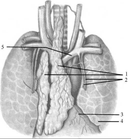
Fig. 226. Thymus gland (thymus)
1 — thymus gland (right and left lobes); 2 — internal thoracic artery and vein; 3 — pericardium; 4 — left lung; 5 — left brachiocephalic vein
The thymus consists of two elongated lobes of asymmetric size: the right and left lobes (lobus dexter and lobus sinister). In their middle portion, the lobes fuse or lie in close contact with each other. The lower part of each lobe is broadened, while the upper part is narrowed. The apices of both lobes are directed superiorly, extending into the neck region like a two-pronged fork (hence the Ukrainian name for the gland). In approximately half of cases, the left lobe of the thymus is longer than the right.
Externally, the thymus is covered by a thin Connective Tissue capsule (capsula thymi), from which interlobular septa (septa interlobularia) extend into the interior of the organ, dividing the thymic tissue into lobules (lobuli thymi) ranging from 1 to 10 mm in size. Each lobule consists of a cortical and a medullary substance. The cortex of the thymus (cortex thymi) is darker in color and situated at the periphery of the lobule, whereas the medulla of the thymus (medulla thymi) is lighter and occupies the central portion of the lobule. The boundary between the cortex and medulla is not always sharply defined.
The stroma of the thymus is formed by a network of reticular Cells and reticular fibers, as well as star-shaped epithelial cells—epithelioreticulocytes—interconnected by cellular processes. The meshes of this network contain thymic lymphocytes (thymocytes), which are packed more densely in the cortex than in the medulla. The medulla contains thymic corpuscles (Hassall's corpuscles) formed by concentric layers of epithelial cells, in the Cytoplasm of which large vacuoles, carotene granules, and coarse bundles of fibrils are found.
The thymus performs multiple Functions. It produces a hormone, thymosin, which controls carbohydrate and calcium METABOLISM in the Body and regulates human skeletal growth (especially During the first 10–15 years of life). Furthermore, it is believed that during childhood, the thymus suppresses The Development of the reproductive Organs. However, The primary function of the gland is its involvement in establishing Immunity against numerous diseases. The thymus is the site of differentiation for T-lymphocytes (thymus-dependent lymphocytes), which originate from hematopoietic stem Cells of the Cytology/practical/86.html">Red Bone Marrow and enter the organ via the bloodstream. Within the thymus, T-lymphocytes acquire properties that provide immune defense against cells rendered foreign to the Organism due to various types of damage. From the thymus, T-lymphocytes are carried by the Blood flow to the Peripheral Organs of The Immune System (Tonsils, Lymph Nodes, lymphoid nodules located in the walls of the hollow Organs of the digestive and respiratory systems, and the spleen). The thymus is the central organ of the immune system; early loss of thymic function leads to immunodeficiency.
Development and Age-Related Features of the Thymus. The thymus develops as a paired organ from the epithelium of the foregut. In humans, the thymus begins as paired epithelial evaginations of the 3rd and 4th pharyngeal pouches at the end of the first to the beginning of the second month of intrauterine development. The epithelial elements originating from the 4th pouches soon regress and serve as the basis for unencapsulated accessory thymic nodules located adjacent to the main thymic primordium. The thymic rudiments grow in a caudal direction, elongating, thickening, and approaching one another. The elongated, slender upper part of the gland's primordium gradually disappears, while the lower, thickened part forms the lobe of the thymus. By the 5th month of intrauterine development, the thymus exhibits a lobular Structure with clearly distinguishable cortical and medullary zones. The thymus forms earlier than other immune system organs and, by the time of birth, has a significant mass—averaging 13.3 g. The thymus reaches its maximum size by Puberty. At ages 10–15, the mass of the gland averages 37.5 g. After 16 years of age, the mass of the thymus gradually decreases due to age-related involution, averaging 25.5 g at 16–20 years, 23.3 g at 21–35 years, and 13.4 g at 50–90 years.
As age increases, the microscopic STRUCTURE OF THE thymus also changes. After birth (approximately up to the age of 10), the cortical substance predominates in the thymus. By age 10, the Proportions of the cortex and medulla are roughly equal (a 1:1 ratio). Thereafter, the cortical zone thins, the number of thymocytes decreases, and the medulla gradually begins to outweigh the cortex. Along with this reorganization and shift in The ratio of cortical to medullary tissue, adipose tissue appears early in the parenchymal stroma of the thymus. Isolated fat cells can be detected in the thymus of children aged 2–3 years. Later, adipose tissue proliferates alongside connective tissue. By ages 30–50, adipose tissue replaces the majority of the thymic parenchyma, and in individuals over 50, it accounts for 90%. However, the glandular parenchyma (lymphoid tissue) does not completely disappear during age-related involution, even in old age, but persists as islands surrounded by adipose tissue.
The thymus is supplied with blood by arterial branches originating from the internal thoracic artery, the concave (inferior) surface of the aortic arch, and the brachiocephalic trunk, which branch into smaller vessels within the interlobular septa. These penetrate into the interior of the lobules, branching into capillaries. The VEINS OF THE thymus drain into the brachiocephalic veins and the internal thoracic veins. The Lymphatic vessels of the thymus form deep parenchymal and superficial subcapsular networks. The parenchymal network is better developed in the cortex of the lobules. Running alongside the Blood Vessels, the lymphatic vessels exit the gland and course toward the anterior mediastinal and tracheobronchial lymph nodes.
Innervation of the thymus is provided by parasympathetic Branches of the Vagus nerve and sympathetic fibers from the stellate and upper thoracic ganglia of the Sympathetic trunk.
Last update: 08/08/2026
Editorial and Educational Adaptation: This material has been compiled based on the primary/original source text. The project team performed an editorial review, corrected technical inaccuracies, structured sections, and adapted the content for an educational format.
What was processed:
- elimination of formatting defects (OCR errors, structural breaks, corrupted characters);
- editorial organization of content;
- standardization of terminology in accordance with academic sources;
- verification of factual statements against the original source text.
All mentions of the author, publication year, and origin of the primary text have been preserved in accordance with the source.