Special Histology and Embryology: Practical Course - V. K. Napkhanyuk 2001
Cardiovascular system
Lymphatic vessels
Lymphatic vessels are subdivided into lymphatic capillaries, intra- and extra-organ lymphatic vessels, and main lymphatic trunks: the Thoracic duct and the Right lymphatic duct.
The lymphatic capillary (vas lymphocapillare) begins blindly in Tissues. Its wall consists of endothelial Cells. There is no basement membrane. The endothelium is connected to the adjacent tissue by anchoring filaments. Among intra- and extra-organ vessels, muscular-type Lymphatic vessels and fibrous-type lymphatic vessels are distinguished. The tunica intima of a lymphatic vessel consists of the endothelium, the subendothelial layer, and a valve formed by it. Large lymphatic vessels resemble muscular-type Veins in their Structure.
Slides for Study
Slide 1. Small Blood Vessels and capillaries of the pia mater (Fig. 20).
Low magnification. Examine the slide. At this magnification, select an area of the slide where small vessels predominate.
High magnification. Study The structure of the walls of an arteriole, venule, and capillary. The diameter of the capillary only slightly exceeds that of erythrocytes, so erythrocytes are arranged in the capillary in a single-file line. In the Capillary Wall, longitudinally positioned pale oval nuclei of endothelial cells and elongated darker nuclei of pericytes are visible. Slightly superficial to them, adventitial cells are also positioned longitudinally. The STRUCTURE OF THE venule wall in the pia mater does not differ from that of the capillary. The difference lies in the width of the lumen. In the venule, erythrocytes are arranged in several rows, giving the vessel an orange color. The arteriole differs from the capillary and venule by the presence of circularly arranged smooth myocytes in its wall. The rod-shaped nuclei of myocytes on the anterior and posterior surfaces of the vessel wall project into the lumen, giving the arteriole a striated appearance. The myocytes on the lateral surface appear as basophilic dots.
Class="center">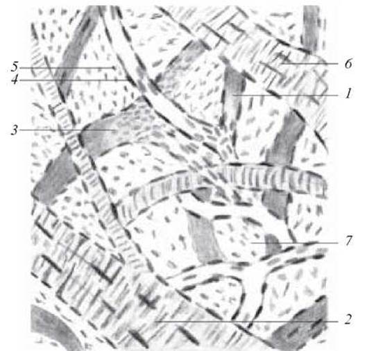
Fig. 20. Small blood vessels and capillaries of the pia mater. Hematoxylin and eosin staining. x 400:
1 — blood capillary; 2 — arteriole; 3 — venule; 4 — Nucleus of an endothelial Cell; 5 — nucleus of an adventitial cell; 6 — nucleus of a smooth Muscle cell; 7 — cells of loose Fibrous Connective Tissue
The nuclei of connective tissue cells are visible between the vessels.
Label on the figure: 1) blood capillary; 2) arteriole; 3) venule; 4) nucleus of an endothelial cell; 5) nucleus of an adventitial cell; 6) nucleus of a smooth muscle cell; 7) cells of loose fibrous connective tissue.
Slide 2. Cytology/practical/72.html">Cross section of a muscular-type artery (Fig. 21).
Low magnification. Examine the slide. At this magnification, select areas of the vessel with the most precise cross section of their wall.
High magnification. Three clearly distinguishable tunics are visible in the artery wall. The tunica intima is formed by the endothelium lining the vessel from the inside—which in cross section appears as a thin line with nuclei protruding into the lumen—and the subendothelial layer of Connective tissue with a faint purple tint due to its basophilia. It is separated from the tunica media by a thin, shiny, transparent internal elastic membrane with a scalloped appearance. The thickest tunic, the tunica media, consists primarily of circularly arranged smooth Muscles, among which individual elastic fibers can occasionally be spotted. At the border between the tunica media and tunica adventitia lies a transparent external elastic membrane, followed by the connective tissue of the tunica adventitia and vasa vasorum.
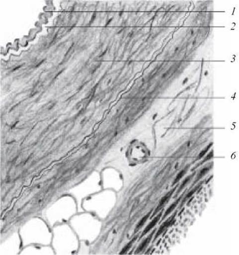
Fig. 21. Cross section of a muscular-type artery. Hematoxylin-eosin staining. x 400:
1 — tunica intima; 2 — internal elastic membrane; 3 — tunica media; 4 — external elastic membrane; 5 — tunica adventitia; 6 — vasa vasorum
Label on the figure: 1) tunica intima; 2) internal elastic membrane; 3) tunica media; 4) external elastic membrane; 5) tunica adventitia; 6) vasa vasorum.
Slide 3. Wall of the aorta (Fig. 22).
Low magnification. Examine the slide. At this magnification, the tunica intima is visible with the endothelium and a thick subendothelial layer. A characteristic feature is the presence in the tunica media of multiple (40–50) fenestrated elastic membranes, which appear transparent because they do not stain with hematoxylin-eosin.
High magnification. Elastic membranes are best studied under high magnification. It is clearly visible that layers of circularly arranged smooth muscle cells lie between the membranes. The tunica adventitia has a standard structure. Vasa vasorum can be seen in both the tunica adventitia and tunica media.
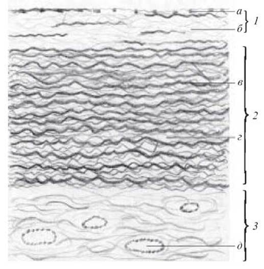
Fig. 22. Wall of the aorta. Hematoxylin-eosin staining. x 80:
1 — tunica intima (a — endothelium; b — subendothelial layer); 2 — tunica media (c — fenestrated elastic membranes; d — smooth myocytes); 3 — tunica adventitia (e — vasa vasorum)
Label on the figure: 1) tunica intima: a) endothelium; b) subendothelial layer; 2) tunica media; c) fenestrated elastic membranes; d) smooth myocytes; e) vasa vasorum; 3) tunica adventitia.
Slide 4. Cross-section of the aorta (Fig. 23).
Low magnification. Examine and draw the specimen. It is easier to capture the general layout at low magnification, using high magnification for studying finer details. Orcein staining selectively highlights elastic fibers, which appear dark brown. Muscle elements in this specimen appear light-colored. In the tunica interna, the sections of the longitudinal plexus of elastic fibers are clearly visible. The elastic plexus at the border between the tunica interna and tunica media is more distinct than in the previous specimen. In the tunica media, dark elastic lamellae stand out sharply, alternating with light layers of smooth muscle cells. The elastic plexus in the adventitia is clearly discernible.
Label the following on the drawing: 1) tunica interna; 2) tunica media with elastic membranes; 3) tunica externa; 4) vasa vasorum: a) artery; b) vein; 5) fat cells.
Slide 5. Cross-section of the brachial vein (Fig. 24).
Low magnification. Examine and draw the tunics, which are indistinctly demarcated. Elastic membranes are absent.
High magnification. The tunica interna is lined with endothelium and features a poorly developed subendothelial layer. The tunica interna forms Valves. The tunica media is significantly thinner than that of an artery and consists of smooth muscle cells. The tunica externa is highly developed, being 2–3 times thicker than the tunica media. It is composed of fibrous connective tissue, with occasional smooth muscle elements interspersed among the fibers.
Label the following on the drawing: 1) fat cell; 2) tunica externa; 3) tunica media; 4) tunica interna; 5) blood in the vascular lumen; 6) venous valve; 7) endothelium; 8) small vein.
Slide 6. Cross-section of the SUPERIOR VENA CAVA (Fig. 25).
Low magnification. Examine and draw the specimen.
At this magnification, the Microscope reveals the endothelium lining the vein from the inside; the subendothelial layer, which appears as a thin sheet of connective tissue; the tunica media containing 2–3 layers of smooth muscle cells; and a well-developed tunica externa composed of fibrous connective tissue with vasa vasorum.
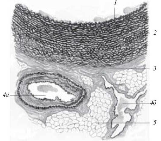
Fig. 23. Cross-section of the aorta. Orcein staining. x 200:
1 — tunica interna; 2 — tunica media with elastic membranes; 3 — tunica externa; 4 — vasa vasorum (a — artery; b — vein); 5 — fat cells
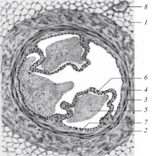
Fig. 24. Cross-section of the brachial vein. Hematoxylin and eosin staining. x 40:
1 — fat cell; 2 — tunica externa; 3 — tunica media; 4 — tunica interna; 5 — blood in the vascular lumen; 6 — venous valve; 7 — endothelium; 8 — small vein
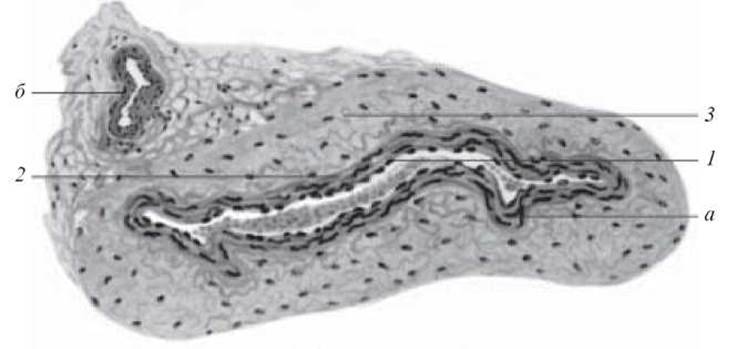
Fig. 25. Cross-section of the superior vena cava. Hematoxylin and eosin staining. x 80:
1 — tunica interna; 2 — tunica media (a — smooth muscle cell); 3 — tunica externa (b — vasa vasorum)
Label the following on the drawing: 1) tunica interna; 2) tunica media: a) smooth muscle cell; 3) tunica externa; b) vasa vasorum.
Last update: 10/08/2026
Editorial and Educational Adaptation: This material has been compiled based on the primary/original source text. The project team performed an editorial review, corrected technical inaccuracies, structured sections, and adapted the content for an educational format.
What was processed:
- elimination of formatting defects (OCR errors, structural breaks, corrupted characters);
- editorial organization of content;
- standardization of terminology in accordance with academic sources;
- verification of factual statements against the original source text.
All mentions of the author, publication year, and origin of the primary text have been preserved in accordance with the source.