Special Histology and Embryology: Practical Course - V. K. Napkhanyuk 2001
Cardiovascular System
Heart
The Heart (cor) is a hollow muscular organ that pumps Blood. The heart wall consists of three layers:
1) the inner layer (endocardium); 2) the middle layer (myocardium); 3) the outer layer (epicardium).
The endocardium (endocardium) lines the interior of the heart chambers, papillary Muscles, chordae tendineae, and heart Valves. The thickness of the endocardium varies across different regions. It is thicker in the left heart chambers and, notably, on the interventricular septum and at the orifices of the major arterial trunks.
The endocardium consists of the following layers:
— endothelium, which contains polygonal Cells resting on a thick basement membrane;
— subendothelial layer, formed by Connective Tissue;
— myoelastic layer, in which smooth Muscle fibers intertwine with elastic fibers;
— connective tissue stroma, containing thick elastic, Collagen, and reticulin fibers.
The valves are located between the atria and ventricles of the heart, as well as the great vessels.
The atrioventricular valve is bicuspid (mitral) in the left half and tricuspid in the right. These are endothelial-covered thin plates of dense Fibrous connective tissue containing very few cells. The subendothelial layer reveals fine collagen fibers that gradually transition into the fibrous plate of the valve cusp, and at the sites of attachment of the bicuspid and tricuspid valves, into the fibrous rings.
The atrial side of the valves has a smooth surface and contains a dense plexus of elastic fibers and bundles of smooth muscle cells within its subendothelial layer.
The ventricular side has an uneven surface due to projections from which the chordae tendineae originate. In this area, only a few elastic fibers lie beneath the endothelium.
The aortic valves are positioned between the ascending part of the aortic arch and the left ventricle.
A vertical section reveals three layers:
1) the inner layer, which is a continuation of the endocardium. The endothelium of this layer contains bundles of submicroscopic filaments 5-8 nm thick; the subendothelial layer contains fibroblasts with long, thin processes that act as consoles supporting the endothelial bundles of longitudinally and transversely running collagen fibrils;
2) the middle layer, which is thin and consists of loose fibrous connective tissue;
3) the outer layer, which, In addition to endothelium, contains collagen fibers originating from the fibrous ring of the aorta.
The myocardium (myocardium) is the muscular layer consisting of closely interconnected striated muscle cells. They form functional muscle fibers arranged in layers. Interstitial layers of loose connective tissue, Blood Vessels, and nerves are located between the muscular elements of the myocardium.
Several types of muscle cells are distinguished. The length of cardiac contractile myocytes ranges from 50 to 120 µm, with a width of 15-20 µm. One to two oval or elongated nuclei are located in the central part of the myocyte. Myofibrils are arranged longitudinally. The Cytoplasm contains a well-developed granular reticulum and a prominent Golgi apparatus. Cardiomyocytes that form muscle fibers interconnect in the region of intercalated discs. Under a Microscope, they appear as dark bands (microphoto 19).
Cardiac conducting myocytes are specialized muscle cells that generate and conduct impulses to the contractile Cells of the heart. These cells form the Conduction system of the Heart, which includes the sinoatrial node, atrioventricular node, atrioventricular bundle, trunk, right and left bundle branches, their ramifications, and Purkinje fibers.
Conducting myocytes differ not only from contractile myocytes but also from one another. Nodal myocytes are smaller than typical myocytes and form a meshwork. The myocytes of the Purkinje conducting muscle fibers are larger than typical ones. The cytoplasm of conducting myocytes stains paler than that of contractile myocytes. Myofibrils within them are sparse and do not form a general striation pattern. The cytoplasm is rich in Cell/35.html">Mitochondria and Glycogen. Intercalated discs are not visible (microphoto 20).
The epicardium (epicardium) is a serous membrane. It consists of mesothelium and a subepicardial foundation.
Slide 7. Section of the human heart (Fig. 26).
Low magnification. Examine the slide. At this magnification, identify the layers of the heart. The endocardium stands out on the inner surface of the section as a more darkly stained layer covered by endothelium. The bulk of the heart wall is composed of the myocardium, formed by cardiac muscle trabeculae cut at various levels. Connective tissue septa permeate the myocardial mass. The epicardium consists of loose connective tissue containing numerous adipose cell clusters and cross-sections of large blood vessels. Externally, the myocardium is covered by mesothelium.
High magnification. The heart muscle forms a network of anastomosing trabeculae. The nuclei of cardiomyocytes are oval-shaped, located along the axis of the muscle trabeculae, and surrounded by areas of sarcoplasm. The longitudinal striation of the heart muscle is due to the arrangement of its myofibrils, while the transverse striation results from the differentiation of myofibrils into discs and bands, similar to the differentiation in skeletal muscles. In addition to regular striations, the heart muscle features characteristic thicker intercalated discs that intersect the trabeculae.
Class="center">
Fig. 26. Section of the human heart. Hematoxylin and eosin staining. х 400:
1 — endocardium (a — endothelial cells; b — nuclei of connective tissue and smooth muscle cells); 2 — Purkinje fibers; 3 — cardiac muscle cells (c — intercalated discs); 4 — blood capillaries
Label the following on the figure: 1) endocardium: a) endothelial cells; b) nuclei of connective tissue and smooth muscle cells; 2) Purkinje fibers; 3) cardiac muscle cells; c) intercalated discs; 4) blood capillaries.
Slide 8. Section of a sheep's heart (Fig. 27).
Low magnification. At this magnification, the layers of the heart wall can be identified: endocardium, myocardium, and epicardium. Directly beneath the endocardium and between the myocardial trabeculae, atypical fibers, or Purkinje fibers, stand out by their thickness and lighter staining. Both longitudinal sections of atypical fibers and transverse or oblique sections of Purkinje fibers can be observed. Their characteristic feature is the dominance of sarcoplasm over myofibrils, which makes the fibers appear lighter than typical myocardial fibers. Myofibrils in atypical fibers do not run parallel, but form fine, intersecting bundles. The nuclei of these fibers are larger than myocardial nuclei, flattened, and stain poorly. Bundles of atypical fibers are surrounded by connective tissue.
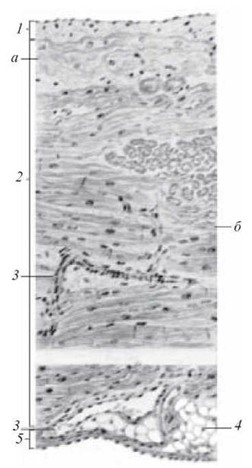
Fig. 27. Section of a sheep's heart. Hematoxylin and eosin staining. х 120:
1 — endocardium; 2 — myocardium (a — atypical cardiomyocytes; b — nuclei of atypical cardiomyocytes); 3 — blood vessels; 4 — fibrous connective tissue; 5 — epicardium
Label the following on the figure: 1) endocardium; 2) myocardium: a) atypical cardiomyocytes; b) nuclei of atypical cardiomyocytes; c) typical cardiomyocytes; d) nuclei of typical cardiomyocytes; 3) blood vessels; 4) fibrous connective tissue; 5) epicardium; 6) intercalated discs; 7) myofibrils of atypical cardiomyocytes; 8) myofibrils.
Control Questions
1. Sources of Development of blood vessels.
2. What distinguishes the Structural Features of blood vessels in different PARTS OF THE vascular bed?
3. General structural plan of the vascular wall. Main Components of each tunic.
4. Types of blood capillaries and their Structure.
5. Classification of blood vessels.
6. Types of Arteries and their structural features.
7. Types of Veins and the structural features of their walls.
8. Which vessels belong to the microcirculatory bed?
9. Arteriovenous anastomoses (AVAs): classification and structural features.
10. Structural Features of the INFERIOR VENA CAVA.
11. STRUCTURE OF THE SUPERIOR VENA CAVA.
12. Lymphatic vessels. Classification. Structure.
13. Sources of Cytology/cytology/67.html">Development and Structure of the endocardium.
14. Sources of Development of the myocardium. Structure of various types of cardiomyocytes.
15. Sources of development and structure of the epicardium.

Electron micrograph 14. Blood capillary. x 20,000:
1 — erythrocyte in the lumen of the blood capillary; 2 — platelet; 3 — Nucleus of the endothelial cell; 4 — intracellular reticular apparatus; 5 — Endoplasmic reticulum; 6 — pinocytotic vesicles; 7 — mitochondria; 8 — Pores in the endothelium; 9 — basement membrane; 10 — adventitial cell (from Rodin's atlas)
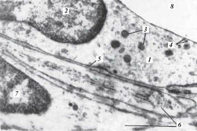
Electron micrograph 15. Arteriolar. x 27,000:
1 — endothelial cell; 2 — nucleus of the endothelial cell; 3 — mitochondria; 4 — pinocytotic vesicles (indicated by arrows); 5 — basement membrane; 6 — two smooth muscle cells; 7 — nucleus of the smooth muscle cell; 8 — lumen of the arteriole (after Cliff)
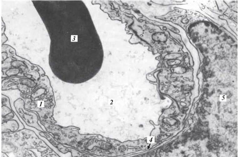
Electron micrograph 16. Venule. x 19,200:
1 — endothelium; 2 — lumen of the venule; 3 — erythrocyte in the lumen of the venule; 4 — basement membrane; 5 — adventitial cell (I. I. Dedov)

Electron micrograph 17. Tunica intima of the aorta. x 12,000:
1 — endothelium; 2 — nucleus of the endothelial cell; 3 — cell boundary; 4 — subendothelial layer (a — collagen and elastic fibrils; b — connective tissue stellate cells); 5 — fenestrated elastic membrane of the tunica media; 6 — opening in the fenestrated elastic membrane (after Seifert)
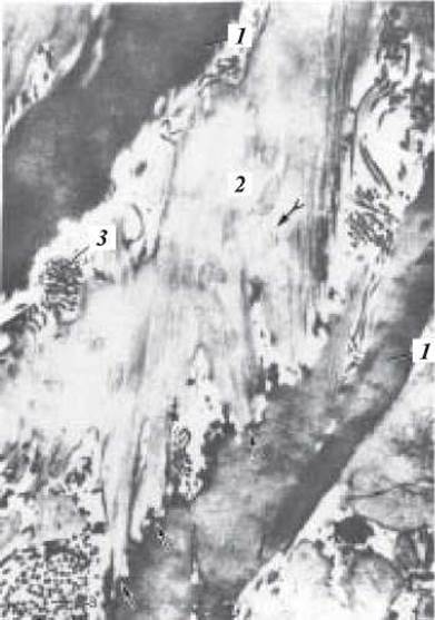
Electron micrograph 18. Tunica media of the aorta. x 12,000:
1 — fenestrated elastic membranes; 2 — cytoplasm of the smooth muscle cell; 3 — collagen protofibrils (after Seifert)
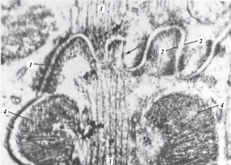
Electron micrograph 19. Intercalated disc. x 76,000:
1 — intercalated disc (boundary between muscle cells); 2 — sarcolemma; 3 — myofibrils; 4 — mitochondria

Electron micrograph 20. Atypical cardiac muscle cells of the atrioventricular bundle. x 26,000:
1 — cell membrane; 2, 3 — two atypical cardiac muscle cells; 4 — desmosomes at the border between two adjacent muscle cells; 5 — mitochondria; 6 — longitudinally and transversely sectioned myoprotofibrils; 7 — glycogen (from Rodin's atlas)
Situational tasks
1. Two artery slides are demonstrated. In the first one, the internal elastic membrane is clearly pronounced at the border between the tunica intima and tunica media, whereas In the second one, the elastic membrane is absent, but the tunica media contains numerous elastic elements (membranes). What types of arteries do the First and Second slides belong to?
2. The slides clearly show a dense network of capillaries located between arterioles. Name this structure. In which organ can this network be found?
3. An electron micrograph shows a cross-section of a vessel whose lumen is filled with a red blood cell, and its wall consists of three layers. The first layer is a thickened cell with pinocytotic vesicles clearly visible in the cytoplasm. The Cell rests on a basal lamina. The second layer is formed by the basal lamina and a pericyte, and the third by an adventitial cell. What is the name of such a vessel?
4. The description of a blood capillary's structure indicates that the endothelial Cell Cytoplasm contains thin areas, and the basement membrane is continuous. Determine the type of this capillary. In which Organs are such capillaries located?
5. It is known that I. M. Sechenov metaphorically referred to arterioles as the "taps" of the body's Circulatory system. What histological and functional features of arterioles provided the basis for such a comparison?
6. When examining a specimen under a Light Microscope, a muscular-type artery and a corresponding vein stained with orcein are visible. Which Structural elements of the vessels will be stained with this dye? By what features can arteries be unmistakably identified?
7. A muscular-type vein is located in the lower half of the trunk. Are the muscular elements in its wall strongly or weakly developed? What determines the degree of development of the muscular elements in its wall?
8. The walls of arteries and veins consist of three tunics. In the description of two of these tunics, it was noted that they contain vasa vasorum. Which tunics are these?
9. On a heart specimen stained with hematoxylin and eosin, muscle fibers of two types are visible: the cytoplasm of one type has an intense pink color with clear cross-striations and intercalated discs; the cytoplasm of the other fibers is paler, the fiber diameter is larger, and cross-striations are not visible. Which types of myocytes make up these fibers?
10. There are two specimens of Striated Muscle tissue. In one of them, numerous nuclei are located beneath the fiber membrane, while in the other, cells with centrally located nuclei are visible. Which of these specimens belongs to the myocardium?
Sample Examination Questions
1. Cardiovascular system. General characteristics, sources of development. Influence of hemodynamic conditions on vessel structure. Vessel regeneration. Age-related changes.
2. Microcirculatory bed, its Structure and Functional characteristics. METABOLISM/2.html">THE CONCEPT OF the histohematic barrier.
3. Arteries. Classification, function, structure, and development of arteries. Interrelationship between arterial structure and hemodynamic conditions. Age-related changes.
4. Veins. Classification. Development, function, structure. Influence of hemodynamic conditions. Age-related changes.
5. Lymphatic capillaries and lymphatic vessels, their structure and functional significance.
6. Heart. Sources of development, histogenesis. Structure of the Heart Wall tunics. Age-related changes.
Last update: 10/08/2026
Editorial and Educational Adaptation: This material has been compiled based on the primary/original source text. The project team performed an editorial review, corrected technical inaccuracies, structured sections, and adapted the content for an educational format.
What was processed:
- elimination of formatting defects (OCR errors, structural breaks, corrupted characters);
- editorial organization of content;
- standardization of terminology in accordance with academic sources;
- verification of factual statements against the original source text.
All mentions of the author, publication year, and origin of the primary text have been preserved in accordance with the source.