Anatomy of the Vessels and Nerves of the Trunk (Angioneurology) - Chornokulskyi S. T. 2011
Veins of the Systemic Circulation. Veins of the Trunk
Inferior Vena Cava
Portocaval and Cavacaval Anastomoses
Only the cardiac Veins are autonomous within the human Venous system. There are clinically significant anastomoses between the origins (roots and tributaries) of the HEPATIC PORTAL VEIN, SUPERIOR VENA CAVA, and INFERIOR VENA CAVA:
I. Cava-caval anastomoses;
II. Portocaval anastomoses:
Class="center">
Fig. 36. VEINS OF THE pelvis:
1 - external iliac vein (v. iliaca externa);
2 - internal iliac vein (v. iliaca interna);
3 - inferior epigastric vein (v. epigastrica inferior);
4 - internal pudendal vein (v. pudenda interna);
5 - levator ani Muscle (m. levator ani);
6 - superior rectal vein (v. rectalis superior)
1. Portosuperior caval;
2. Portoinferior caval;
III. Cava-porto-caval anastomoses.
These anastomoses are located within the walls of the Abdominal cavity (anterior and posterior) and its Organs (superiorly and inferiorly), which can be conceptually envisioned as projected onto the walls of a cube:
— anterior;
— posterior;
— superior;
— inferior.
Study outline for anastomoses
1. State the Location OF THE anastomosis.
2. Name the veins belonging to the anastomosing systems.
3. Mode of anastomosis (via a venous plexus).
4. Sequentially list all veins draining Blood from the anastomosis to the major venous trunks (v. cava superior, v. cava inferior, v. hepatis).
— There are 9 primary anastomoses.
— On the anterior abdominal wall (conventionally a cube) — 3;
— On the posterior wall — 3;
— On the superior wall — 2;
— On the inferior wall — 1.
I. Anastomoses of the anterior abdominal wall (Fig. 35).
A. Cava-caval anastomoses:
1. Via the superior and inferior epigastric veins on the right and left, which anastomose with the subcutaneous veins of the umbilical region, connecting v. cava superior and v. cava inferior.
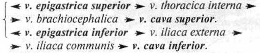
2. Between the thoracoepigastric and superficial epigastric veins:

B. Porto-cava-caval anastomosis:
3. Within the subcutaneous veins of the umbilical region, the paraumbilical veins anastomose with the superior and inferior epigastric veins. This forms a porto-superior- and inferior-caval anastomosis. Blood reaches the portal vein of the Liver via vv. paraumbilicales, while the cava-caval anastomosis is described above. In cases of portal Hypertension of various etiologies (malignancy, liver cirrhosis), the umbilical region presents with the appearance known as "HEAD of Medusa" (caput medusae), marked Splenomegaly, and an increased amount of serous fluid in the peritoneal cavity (ascites); esophageal varices and hemorrhoids are also observed.
Anastomoses of the posterior abdominal wall (Fig. 37).
A. Cava-caval anastomoses:
4. Between the lumbar veins and the ascending lumbar veins:
vv. lumbales → v. cava inferior.
vv. lumbales ascendentes → v. azygos (v. hemiazygos) v. cava superior.
5. Between the vertebral vein, posterior intercostal veins, lumbar veins, and lateral sacral veins via the external and internal vertebral venous plexuses. These plexuses are located along the entire length of THE Vertebral Column.
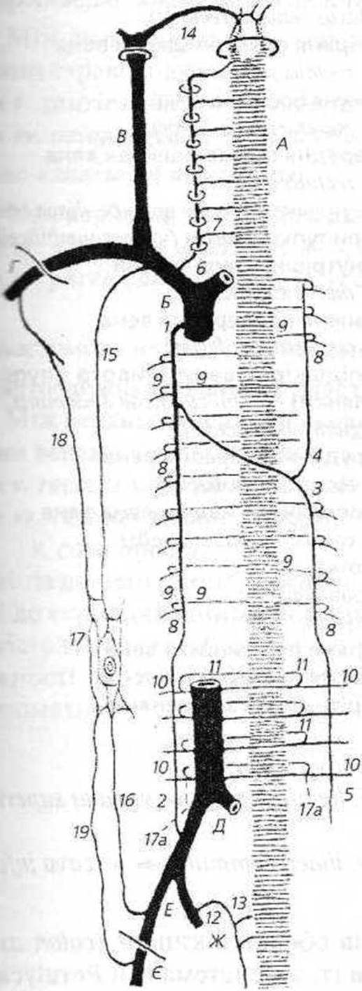
Fig. 37. Diagram of cava-caval anastomoses (according to Prof. I. Ye. Kefeli).
1 - azygos vein (v. azygos);
2,5 - ascending lumbar vein (v. lumbalis ascendens);
3 - hemiazygos vein (v. hemiazygos);
4 - accessory hemiazygos vein
(v. hemiazygos accessoria);
6 - vertebral vein (v. vertebralis);
7, 8, 9, 10, 11 - intervertebral veins (vv. intervertebrales);
12, 13 - lateral sacral veins (vv. sacrales laterales);
14 - Dural Venous Sinuses (sinus durae matris);
15 - internal thoracic vein (v. thoracica interna);
16 - inferior epigastric vein (v. epigastrica inferior);
17 - subcutaneous abdominal veins of the umbilical region (vv. subcutaneae abdominis [regionis umbilicalis]);
18 - thoracoepigastric vein (v. thoracoepigastrica);
19 - superficial epigastric vein (v. epigastrica superficialis);
A - internal vertebral venous plexus;
B - superior vena cava;
D - subclavian vein;
E - inferior vena cava;
F - external iliac vein;
G - femoral vein;
H - internal iliac vein
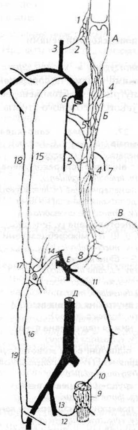
Fig. 38. Diagram of portocaval anastomoses (according to Prof. I. Ye. Kefeli).
1 - pharyngeal plexus (plexus pharyngeus);
2 - pharyngeal veins (vv. pharyngeae);
3 - internal jugular vein (v. jugularis interna);
4, 5 - esophageal veins (vv. oesophageales);
6 - azygos vein (v. azygos);
7 - hemiazygos vein (v. hemiazygos);
8 - left gastric vein (v. gastrica sinistra);
9 - rectal venous plexus (plexus venosus rectalis);
10 - superior rectal vein (v. rectalis superior);
11 - inferior mesenteric vein (v. mesenterica inferior);
12 - middle rectal vein (v. rectalis media);
13 - internal iliac vein (v. iliaca interna);
14 - paraumbilical veins (vv. paraumbilicales);
15 - internal thoracic vein (v. thoracica interna);
16 - inferior epigastric vein (v. epigastrica inferior);
17 - subcutaneous veins of the abdomen [umbilical region] (vv. subcutaneae abdominis [regionis umbilicalis]);
18 - thoracoepigastric vein (v. thoracoepigastrica);
19 - superficial epigastric vein (v. epigastrica superficialis);
A - Pharynx;
B - Esophagus;
C - Stomach;
D - superior vena cava;
E - inferior vena cava;
F - hepatic portal vein with its tributaries

B. Porto-cava-caval anastomosis:
6. Between the veins of the mesoperitoneal segments of the colon (colon ascendens, colon descendens) and the lumbar veins (the so-called Retzius venous system).
v. colica dextra → v. mesenterica superior → v. portae hepatis.
v. colica sinistra → v. mesenterica inferior → v. splenica → v. portae hepatis.
vv. lumbales → v. cava inferior.
This is a porto-inferior caval anastomosis. Considering the cavacaval anastomosis of the posterior wall (4), one can easily envision the entire porto-cava-caval anastomosis there as well.
Anastomoses of the upper abdominal wall (Fig. 38).
A. Porto-superior caval anastomosis:
Between the left gastric vein and the esophageal veins in the abdominal region of the esophagus.
v. gastrica sinistra → v. portae hepatis.
vv. oesophageales → v. azygos → v. cava superior.
B. Cava-caval anastomosis:
8. Between the superior and inferior phrenic veins.
vv. phrenicue superiores → v. azygos → v. cava superior.
vv. phrenicue inferiores → v. cava inferior.
Anastomoses of the lower abdominal wall.
Porto-inferior caval anastomosis:
Between the superior rectal vein and the middle and inferior rectal veins via the rectal venous plexus.
v. rectalis superior → v. mesenterica inferior → v. lienalis → v. portae hepatis.
vv. rectales mediae et inferiores → v. iliaca interna → v. iliaca communis → v. cava inferior.
All of the aforementioned intervenous anastomoses provide collateral venous return to The Heart in the event of blood flow obstruction in one of the major veins (superior and inferior venae cavae, portal vein). Depending on the stage of the condition, this manifests with specific morphological and functional symptoms.
Last update: 11/08/2026
Editorial and Educational Adaptation: This material has been compiled based on the primary/original source text. The project team performed an editorial review, corrected technical inaccuracies, structured sections, and adapted the content for an educational format.
What was processed:
- elimination of formatting defects (OCR errors, structural breaks, corrupted characters);
- editorial organization of content;
- standardization of terminology in accordance with academic sources;
- verification of factual statements against the original source text.
All mentions of the author, publication year, and origin of the primary text have been preserved in accordance with the source.