Human Anatomy - Zinoviy Mirchuk, Maryna Mirchuk 2007
Axial skeleton
Vertebral column
THE Vertebral Column (Fig. 2) consists of 33–34 vertebrae. In an adult human, 24 of these are free (7 cervical, 12 thoracic, and 5 lumbar), while the remaining vertebrae are fused together to form the sacrum (comprising 5 sacral vertebrae) and the coccyx (comprising 4–5 rudimentary coccygeal vertebrae). Occasionally, the sacrum is formed by 4 or 6 vertebrae. In rare cases, the total number of vertebrae reaches 35–37 due to an increased number of coccygeal segments.
Regardless of their anatomical region, all vertebrae share a common structural plan. A vertebra (with the exception of the first cervical vertebra, the atlas) consists of an anterior body and a posterior vertebral arch connected to the body by two pedicles, which bound the vertebral foramen. The foramina of all vertebrae align to form the vertebral canal, housing the Spinal Cord and its Meninges. The vertebral arch bears seven processes for Muscle attachment. A single, unpaired spinous process projects posteriorly along the midline. Paired transverse processes extend laterally in the frontal plane. Paired superior and inferior articular processes project upward and downward from the arch, enabling adjacent vertebrae to articulate with one another. The bases of these articular processes bound the superior and inferior vertebral notches; when vertebrae stack together, these notches form the intervertebral foramina, through which Spinal Nerves and Blood Vessels pass.
Class="center">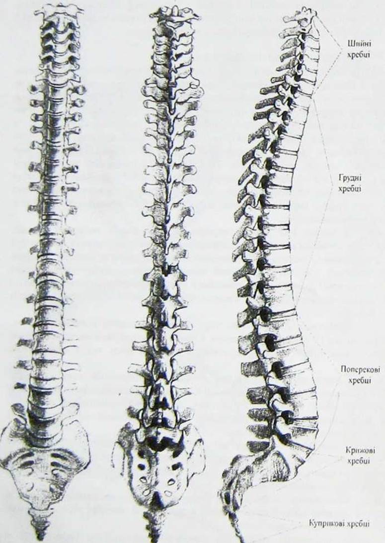
Fig. 2. The vertebral column
The transverse process of a cervical vertebra is also referred to as the costotransverse process. It terminates in two tubercles: an anterior and a posterior tubercle. The anterior tubercle of the sixth cervical vertebra is more prominent than those of the other vertebrae and is known as the carotid tubercle due to its close proximity to the carotid artery. This tubercle allows the carotid artery to be compressed against it to control Hemorrhage in the HEAD and Neck regions.
Adjacent vertebrae articulate via intervertebral discs, intervertebral symphyses, and joints, enabling the vertebral column to perform a wide range of movements. Driven by skeletal Muscles, Movements of the vertebral column occur around three axes: frontal, sagittal, and vertical (longitudinal). Flexion and extension take place around the frontal (transverse) axis, adduction and abduction (lateral bending to the right and left) occur around the sagittal axis, and rotational movements, or rotation (turning to the right and left), occur around the vertical axis. The cervical vertebrae are the most mobile, whereas the thoracic vertebrae exhibit the least mobility.
Last update: 10/08/2026
Editorial and Educational Adaptation: This material has been compiled based on the primary/original source text. The project team performed an editorial review, corrected technical inaccuracies, structured sections, and adapted the content for an educational format.
What was processed:
- elimination of formatting defects (OCR errors, structural breaks, corrupted characters);
- editorial organization of content;
- standardization of terminology in accordance with academic sources;
- verification of factual statements against the original source text.
All mentions of the author, publication year, and origin of the primary text have been preserved in accordance with the source.