Human Anatomy - Kotsan I. Ya. 2009
Brain
Meninges
The Brain, much like the Spinal Cord, is enclosed by three meninges: the dura mater, the arachnoid mater, and the pia mater (Fig. 250).
Class="center">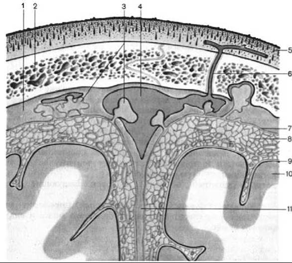
Fig. 250. Diagram of the relationships between the meninges, the superior sagittal sinus, the cranial vault, and the brain surface in a frontal section (after M. R. Sapin)
1 — dura mater; 2 — cranial vault; 3 — arachnoid granulations; 4 — superior sagittal sinus; 5 — scalp; 6 — emissary vein; 7 — arachnoid mater of the brain; 8 — subarachnoid space; 9 — pia mater of the brain; 10 — brain; 11 — falx cerebri
The dura mater of the brain (dura mater encephali) is a tough Connective Tissue membrane located externally to the other meninges. It lines the cranial cavity from the inside and simultaneously serves as the periosteum for the inner surface of the cranial bones. The dura mater consists of external and internal layers (leaflets) and contains a rich network of Blood Vessels and nerves. It is tightly fused with the BONES OF THE cranial base, but loosely attached to the bones of the vault. In the region of the foramen magnum, it merges with its edges and continues into the spinal dura mater. The inner surface of the dura mater, facing the brain, is lined with endothelium, making it smooth and glossy. In certain areas, the cerebral dura mater splits, and its inner layer extends deeply into the cerebral fissures to form the so-called dural folds. These folds separate specific PARTS OF THE brain from one another and, together with the CEREBROSPINAL FLUID, protect the brain from mechanical shocks. Furthermore, the dura mater forms venous sinuses (through the splitting of its layers) filled with venous blood, which ensure the outflow of venous blood from the brain.
The largest folds of the cranial dura mater include:
The falx cerebri is situated in the sagittal plane. It is a thin, sickle-shaped fold of the dura mater that extends into the longitudinal cerebral fissure, without reaching the corpus callosum, and separates the right and left cerebral hemispheres. The falx cerebri stretches from the crista galli to the internal occipital protuberance.
The tentorium cerebelli is a horizontal plate that arches like a tent over the posterior cranial fossa, which houses the Cerebellum. Extending into the transverse cerebral fissure, the tentorium cerebelli separates the latter from the cerebellar hemispheres. Its posterolateral margin is attached to the superior border of the petrous part of the Temporal bone and the transverse sinus sulcus of the Occipital bone, while its anterior free edge forms the tentorial notch (incisura tentorii).
The falx cerebelli appears as a continuation of the falx cerebri, extending from the internal occipital protuberance to the posterior margin of the foramen magnum. It divides the cerebellar hemispheres.
The Diaphragm of the sella (diaphragma sellae) is a horizontally positioned membrane with a central aperture, stretched over the hypophysial fossa to form its roof. The Pituitary Gland (hypophysis) lies within the fossa beneath this diaphragm. The infundibulum of the Hypothalamus passes through the central opening of the sella diaphragm, connecting the hypothalamus to the pituitary gland.
At the sites where the folds originate, as well as in areas where the dura mater attaches to the bones of the internal cranial base, Dural Venous Sinuses (sinus durae matris) are formed within the splits of the dura mater. These dural sinuses act as channels through which venous blood drains from the brain into the internal jugular Veins. The layers of the dura mater that form the sinuses are tightly stretched and do not collapse. Consequently, when cut, the sinuses remain patent and contain no Valves. This structural adaptation allows venous blood to drain freely from the brain regardless of fluctuations in intracranial pressure. Corresponding sulci are present on the inner surfaces of the cranial bones where these dural sinuses are located.
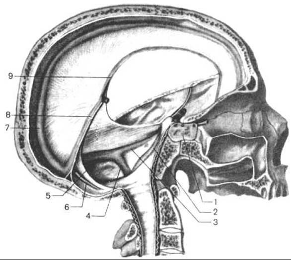
Fig. 251. Dural venous sinuses (after M. R. Sapin)
1 — cavernous sinus; 2 — inferior petrosal sinus; 3 — superior petrosal sinus; 4 — sigmoid sinus; 5 — transverse sinus; 6 — occipital sinus; 7 — superior sagittal sinus; 8 — straight sinus; 9 — inferior sagittal sinus
The dural venous sinuses of the brain are classified as follows (Fig. 251):
The superior sagittal sinus (sinus sagittalis superior) runs along the entire outer edge of the falx cerebri, extending from the crista galli of the Ethmoid bone to the internal occipital protuberance. In its anterior region, this sinus forms anastomoses with the VEINS OF THE Nasal cavity. The posterior end of the sinus empties into the transverse sinus. Lateral lacunae (lacunae laterales) are located to the right and left of the superior sagittal sinus. The cavities of these lateral lacunae communicate with the lumen of the superior sagittal sinus and receive veins from the cerebral dura mater, the brain itself, and the diploic veins.
The inferior sagittal sinus (sinus sagittalis inferior) is situated within the lower free margin of the falx cerebri and is considerably smaller than the superior sagittal sinus. Its posterior end empties into the straight sinus.
The straight sinus (sinus rectus) lies sagittally within the splitting of the tentorium cerebelli along the line of attachment of the falx cerebri. In addition to the inferior sagittal sinus, the great cerebral vein (vein of Galen) empties into the anterior end of the straight sinus. Posteriorly, the straight sinus drains into the middle portion of the transverse sinus.
The transverse sinus (sinus transversus) is located along the line where the tentorium cerebelli detaches from the cerebral dura mater. On the inner surface of the squamous occipital bone, this sinus corresponds to the transverse sulcus. The confluence of the superior sagittal, occipital, and straight sinuses into the transverse sinus is known as the confluence of sinuses (confluens sinuum). To the right and left, the transverse sinus continues as the sigmoid sinus of the respective side.
The sigmoid sinus (sinus sigmoideus) is a paired Structure located within the corresponding sulcus on the inner surface of the cranium, featuring a curved S-shape. In the region of the jugular foramen, the sigmoid sinus continues as the internal jugular vein.
The occipital sinus (sinus occipitalis) lies at the Base of the falx cerebelli. Descending along the internal occipital crest, it reaches the posterior margin of the foramen magnum, where it divides into two branches that embrace the foramen posteriorly and laterally. Each branch of the occipital sinus empties into the ipsilateral sigmoid sinus. The superior end of the occipital sinus drains into the transverse sinus.
The cavernous sinus (sinus cavernosus) is a paired structure located at the base of the Skull, lateral to the sella turcica. The Internal Carotid Artery and several Cranial Nerves pass through this sinus, which features a complex structure of interconnected cavernous spaces—hence its name. The right and left cavernous sinuses are connected by anastomoses known as the anterior and posterior intercavernous sinuses (sinus intercavernosi), located within the diaphragm of the sella turcica, anterior and posterior to the pituitary stalk. The sphenoparietal sinus and the superior ophthalmic vein empty into the anterior parts of the cavernous sinus.
The sphenoparietal sinus (sinus sphenoparietalis) is a paired sinus running along the free posterior margin of the lesser wings of the Sphenoid bone, extending from the inner surface of the Parietal bone to the cavernous sinus.
The superior and inferior petrosal sinuses (sinus petrosus superior et sinus petrosus inferior) are paired structures situated along the upper and lower borders of the petrous part of the temporal bone. Both sinuses contribute to the drainage pathways of venous blood from the cavernous sinus into the sigmoid sinus.
The right and left petrosal sinuses are connected by veins located within the dural split in the region of the basilar part of the occipital bone, collectively known as the basilar plexus (plexus basilaris). Through the foramen magnum, this plexus communicates with the internal vertebral venous plexuses.
In some areas, the dural venous sinuses form anastomoses with the superficial veins of the HEAD via emissary veins (vv. emissariae). Furthermore, the dural sinuses communicate with the diploic veins (vv. diploicae), which reside within the spongy substance of the cranial vault bones and drain into the superficial veins of the head.
Thus, venous blood from the brain drains through its superficial and deep venous systems into the dural sinuses, and subsequently into the internal jugular vein. Thanks to the emissary veins, venous plexuses, and anastomoses between the sinuses and diploic veins, cerebral venous blood can also drain into the superficial Veins of the Head and Neck.
The Blood supply to the dura mater is provided by Branches of the ophthalmic, maxillary, ascending pharyngeal, and occipital Arteries.
Venous drainage occurs via correspondingly named veins, which empty partly into the venous sinuses and partly into the pterygoid plexus.
The Innervation of the dura mater is supplied by branches of the trigeminal and vagus nerves.
The arachnoid mater of the brain (arachnoidea mater encephali) is a delicate, translucent connective tissue membrane situated between the dura mater and pia mater. It envelops the brain entirely, yet does not dip into the sulci. Composed of dense Fibrous connective tissue, the arachnoid mater is devoid of Blood Vessels and is nourished by the cerebrospinal fluid. A subdural space (spatium subdurale), containing a small amount of clear fluid, lies between the arachnoid and the dura mater. The arachnoid mater is separated from the pia mater by the subarachnoid space (spatium subarachnoidereum), which contains the cerebrospinal fluid. Where the arachnoid spans broad and deep sulci, the subarachnoid space expands to form subarachnoid cisterns (cisternae subarachnoidales) of varying sizes. Over the convexities of the brain and the surfaces of the gyri, the arachnoid and pia maters closely appose one another, significantly narrowing the subarachnoid space in these regions into a capillary-like cleft.
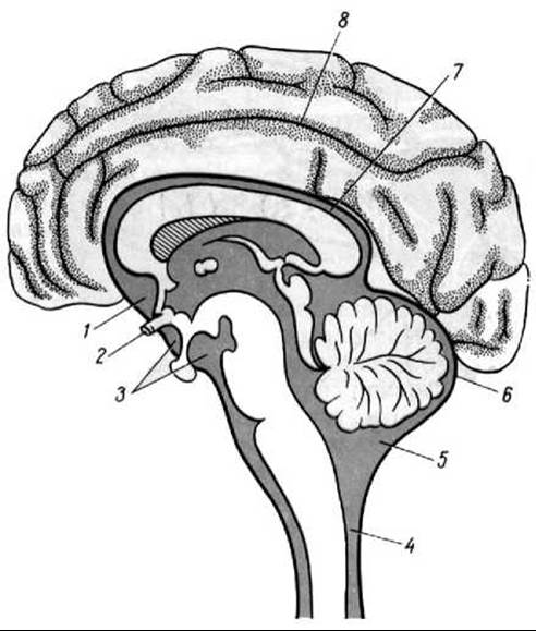
Fig. 252. The subarachnoid space (after M. G. Prives)
1 — chiasmatic cistern; 2 — optic chiasm; 3 — interpeduncular cistern; 4 — subarachnoid space of the spinal cord; 5 — cerebellomedullary cistern; 6 — arachnoid mater; 7 — subarachnoid space over the corpus callosum; 8 — subarachnoid space within the sulci
The largest subarachnoid cisterns are the following (Fig. 252):
The cerebellomedullary cistern (cisterna cerebellomedullaris) is an unpaired cistern located between the Medulla Oblongata ventrally and the cerebellum dorsally. Cerebrospinal fluid is sampled from this cistern during suboccipital puncture. It is the largest of all the subarachnoid cisterns.
The cistern of the lateral recess of the cerebrum (cisterna fossae lateralis cerebri) is a paired cistern situated on the inferolateral surface of the cerebrum within the corresponding fossa, which corresponds to the anterior parts of the lateral sulcus of the cerebral hemisphere.
The chiasmatic cistern (cisterna chiasmatica) is an unpaired cistern located at the base of the brain, anterior to the optic chiasm.
The interpeduncular cistern (cisterna interpeduncularis) is an unpaired cistern occupying the interpeduncular fossa and the space anterior to the cerebral peduncles, bounded anteriorly by the optic chiasm.
A distinctive structural feature of the arachnoid mater is the arachnoid granulations (granulationes arachnoideae). These are grayish-pink, rounded projections of the arachnoid mater that protrude into the lumen of the dural venous sinuses or into adjacent blood lakes. Present in both children and adults, they reach their greatest size and number in old age. As they enlarge, their pressure on the cranial bones creates depressions on the inner surface of the latter, known in Osteology as granular pits (foveolae granulares). The granulations serve to drain cerebrospinal fluid into the bloodstream via filtration.
The arachnoid mater connects to the pia mater through numerous bundles of Collagen and elastic fibers. These connective tissue trabeculae and the cerebrospinal fluid prevent the compression of cerebral vessels coursing within the subarachnoid space.
The pia mater of the brain (pia mater encephali) is the innermost meningeal layer. It closely adheres to the external surface of the brain, extending into all its sulci and fissures. Composed of loose connective tissue, the pia mater forms two layers (inner and outer), between which blood vessels run. These vessels penetrate the brain tissue to nourish it. In certain areas, the pia mater invaginates into the ventricular cavities to form the choroid plexuses, which produce cerebrospinal fluid.
Last update: 08/08/2026
Editorial and Educational Adaptation: This material has been compiled based on the primary/original source text. The project team performed an editorial review, corrected technical inaccuracies, structured sections, and adapted the content for an educational format.
What was processed:
- elimination of formatting defects (OCR errors, structural breaks, corrupted characters);
- editorial organization of content;
- standardization of terminology in accordance with academic sources;
- verification of factual statements against the original source text.
All mentions of the author, publication year, and origin of the primary text have been preserved in accordance with the source.