Human Anatomy, Part 1 - K. A. Dyubenko, A. K. Kolomiytsev, Yu. B. Chaykovsky 2002
Special Part
Axial skeleton, sceleton axiale - Bones of the neurocranium
Parietal bone
The parietal bone, os parietale, is a paired bone (Fig. 39) of quadrilateral shape, bowl-like in appearance, forming the upper and lateral PARTS OF THE cranial vault. It develops intramembranously from Connective Tissue. It features two surfaces—the external surface, facies externa, and the internal surface, facies interna—and four borders: the superior (sagittal) border, margo sagittalis, the inferior (squamous) border, margo squamosus, the anterior (frontal) border, margo frontalis, and the posterior (occipital) border, margo occipitalis.
Class="center">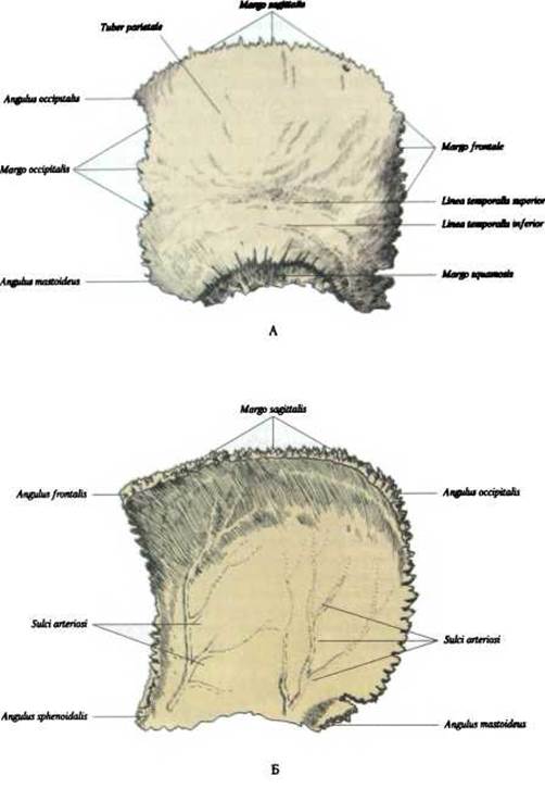
Fig. 39. Parietal bone, os parietale:
A - external surface; B - internal surface
Corresponding to its four borders, the parietal bone has four angles: the frontal angle, angulus frontalis; the occipital angle, angulus occipitalis; the sphenoidal angle, angulus sphenoidalis; and the mastoid angle, angulus mastoideus.
The external surface of the parietal bone is smooth and convex. The area of greatest convexity is called the parietal eminence, tuber parietale. Below this eminence, the superior and inferior temporal lines, linea temporales superior et inferior, run horizontally. The superior temporal line serves as the attachment site for the temporal fascia, while the inferior serves for the temporal Muscle.
The internal surface is concave. It exhibits cerebral impressions reflecting the Brain's relief, impressiones digitatae, as well as arterial grooves, sulci arteriosi, for the middle meningeal artery, sul. a. meningeae mediae.
Along the upper border of the cerebral surface runs a partial groove for the superior sagittal sinus, sul. sinus sagittalis superior. In the posterior part of this same upper border lies a small parietal foramen, foramen parietale, which serves as a venous emissary outlet, emissarium, transmitting the parietal emissary vein that connects the superficial temporal vein with the superior sagittal sinus. Deep within and adjacent to the sagittal groove, numerous granular foveolae (arachnoid granulations), foveolae granulares, are observed. On the cerebral surface, near the mastoid angle, There is a short, deep sigmoid sulcus, sul. sinus sigmoidei, one end of which continues into the corresponding sulcus of the Temporal bone, and the other into the sulcus of the occipital sinus of the Occipital bone.
The superior (sagittal) border is deeply serrated, longer than the others, and contributes to The formation of the sagittal suture, sutura sagittalis.
The inferior (squamous) border is arched in shape, participating in the Formation of the squamous, parietomastoid, and sphenoparietal sutures.
The anterior (frontal) border articulates with the parietal margin of the squamous part of the Frontal bone, forming the coronal suture, sutura coronalis.
The posterior (occipital) border articulates with the lambdoid margin of the occipital bone, forming the lambdoid suture, sutura lambdoidea.
Ossification. Centers of ossification appear in the 2nd month of intrauterine development within the region of the parietal eminence. Ossification of the parietal bone is completed by the 2nd year of life.
Last update: 08/08/2026
Editorial and Educational Adaptation: This material has been compiled based on the primary/original source text. The project team performed an editorial review, corrected technical inaccuracies, structured sections, and adapted the content for an educational format.
What was processed:
- elimination of formatting defects (OCR errors, structural breaks, corrupted characters);
- editorial organization of content;
- standardization of terminology in accordance with academic sources;
- verification of factual statements against the original source text.
All mentions of the author, publication year, and origin of the primary text have been preserved in accordance with the source.