HUMAN ANATOMY ATLAS - G.L. Bilich - 2014
THE LOCOMOTOR SYSTEM
THE SKELETAL SYSTEM
BONES OF THE SKULL
Ossification of the Skull Bones
The BONES OF THE skull develop from mesenchyme in two ways (Table 10):
✵ Directly from mesenchyme — direct osteogenesis — forming the bones of the cranial vault and facial Skeleton. They pass through two stages: membranous (Connective Tissue) and bony;
✵ With intermediate Cartilage formation — indirect osteogenesis, or endochondral ossification — forming the bones of the skull base. In this process, they pass through three stages: membranous (connective tissue), cartilaginous, and bony.
Elements formed through Direct and Indirect osteogenesis (desmocranium and chondrocranium) can fuse into a single bone (occipital, temporal, and sphenoid bones). The clavicle is the only tubular bone formed via direct osteogenesis; consequently, congenital defects in direct osteogenesis simultaneously affect both the skull bones and the clavicle.
The facial skeleton lies inferior to the neurocranium. A significant part of the facial skeleton is occupied by the Skeleton of the masticatory apparatus. This includes the paired Maxilla, as well as the Mandible, which articulates movably with the skull. The remaining bones of the facial region are small in size, contribute to the walls of the orbital, nasal, and oral cavities, and determine facial configuration. Some skull bones contain air-filled cavities that communicate with the Nasal cavity. Pneumatization of these bones reduces skull weight while maintaining structural strength. These cavities also act as voice resonators. A special place is occupied by the Hyoid bone, located in the anterior neck region and connected to the skull bones by Ligaments and Muscles.
Class="center">
Fig. 42. Bones of the neurocranium (grey) and facial skeleton (blue), lateral view, right side

Fig. 43. Ossification of the neurocranium bones, lateral view, right side (endochondral — blue, endesmal — grey)
Table 9. Bones of the Neurocranium and Facial Skeleton
Neurocranium |
Facial skeleton |
✵ Frontal bone (unpaired) ✵ Sphenoid bone (unpaired) ✵ Temporal bone (paired) ✵ Parietal bone (paired) ✵ Occipital bone (unpaired) ✵ Ethmoid bone (unpaired) |
✵ Nasal bone (paired) ✵ Lacrimal bone (paired) ✵ Ethmoid bone ✵ Vomer (unpaired) ✵ Sphenoid bone (pterygoid process) ✵ Inferior nasal concha (paired) ✵ Maxilla (paired) ✵ Mandible (unpaired) ✵ Zygomatic bone (paired) ✵ Palatine bone (paired) ✵ Hyoid bone (unpaired) |
Table 10. Bones of the Desmocranium and Chondrocranium
Desmocranium |
Chondrocranium |
✵ Nasal bone ✵ Lacrimal bone ✵ Zygomatic bone ✵ Maxilla ✵ Mandible ✵ Occipital bone (superior part of the squamous part) ✵ Temporal bone (squamous and tympanic parts) ✵ Palatine bone ✵ Vomer ✵ Parietal bone ✵ Frontal bone ✵ Zygomatic bone |
✵ Sphenoid bone (except for the medial plate of the pterygoid process) ✵ Temporal bone (petrous and mastoid parts, and styloid process) ✵ Occipital bone (except for the superior part of the squamous part) ✵ Inferior nasal concha ✵ Hyoid bone ✵ Ethmoid bone |
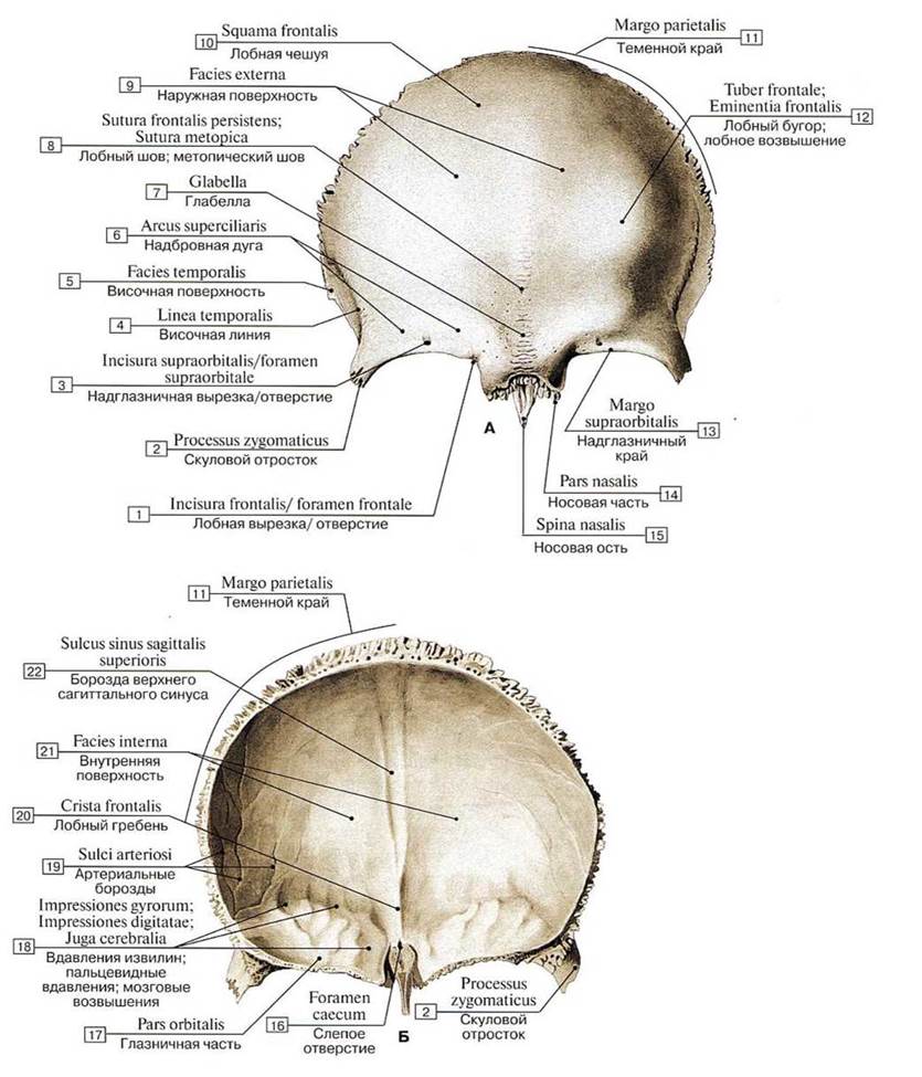
Fig. 44. Frontal bone (A — external view, B — internal view):
1 — Frontal notch/foramen; 2 — Zygomatic process; 3 — Supra-orbital notch/foramen; 4 — Temporal line; 5 — Temporal surface; 6 — Superciliary arch; 7 — Glabella; 8 — Frontal suture; Metopic suture; 9 — External surface; 10 — Squamous part; 11 — Parietal margin; 12 — Frontal tuber; Frontal eminence; 13 — Supra-orbital margin; 14 — Nasal part; 15 — Nasal spine; 16 — Foramen caecum; 17 — Orbital part; 18 — Impressions of cerebral gyri; 19 — Grooves for Arteries; 20 — Frontal crest; 21 — Internal surface; 22 — Groove for superior sagittal sinus
Last update: 08/08/2026
Editorial and Educational Adaptation: This material has been compiled based on the primary/original source text. The project team performed an editorial review, corrected technical inaccuracies, structured sections, and adapted the content for an educational format.
What was processed:
- elimination of formatting defects (OCR errors, structural breaks, corrupted characters);
- editorial organization of content;
- standardization of terminology in accordance with academic sources;
- verification of factual statements against the original source text.
All mentions of the author, publication year, and origin of the primary text have been preserved in accordance with the source.