Human Anatomy and Physiology (with Age-Related Features of the Child's Body) - Sapin M.R., Sivoglazov V.I. 2002
Musculoskeletal System
The Doctrine of Bones and Their Joints (Osteoarthrology)
Bones of the Lower Extremities and Their Joints
The Skeleton of the lower extremities consists of the BONES OF THE lower extremity girdle (Pelvic Girdle) and the free lower extremities (Fig. 23). The pelvic girdle consists of two hip bones connected to the sacrum, to which the right and left free lower extremities attach.
Bones and joints of the pelvic girdle.
The pelvic girdle is formed by two hip bones. The hip bone is flat in shape and consists of the ilium, ischium, and pubis, which fuse in adults in the region of the acetabulum—a deep socket that articulates with the HEAD of the Femur. The ilium is located above the acetabulum, the pubis anteriorly and inferiorly, and the ischium inferiorly and posteriorly to it. The ischium and pubis bound a large, oval-shaped obturator foramen, which is covered by the fibrous obturator membrane.
Class="center">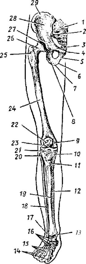
Fig. 23. Bones of the lower extremity. Anterior view:
1 — sacrum, 2 — sacroiliac joint, 3 — superior ramus of pubic bone, 4 — symphysial surface of pubic bone, 5 — inferior ramus of pubic bone, 6 — ramus of ischium, 7 — ischial tuberosity, 8 — body of ischium, 9 — medial epicondyle of femur, 10 — medial condyle of Tibia, 11 — tibial tuberosity, 12 — body of tibia, 13 — medial malleolus, 14 — Phalanges, 15 — Metatarsal Bones, 16 — Tarsal Bones, 17 — lateral malleolus, 18 — Fibula, 19 — anterior border of tibia, 20 — head of fibula, 21 — lateral condyle of tibia, 22 — Patella, 23 — lateral epicondyle of femur, 24 — femur, 25 — greater trochanter of femur, 26 — neck of femur, 27 — head of femur, 28 — ala of ilium, 29 — iliac crest
The ilium consists of a massive body and a thin, broad ala that terminates superiorly in the iliac crest. The ends of the crest project anteriorly and posteriorly as the anterior and posterior superior and inferior iliac spines. Below the posterior inferior iliac spine lies the greater sciatic notch, bounded inferiorly by the ischial spine. The concave internal surface of the ala forms the iliac fossa. Above the greater sciatic notch, the auricular surface is visible, articulating with the corresponding surface of the sacrum.
The ischium has a body, which participates in The formation of the acetabulum, and a ramus that bounds the obturator foramen and forms the ischial tuberosity. Posterior and superior to the ischial tuberosity lies the lesser sciatic notch, which the ischial spine separates from the greater sciatic notch.
The pubic bone also has a body, which participates in the Formation of the acetabulum, and two rami—superior and inferior—connecting at an angle. On the Medial surface of the angle lies the symphysial surface, forming the Pubic Symphysis with the corresponding surface of the opposite bone. The posterior margin of the superior ramus forms the pubic tubercle and pubic crest, which transitions into the arcuate line of the ilium located at the boundary of the greater and lesser pelvis.
JOINTS OF THE pelvic girdle. The hip bones articulate posteriorly with the sacrum via the paired sacroiliac joints, and anteriorly they form the pubic symphysis.
The sacroiliac joint is flat, virtually immovable, and formed by the articulating auricular surfaces of the hip bone and sacrum. The joint is reinforced by strong sacroiliac and iliolumbar ligaments. It is further reinforced by the extrinsic sacrot事實 (sacrospinous and sacrotuberous) ligaments, which convert the greater and lesser sciatic notches into the greater and lesser sciatic foramina.
The pubic symphysis, formed by the opposing symphysial surfaces of the pubic bones, is reinforced by the superior pubic ligament and the arcuate pubic ligament (inferiorly).
The Pelvis as a whole. The hip bones and sacrum, connecting via the sacroiliac joints and the pubic symphysis, form the pelvis (Fig. 24). The pelvis is a bony ring enclosing a cavity that contains visceral Organs. The pelvis also serves as a support for the trunk and lower extremities.
The terminal line, formed by the arcuate line of the ilia, the pubic crests, and the sacral promontory, divides the pelvis into the greater pelvis and the lesser pelvis. The lesser pelvis represents the birth canal in females. The shape and size of the pelvis exhibit distinct Sexual Dimorphism. The female pelvis is broader and shallower; the male pelvis is narrower and deeper. The pubic arch (subpubic angle) is acute in males (about 70–75°), while in females it approaches a right angle or is even obtuse (90–100°). The ischial tuberosities and iliac alae in the female pelvis are spaced further apart and more flared. For instance, the distance between both anterior superior iliac spines is 25–27 cm in females and 22–23 cm in males. The superior aperture (inlet) of the female lesser pelvis is wider and transversely oval in shape (longitudinal oval in males). The main dimensions of the lesser pelvis are given in Table 5. The conjugate (direct diameter) of the superior aperture is the distance between the sacral promontory and the upper margin of the symphysis; that of the inferior aperture is the distance between the tip of the coccyx and the lower margin of the pubic symphysis. The transverse diameter of the superior aperture is the distance between the most distant points of the terminal line; that of the inferior aperture is the distance between the inner margins of the ischial tuberosities. The oblique diameter of the superior aperture is the distance between the sacroiliac joint on one side and the iliopubic eminence on the other.

Fig. 24. Pelvis. Lines indicating the dimensions of the greater pelvis and the pelvic inlet are shown:
1 — greater pelvis, 2 — ala of ilium, 3 — iliac crest, 4 — anterior superior iliac spine, 5 — anterior inferior iliac spine, 6 — lesser pelvis, 7 — acetabulum, 8 — pubic crest, 9 — pubic tubercle, 10 — ischium, 11 — ischial tuberosity, 12 — inferior ramus of pubic bone, 13 — subpubic arch, 14 — obturator foramen, 15 — right sacroiliac joint, 16 — sacrum.
I — distance between the two most distant points of the iliac alae; II — distance between the two anterior superior spines; III — transverse diameter of the pelvic inlet, IV — oblique diameter of the pelvic inlet
Thus, the sexual differences of the female pelvis primarily amount to its larger dimensions, greater volume, and widened inferior aperture. This is due to its primary function—the pelvis serves as a container for the developing fetus, which during childbirth exits the pelvic cavity through its inferior aperture.
Table 5 Dimensions of the lesser pelvis in females (f) and males (m)
|
Pelvic apertures (inlet/outlet) |
Dimensions in cm |
|||||
|
con |
jugate |
oblique |
transverse |
|||
|
f |
m |
f |
m |
f |
m |
|
|
Superior |
11.0 |
10.5 |
12.0 |
12.0 |
13.0 |
12.5 |
|
Inferior |
9.5 |
7.5 |
11.0 |
8.0 |
||
The pelvis of a newborn infant is funnel-shaped. Its anteroposterior diameter exceeds the transverse diameter, the sacral promontory is poorly defined, the superior aperture is rounded, and the ilium is positioned more vertically. After birth, the shape and dimensions of the pelvis gradually change. The ischial tuberosities move outward, the obturator foramina enlarge and assume an oblique position, and the lesser pelvis acquires a cylindrical shape. Rapid pelvic growth occurs during the prepubertal period.
Bones and joints of the free lower extremity. The skeleton of the free lower extremity consists of the femur, patella, two leg bones (tibia and fibula), and the Bones of the FOOT.
The femur is the largest tubular bone in The Human Body. It consists of a body (shaft) and two epiphyses. The upper (proximal) epiphysis features the femoral head for articulation with the hip bone, separated from the shaft by a long neck. At the Base of the neck are the greater and lesser trochanters (tuberosities). The bone shaft bears ridges and rough lines for Muscle attachment.
The thickened lower epiphysis bears large medial and lateral condyles, which articulate with the tibia of the leg, and two projecting epicondyles—medial and lateral. On the anterior surface of the epiphysis between the condyles lies a smooth area—the patellar surface.
The patella is a large sesamoid bone embedded within the tendon of the quadriceps femoris muscle. Its apex points inferiorly, its base superiorly, and its articular surface, covered with Cartilage, faces posteriorly. The patella can be easily palpated in a living person.
The bones of the leg consist of the medially situated tibia and the laterally placed fibula. An interosseous connective-tissue membrane of the leg is stretched between these two bones.
The tibia is a massive bone and the only one of the two leg bones that articulates with the femur. The proximal epiphysis of the tibia is broad and thick, featuring two condyles (medial and lateral) that bear slightly concave articular surfaces separated by the intercondylar eminence. On the lateral surface of the corresponding tibial condyle, There is a fibular articular facet for articulation with the head of the fibula.
The shaft of the tibia is triangular in shape. The sharp anterior border near the proximal epiphysis widens to form the tibial tuberosity, which serves as the attachment site for the tendon of the powerful quadriceps femoris muscle. The interosseous membrane of the leg attaches to the lateral (interosseous) border. The distal epiphysis bears the inferior articular surface for articulation with the talus of the foot. The medial end of the epiphysis extends downward to form the medial malleolus. On the lateral side of the lower epiphysis, there is a fibular notch for articulation with the fibula.
The fibula is a slender, long bone, featuring a head proximally with an articular facet for articulation with the proximal epiphysis of the tibia. The triangular-shaped body of the bone terminates distally in a thickened lateral malleolus equipped with an articular surface. The inferior articular surface of the tibia and the articular surfaces of the malleoli form a mortise that grips the trochlea of the talus from above and laterally.
The bones of the foot comprise the tarsal bones, metatarsal bones, and phalanges of the digits. The human foot performs a highly specialized function of locomotion and support. This is reflected in the architecture of its skeleton, which is built like a sturdy and resilient arched vault with short toes.
The tarsal bones (seven short bones) are arranged in two rows. The proximal (posterior) row contains the large talus and calcaneus. In the distal (anterior) row, the cuboid bone lies laterally, the narrow navicular bone lies medially, and anterior to it are the three cuneiform bones: medial, intermediate, and lateral.
The body of the talus bears superior and two lateral surfaces for articulation with the corresponding articular surfaces of the leg bones.
The calcaneus, which is the largest tarsal bone, is located beneath the talus and terminates posteriorly in a robust calcaneal tuberosity. Anterior to the calcaneus lies the cuboid bone, which forms the lateral border of the tarsus.
The navicular bone is situated medially and anterior to the head of the talus. The anterior surface of the navicular bears three flat articular surfaces for articulation with the three cuneiform bones (medial, intermediate, and lateral).
The metatarsal bones are five short tubular bones, each comprising a base, a body, and a head. By their bases, the metatarsal bones articulate with the cuneiform and cuboid bones, and by their heads, with the bases of the corresponding proximal phalanges.
The bones of the toes (phalanges) are short tubular bones. The first digit (hallux) has two phalanges, while the remaining digits (II–V) each have three. Each proximal bone phalanx articulates by its base with the corresponding metatarsal bone and by its head with the middle phalanx. The middle phalanges articulate with the bases of the distal phalanges.
Articulations of the bones of the free lower limb. The Structure of the joints in the free lower limb is determined by the specific nature of their Functions, their involvement in spatial locomotion, and the maintenance of balance.
The hip joint is a ball-and-socket (spheroidal), multiaxial joint formed by the acetabulum of the hip bone, which is supplemented by a fibrocartilaginous labrum, and the head of the femur. The capsule of the hip joint is strong and also encompasses the femoral neck. The Joint Capsule is reinforced by thick, robust ligaments: the iliofemoral, pubofemoral, ischiofemoral, and others. The ligament of the head of the femur is located intracapsularly; it transmits Vessels and nerves to the femoral head. The deep socket of the joint and tightly taut ligaments not only reinforce the hip joint but also restrict its mobility, which is crucial for body stability in space.
The knee joint is a large, complexly structured joint that functions as a hinge-pivot (modified hinge) joint. Flexion and extension are possible around the frontal (transverse) axis, while rotational movements can occur when the leg is semi-flexed at the knee and the collateral ligaments are relaxed. The joint is formed by the femur, tibia, and patella. Within the joint, There are two crescent-shaped menisci—medial and lateral—which mitigate incongruities between the articular surfaces. The joint capsule is strong, and its synovial membrane forms folds within the joint. Interwoven into the capsule are collateral (lateral and medial) and other ligaments that reinforce the joint and prevent hyperextension. Two ligaments, the anterior and posterior cruciate ligaments, covered by the synovial membrane, are located intracapsularly. They also reinforce the joint and restrict tibial rotation within the knee joint.
Articulations of the leg bones. The proximal epiphyses of the tibia and fibula form a flat, slightly movable tibiofibular joint. The shafts of these bones are united by a strong interosseous membrane, whereas their distal epiphyses are connected by ligaments that form the tibiofibular syndesmosis.
The ankle joint and articulations of the foot. The ankle joint, also referred to as the talocrural joint, is complex in STRUCTURE AND FUNCTIONS as a hinge joint; it is formed by the articular surfaces of the tibia and fibula, and the talus. Joined together, the tibia and fibula grip the trochlea of the talus with their malleoli like a mortise. The joint capsule is reinforced by collateral ligaments. In this joint, plantarflexion and dorsiflexion of the foot are possible around a transverse axis passing through the trochlea of the talus.
The tarsal bones, by articulating with one another, form the slightly movable subtalar, talocalcaneonavicular, calcaneocuboid, transverse tarsal (Chopart's), and tarsometatarsal joints.
The subtalar joint is formed by the adjoining surfaces of the talus and calcaneus, and is reinforced by the strong, tightly taut interosseous talocalcaneal and other ligaments. The other tarsal joints are likewise reinforced by short ligaments. Movements in these morphologically diverse joints are combined with movements in the ankle joint, endowing the foot with a relatively high degree of mobility.
The tarsometatarsal joints are flat in shape and slightly movable, and are reinforced by dorsal and interosseous ligaments.
For practical purposes, the calcaneocuboid and talonavicular joints, which lie along the same line and share a common ligament (the bifurcated ligament), are grouped together as the transverse tarsal joint (Chopart's joint). When the bifurcated ligament is transected during a surgical Procedure, the foot easily disarticulates. In surgery, the tarsometatarsal joints are also referred to as Lisfranc's joint. Upon transection of the medial interosseous ligament (the "key" of Lisfranc's joint), the distal part of the foot can be separated from its proximal part during surgery.
The metatarsophalangeal joints are spheroidal in shape, whereas the interphalangeal joints are hinge joints. These joints are reinforced by collateral and plantar ligaments, much like the analogous joints of the hand.
The human foot, functioning as an integrated osteoarticular arched structure with its convexity directed superiorly, exhibits a high degree of resilience.
The bones of the foot, by articulating with each other, form arches directed longitudinally and transversely.
There are five longitudinal arches (corresponding to the number of metatarsal bones) and a transverse arch of the foot (Fig. 25). All longitudinal arches (curves) converge posteriorly on the calcaneal tuberosity and rest anteriorly on the heads of the metatarsal bones. The longest and highest is the second longitudinal arch of the foot. The archwise curved transverse arch passes through the highest points of the foot.
The arches of the foot are reinforced by tightly stretched ligaments, including the powerful long plantar ligament. These ligaments act as the passive "ties" of the foot; when they relax, the arches flatten, which can lead to Flatfoot. Muscles also play a vital role in reinforcing the foot arches, functioning as their active "ties".
Development and Age-Related Features of the Appendicular Skeleton
All bones of the limbs, except for the clavicles—which develop from Connective Tissue—go through three Selection/3.html">Stages of development: connective tissue, cartilage, and bone.
The process of ossification in the clavicle begins in the 6th week of embryonic development and is nearly complete by the time of birth.
In the diaphyses of tubular bones, the initial (primary) ossification centers appear at the end of the 2nd to the beginning of the 3rd month of intrauterine development, whereas in the epiphyses and apophyses, they appear after birth. Only a few epiphyses begin to ossify shortly before birth. Fusion of the epiphyses with the diaphyses typically occurs between the ages of 13 and 15, taking place 1–2 years earlier in girls than in boys.
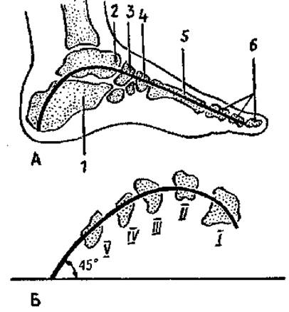
Fig. 25. Diagrams of the foot arches:
A — diagram of the longitudinal (second) arch of the foot: 1 — calcaneus, 2 — talus, 3 — navicular bone, 4 — intermediate cuneiform bone, 5 — second metatarsal bone. B — diagram of the transverse arch of the foot: I — V — cross-section of the metatarsal bones
In the Carpal Bones, ossification centers appear after birth: in the capitate During the first year of life, in the hamate at the end of the first to the beginning of the second year, and in the remaining carpal bones between the ages of 2 and 11.
In the bones of the pelvic girdle (ilium, ischium, and pubis), ossification centers appear between 3.5 and 4.5 months of intrauterine development. Fusion of all three bones into a single hip bone occurs at ages 12–15.
In the tarsal bones (navicular, cuboid, and cuneiforms), ossification centers appear in the period ranging from 3 months after birth up to 5 years. The remaining (secondary) ossification centers form postnatally.
The Development of synovial joints starts in the 6th week of embryonic development. The joint capsules in newborns are tightly stretched, and most ligaments are not yet fully formed. The most intensive development of joints and ligaments occurs up to the age of 2–3 years, driven by the increasing motor activity of the child. In children aged 3–8, the range of motion in all joints increases, while the collagenization of joint capsules and ligaments accelerates simultaneously. The formation of articular surfaces, capsules, and ligaments is generally completed during adolescence (13–16 years).
In newborns, the lower limbs grow more rapidly, eventually becoming longer than the upper limbs. The peak growth rate of the lower limbs is observed in boys at ages 12–15, whereas in girls, the increase in leg length occurs between 13 and 14 years of age.
In postnatal ontogeny, Changes in the shape and dimensions of the pelvis occur under METABOLISM/18.html">The Influence of body weight and abdominal organs, muscular action, and Sex Hormones. As a result of these diverse factors, the anteroposterior diameter of the pelvis increases (from 2.7 cm in a newborn to 9.5 cm at age 12), and the transverse diameter grows, reaching adult dimensions by ages 13–14. Sexual dimorphism in pelvic shape becomes noticeable after age 9. In boys, the pelvis is taller and narrower than in girls.
REVIEW AND SELF-Control Questions:
1. Name the Structural Features of the lower limb bones that distinguish them from the upper limb bones. Explain the functional reasons for these differences.
2. Name the walls and BOUNDARIES OF THE lesser and greater pelvis.
3. Describe the structural CHARACTERISTICS OF THE male and female pelvis. List the known dimensions of the greater and lesser pelvis.
4. Describe the STRUCTURE OF THE sacroiliac, hip, and knee joints. What determines their structure and their differences from the corresponding joints of the upper limbs?
5. What connections exist between the tibia and fibula, and where are these connections located?
6. What do you know about the structure and Functions of the ankle joint?
7. Name the joints connecting the bones of the foot and the ligaments known to reinforce these joints.
8. What do you know about the arches of the foot and their "ties"?
Last update: 10/08/2026
Editorial and Educational Adaptation: This material has been compiled based on the primary/original source text. The project team performed an editorial review, corrected technical inaccuracies, structured sections, and adapted the content for an educational format.
What was processed:
- elimination of formatting defects (OCR errors, structural breaks, corrupted characters);
- editorial organization of content;
- standardization of terminology in accordance with academic sources;
- verification of factual statements against the original source text.
All mentions of the author, publication year, and origin of the primary text have been preserved in accordance with the source.