MEDICAL BIOLOGY, HUMAN ANATOMY, PHYSIOLOGY AND PATHOLOGY - Y.I. Fedoniuk 2010
ANATOMY, PHYSIOLOGY, PATHOLOGY
SECTION 3. ANATOMICAL AND PHYSIOLOGICAL ASPECTS OF BODY FUNCTION SELF-REGULATION
SPINAL CORD
2. FUNCTIONS OF THE SPINAL CORD
Two Main Functions of the Spinal Cord are distinguished: reflex and conduction.
The reflex activity of the spinal cord is associated with reflex arcs that close at the level of the spinal nerve centers. Spinal cord Reflexes are segmental in character and relatively simple. There are both motor and autonomic reflexes of the spinal cord. The spinal cord houses nerve centers that regulate motor functions. For example, in the anterior horns of the cervical segments, there are clusters of motor Neurons from which the phrenic nerve originates, participating in Respiration; in the cervical and thoracic regions lie the motor centers of the upper extremities, chest, back, and Abdominal Muscles; in the lumbar region are the motor centers of the lower extremities. Through reflex arcs closing within these motor centers, the spinal cord regulates motor activity. Tonic and phasic reflexes of the spinal cord are distinguished.
An example of a tonic reflex is the Regulation of Muscle tone by the spinal cord. The reflex nature of muscle tone was proved in an experiment on a spinal frog (with the Brain removed). The spinal frog is suspended from a stand, and attention is drawn to its hind limbs. Although they hang down, they remain in a slightly flexed position. If the dorsal roots are cut on one side, the limb on that side hangs limply As a result of the loss of tone (Fig. 3.9). This indicates damage to the reflex arc. Reflex tone is essential for maintaining body posture and keeping balance during walking. Maintaining a specific body position for a long time, such as tilting the HEAD while writing or reading, is possible only due to reflex muscle tone.
Class="center">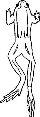
Fig. 3.9. A frog with severed dorsal roots
Phasic spinal reflexes include tendon reflexes. They occur in response to rapid muscle stretching. This stimulates the muscle proprioceptors; the excitation travels along the reflex arc to the effector and causes a contraction. An example is the knee-jerk reflex, which is elicited by a light tap with a neurological hammer on the tendon of the quadriceps femoris muscle. Phasic reflexes also include cutaneous reflexes, which arise from The stimulation of Skin Receptors.
For instance, stimulation of the SOLE OF THE FOOT results in its plantar flexion. This is the plantar reflex.
The reflex activity of the spinal cord is modulated by higher centers located in the brain. The morphological basis for this activity consists of ascending and descending pathways that connect the nerve centers of the spinal cord with the brain and vice versa.
The conduction function is associated with the passage of conducting pathways—ascending and descending—through the spinal cord. They are located in the funiculi and form the White matter OF the spinal cord. Ascending pathways carry information from skin receptors—exteroreceptors (tactile, Temperature, pain)—and muscle receptors (proprioceptors) via the dorsal roots into the spinal cord, where they synapse with its neurons, and further up to the nerve centers of the brain. Thus, the ascending pathways are sensory.
The spinothalamic tract (pathway of tactile, pain, and temperature sensitivity) and spinocerebellar tracts (pathways of proprioceptive muscle-joint sensitivity) are distinguished.
The descending Pathways of the spinal cord transmit excitation from the nerve centers of the brain to the motor neurons of the anterior horns of the spinal cord and further to the skeletal muscles. These are motor tracts.
The primary one is the pyramidal or corticospinal tract, which runs from Betz Cells (motor areas of the Cerebral Cortex) to the motor neurons of the anterior horns of the spinal cord. This is the main pathway for The regulation of conscious, voluntary motor activity.
The vestibulospinal, rubrospinal, and reticulospinal pathways are also distinguished. These tracts help maintain muscle tone.
The spinal cord has a segmental Structure. Two pairs of dorsal (posterior) and ventral (anterior) roots emerge from each spinal segment. According to physiological experiments.
Last update: 08/08/2026
Editorial and Educational Adaptation: This material has been compiled based on the primary/original source text. The project team performed an editorial review, corrected technical inaccuracies, structured sections, and adapted the content for an educational format.
What was processed:
- elimination of formatting defects (OCR errors, structural breaks, corrupted characters);
- editorial organization of content;
- standardization of terminology in accordance with academic sources;
- verification of factual statements against the original source text.
All mentions of the author, publication year, and origin of the primary text have been preserved in accordance with the source.