Principles of Biochemistry Volume 3 - A. Lehninger 1985
Selected Aspects of Human Biochemistry
Hormones
The stimulation of glycogen breakdown in the presence of epinephrine occurs via an amplification cascade
Let us now combine the phenomena described above and trace the chain of events whereby epinephrine stimulates Glycogen breakdown to glucose in the Liver, which is then released into the bloodstream (Fig. 25-11). Epinephrine reaches The surface of liver Cells, where it binds to a specific adrenergic receptor. This binding (with the hormone never entering The Cell) induces a conformational change in the receptor protein. This alteration is somehow transmitted across the membrane and “turns on” adenylate cyclase, which is associated with the inner surface of The Plasma Membrane. Activated adenylate cyclase then begins converting ATP into cAMP, a second messenger, causing the cytosolic concentration of cAMP to rapidly peak at ~10-6 M. The generated cAMP, in turn, binds to the Regulatory Subunits of protein kinase, leading to the release of enzymatically active catalytic subunits. Subsequently, the activated protein kinase catalyzes the phosphorylation (using ATP) of the inactive dephosphorylated form of phosphorylase kinase, converting it into the active phosphorylated form. Active phosphorylase kinase, which requires Ca2+ ions for its function, then catalyzes the phosphorylation of relatively inactive phosphorylase b by means of ATP, yielding active phosphorylase a. The latter rapidly breaks down glycogen to form glucose-1-phosphate, which is further converted into glucose-6-phosphate and subsequently into free glucose that enters the bloodstream (Fig. 25-11). Despite the numerous steps in this sequence of events, Glycogen phosphorylase activity peaks within just a few minutes of epinephrine binding to the liver cells.
The sequence of steps illustrated in Fig. 25-11 can be viewed as an enzyme cascade, where each enzyme activates multiple molecules of the next enzyme in the pathway. This mechanism achieves a massive and rapid Amplification of the incoming signal, estimated to be roughly 25 million-fold. Consequently, the binding of only a few epinephrine molecules to hepatic adrenergic receptors triggers the rapid release of several grams of glucose into the Blood.
The cascade process schematized in Fig. 25-11 proceeds identically in The Liver and Skeletal Muscle up to The formation of glucose-6-phosphate. However, muscle lacks glucose-6-phosphate and therefore cannot produce free glucose. Instead, the elevation in glucose-6-phosphate concentration significantly accelerates Glycolysis to produce lactic acid, thereby generating ATP to fuel Muscle contraction. As relatively recent studies have shown, epinephrine stimulates hepatic glycogen breakdown through an additional amplification cascade that operates in parallel with the one shown in Fig. 25-11. In this second cascade, which under certain conditions becomes predominant, Ca2+ ions serve as the intracellular second messenger.
Class="center">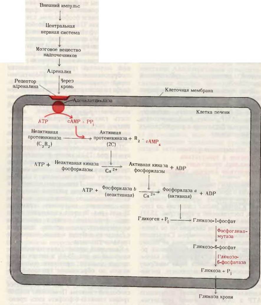
Fig. 25-11. Epinephrine triggers an amplification cascade in liver cells. The binding of a few epinephrine molecules to specific cell-surface receptors initiates a sequence of enzymatic reactions that results in the release of a massive amount of glucose into the bloodstream.
The cascade shown in Fig. 25-11 is triggered in the liver not only by epinephrine but also, as we will see below, by the pancreatic hormone Glucagon.
Last update: 06/08/2026
Editorial and Educational Adaptation: This material has been compiled based on the primary/original source text. The project team performed an editorial review, corrected technical inaccuracies, structured sections, and adapted the content for an educational format.
What was processed:
- elimination of formatting defects (OCR errors, structural breaks, corrupted characters);
- editorial organization of content;
- standardization of terminology in accordance with academic sources;
- verification of factual statements against the original source text.
All mentions of the author, publication year, and origin of the primary text have been preserved in accordance with the source.