Review of Medical Physiology - William F. Ganong 2002
Functions of the Digestive System
Mechanisms of Gastrointestinal Regulation
The Liver and the Biliary System
Bile is secreted by Liver Cells into the bile ducts, which empty into the duodenum. During the interdigestive period, most of these ducts are closed, and bile flows back into the Gallbladder, where it is stored. When food enters the Oral Cavity, the sphincters around the duct orifices relax. If gastric contents enter the duodenum, the intestinal mucosal hormone CCK (cholecystokinin) stimulates contraction of the gallbladder.
The liver is composed of lobules through which Blood flows from the Branches of the portal vein, past hepatocytes and sinusoids, to the central vein of each lobule. Numerous fenestrations are present between endothelial cells, allowing plasma to come into close contact with hepatocytes (Fig. 26-20). Normally, there is only a single layer of hepatocytes between sinusoidal-type blood capillaries, so the total contact area between liver cells and plasma is large. Blood enters the sinusoidal capillaries via the hepatic artery. Central Veins converge to form the hepatic veins, which drain into the INFERIOR VENA CAVA. Hepatic blood flow is described in detail in Chapter 32. The mean transit time for blood through a hepatic lobule from the portal venule to the central vein of the liver is 8.4 s. Numerous macrophages (Kupffer cells) are anchored to the endothelium of the sinusoidal capillaries and project into the lumen. The Functions of these cells are described in detail in Chapter 27.
Another perspective on liver architecture is its division into hepatic acini. As detailed in Chapter 32 regarding hepatic Circulation, the center of each acinus is a vascular stalk containing the terminal branches of the portal vein, hepatic Arteries, and bile ducts (see Fig. 32-16). Blood flows outward from the vascular stalk to the terminal hepatic venules located at the periphery of the acinus. Consequently, the cells closest to the vascular stalk receive well-oxygenated blood, whereas the cells at the periphery of the acinus receive the least oxygenated blood and are therefore most susceptible to hypoxic injury.
Each liver Cell is also connected to multiple bile canaliculi (Fig. 26-21). These drain into interlobular bile ducts, which merge to form the right and left hepatic ducts. These ducts unite outside the liver to form the common hepatic duct. The cystic duct emerges from the gallbladder. The common hepatic duct joins the cystic duct to form the common bile duct, which empties into the duodenum near the duodenal papilla. Its opening, surrounded by the sphincter of Oddi, typically unites with the main pancreatic duct immediately before entering the duodenum (see Fig. 26-17). The walls of the extrahepatic biliary ducts and the gallbladder contain fibrous tissue and smooth Muscle. Their mucous membranes contain mucous glands and are lined with a layer of columnar cells. In the gallbladder, the mucosa is prominently folded, which increases its surface area and gives the internal space of the gallbladder a honeycomb appearance. In primates, the gallbladder mucosa is also folded to form so-called spiral Valves.
Class="center">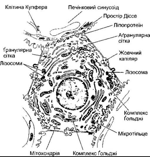
Fig. 26-20. Hepatocyte. Note The Cell's relationship with the bile canaliculus and sinusoids. Also note the wide fenestrations between the endothelial cells adjacent to the hepatocytes (reproduced with permission from Fawcett DW, Bloom and Fawcett, A Textbook of Histology, 11th ed. Saunders, 1986).

Fig. 26-21. Top: Structural Organization of the liver; CV = central vein; PT = portal tract containing branches of the bile duct, portal vein, and hepatic artery. Bottom: Arrangement of hepatocytes, sinusoids, and bile ducts within a liver lobule, illustrating the centripetal flow of blood from the sinusoids to the central vein and the centrifugal flow of bile from the bile canaliculi into the bile ducts (reproduced with permission from Fawcett DW: Bloom and Fawcett, A Textbook of Histology, 11th ed. Saunders, 1986).
Functions of the Liver
The liver is the largest gland in the body and performs a wide range of functions (Table 26-7). Some of these are described in this chapter, while others are covered in separate chapters dedicated to the respective Organ Systems.
The major proteins synthesized by the liver are listed in Table 27-10. Many of these belong to acute-phase proteins—proteins whose Synthesis and Secretion into plasma are upregulated in response to severe stimuli. Other proteins transport Steroids and certain Hormones in the plasma, as well as blood clotting factors.
Bile
The components of bile include bile salts, bile pigments, and other substances dissolved in an alkaline electrolyte solution similar to pancreatic juice (Table 26-8). Approximately 500 mL of bile is secreted daily. Some bile components are reabsorbed in the intestine and subsequently re-excreted by the liver (enterohepatic circulation).
Bile pigment glucuronides—bilirubin and biliverdin—are responsible for the golden-yellow color of bile. The formation of these Hemoglobin breakdown products is described in detail in Chapter 27, and their excretion is discussed below. Bile salts are The sodium and potassium salts of bile acids; they are secreted into bile and conjugated with Glycine or taurine, a Cysteine derivative. Bile acids are synthesized from Cholesterol. Humans have four such acids (Fig. 26-22). Like vitamin D, cholesterol, various Steroid Hormones, and digitalis Glycosides, bile acids contain a cyclopentanoperhydrophenanthrene Nucleus (see Chapter 20). The two major (primary) bile acids produced in the liver are cholic acid and chenodeoxycholic acid. In the colon, Bacteria convert cholic acid to deoxycholic acid and chenodeoxycholic acid to lithocholic acid. Because they are formed through bacterial action, deoxycholic and lithocholic acids are referred to as secondary bile acids.
Table 26-7. Major functions of the liver1

1 Numbers in parentheses indicate the chapter in our book where these functions are described.
Table 26-8. Composition of human hepatic duct bile
97.0% |
|
Bile salts |
0.7% |
Bile pigments |
0.2% |
Cholesterol |
0.06% |
Inorganic salts |
0.7% |
0.15% |
|
Lecithin |
0.1% |
Fat |
0.1% |
Alkaline phosphatase |
Bile acid salts exert several important effects and also reduce surface tension. By conjugating with Phospholipids and monoglycerides, bile salts are responsible for the emulsification of fats, preparing them for Digestion AND ABSORPTION in the Small Intestine (see Chapter 25). Bile salts are amphipathic compounds possessing both hydrophilic and hydrophobic domains. One surface of the molecule is hydrophilic due to the presence of polar peptide bonds, carboxyl groups, and hydroxyl groups, whereas the opposite surface is hydrophobic. Consequently, bile acids tend to form cylindrical disks called micelles, with their hydrophilic surfaces facing outward and their hydrophobic surfaces facing inward (Fig. 26-23). At a specific concentration, known as the critical micelle concentration, all bile acids entering the solution form micelles. Lipids accumulate within the micelles, with cholesterol sequestered in the Hydrophobic core, while amphipathic phospholipids and monoglycerides extend their hydrophilic heads outward and their hydrophobic tails toward the center. Micelles play a crucial role in keeping lipids in solution and transporting them to the brush border of intestinal epithelial cells, where they are absorbed (see Chapter 25).

Fig. 26-22. Human bile acids. Numbers in the formula for cholic acid indicate the positions on the steroid ring.

Fig. 26-23. Cross-section of a mixed (bile acid-lipid) disc-shaped micelle containing free bile acids and cholesterol in a hydrophobic environment. The outward-facing surface of each bile acid is hydrophilic due to polar peptide bonds, carboxyl groups, and OH groups.
About 90-95% of bile salts are absorbed in the small intestine: a portion via non-ionic diffusion, but the majority (Fig. 26-24) through an efficient Na+-bile salt cotransport system mediated by basolateral Na+-K+-ATPase in the terminal ileum, analogous to the Na+-glucose transport system (see Chapter 25). One of the Na+-bile salt cotransporters involved in secondary Active Transport has already been cloned, pointing to the existence of others.
The remaining 5-10% of bile acids reach the ileum, where they are converted into deoxycholic and lithocholic acid salts. Lithocholates are relatively insoluble; most are excreted in the feces, and only 1% is absorbed.
Absorbed bile acids are transported via the vena cava back to the liver and re-excreted into the bile (enterohepatic circulation). The amount lost in the stool is replenished by hepatic synthesis, with a normal synthesis rate of 0.2-0.4 g/day. The total pool of bile acids undergoing recurrent enterohepatic recycling is approximately 3.5 g. It is estimated that the entire pool recycles twice per meal, amounting to six to eight times daily. When bile fails to reach the intestine, over 50% of ingested fat appears in the feces. Fat-soluble Vitamins are absorbed in a similar manner. When reabsorption of bile salts is prevented due to resection of the terminal ileum or pathological conditions within it, fecal fat content increases. This occurs because, with disrupted enterohepatic circulation, the liver cannot sufficiently upregulate bile salt synthesis to compensate for daily losses. Other consequences of terminal ileum resection are described in the section on malabsorption.

Fig. 26-24. Enterohepatic circulation of bile acids. Solid lines entering the portal system indicate bile acids of hepatic origin, while dashed lines represent bile acids formed through bacterial action.
Bilirubin METABOLISM and Excretion
Most of the body's bilirubin is derived from hemoglobin breakdown products (see Chapter 27). Bilirubin binds to albumin in the Circulatory system. While a fraction is bound irreversibly, the majority reaches the liver, where free bilirubin enters hepatocytes and binds to cytoplasmic proteins (Fig. 26-25). The next step is conjugation with glucuronic acid in a reaction catalyzed by the enzyme glucuronyl transferase (UDP-glucuronyltransferase). This enzyme is located primarily in the smooth Endoplasmic reticulum. Each bilirubin molecule reacts with two molecules of uridine diphosphoglucuronic acid (UDPGA) to form bilirubin diglucuronide. This glucuronide, which is more water-soluble than free bilirubin, is thought to be actively transported against a concentration gradient into the bile canaliculi. A small amount of bilirubin glucuronide enters the blood, where it binds less tightly to albumin than free bilirubin, and is subsequently excreted in the urine. Consequently, total plasma bilirubin normally consists of free bilirubin and a small amount of conjugated bilirubin. The major portion of bilirubin glucuronide passes through the bile ducts into the intestine.
The intestinal mucosa is relatively impermeable to conjugated bilirubin, yet permeable to unconjugated bilirubin and urobilinogens—a group of colorless bilirubin derivatives produced by bacterial action in the gut. Evidently, a fraction of the bile pigments and urobilinogens is reabsorbed into the portal circulation. Some reabsorbed substances are re-excreted by the liver (enterohepatic circulation), while small amounts of urobilinogens enter the systemic circulation and are eliminated in the urine.
Jaundice
When free or conjugated bilirubin accumulates in the blood, the Skin, sclerae, and mucous membranes turn yellow. This yellow discoloration is known as jaundice and typically manifests when total plasma bilirubin exceeds 2 mg/dL (34 µmol/L). Hyperbilirubinemia may result from excessive bilirubin production (e.g., hemolytic anemia); reduced hepatic uptake of bilirubin; impaired intracellular protein binding or conjugation; defective secretion of conjugated bilirubin into the bile canaliculi; or intrahepatic/extrahepatic obstruction of the bile ducts. The first three mechanisms lead to elevated levels of unconjugated bilirubin. In cases of impaired conjugated bilirubin secretion or bile duct obstruction, bilirubin glucuronide regurgitates into the blood, resulting in a predominance of elevated conjugated bilirubin in the plasma.
Other Substances Conjugated by Glucuronyl Transferase
In addition to bilirubin, the glucuronyl transferase system in the smooth reticulum catalyzes the glucuronidation of various other substances. These include steroids (see Chapters 20 and 23) and numerous pharmacological agents. These diverse compounds may compete with bilirubin for the enzyme system if present in significant quantities. Furthermore, certain barbiturates, antihistamines, anticonvulsants, and Other Compounds induce marked proliferation of the smooth endoplasmic reticulum in hepatocytes, concomitantly increasing hepatic glucuronyl transferase activity. Phenobarbital can be successfully used to treat congenital disorders characterized by a relative deficiency of glucuronyl transferase (type 2 UDP-glucuronyltransferase deficiency).
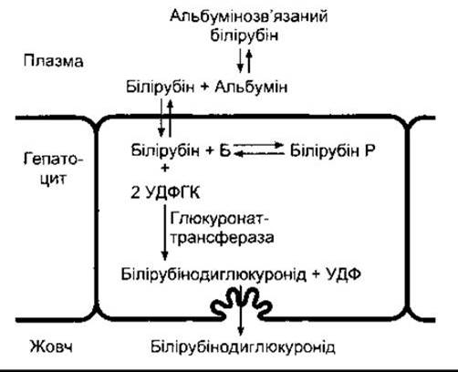
Fig. 26-25. Hepatic bilirubin metabolism; B - intracellular binding proteins; UDPGA - uridine diphosphoglucuronic acid; UDP - uridine diphosphate.
Other Substances Excreted in Bile
Cholesterol and alkaline phosphatase are also excreted into the bile. In patients with jaundice caused by intra- and extrahepatic bile duct obstruction, blood levels of both substances are elevated, whereas a somewhat smaller increase is observed in jaundice resulting from non-obstructive hepatocellular disease. Adrenocortical Hormones, Other Steroids, and numerous drugs are excreted in bile and subsequently reabsorbed (enterohepatic circulation).
Functions of the Gallbladder
Normally, in healthy individuals, bile flows into the gallbladder. As long as the sphincter of Oddi remains closed, bile is concentrated within the gallbladder through water absorption. The degree of concentration is reflected by an increase in solid content (Table 26-9): hepatic bile contains 97% water, whereas gallbladder bile contains only 89%. When the bile and cystic ducts are clamped, intrabiliary pressure rises to 320 mm bile in 30 minutes, and bile secretion ceases. However, when the bile ducts are occluded and the cystic duct remains open, water is reabsorbed in the gallbladder, and intrabiliary pressure increases by only 100 mm bile over several hours. Another function of the gallbladder is the acidification of bile (see Table 26-9).
Regulation of Biliary Secretion
From the moment food enters the oral cavity, the resistance of the sphincter of Oddi decreases. Fatty Acids and Amino Acids in the duodenum release CCK, which causes gallbladder contraction. Substances that stimulate gallbladder contraction are termed cholekinetics.
Bile production is enhanced by vagal stimulation and the hormone secretin, which increases the water and HCO3 content of bile. Substances that enhance bile secretion are known as choleretics. Bile salts are potent and vital physiological choleretics; although reabsorbed from the intestine to inhibit de novo bile acid synthesis, they are rapidly re-secreted and significantly augment bile flow.
Table 26-9. Comparison of Hepatic Duct Bile and Gallbladder Bile in Humans
Hepatic duct bile |
Gallbladder bile |
|
Percentage of solids |
2-4 |
10-12 |
Effects of Cholecystectomy
Intermittent release of bile from the gallbladder facilitates digestion, though it is not strictly essential for the process. Patients who have undergone a cholecystectomy maintain a relatively normal health status, although they exhibit a continuous, slow release of bile directly into the duodenum. Over time, the bile ducts become somewhat dilated, and There is a tendency for an increased volume of bile to enter the duodenum, particularly following a meal. Cholecystectomy patients can even consume fried foods, although they should avoid high-fat food products.
Visualization of the gallbladder
Ultrasound examination of the right upper quadrant of the abdomen (Ultrasonography) and computed tomography have become the most common Methods for visualizing the gallbladder and detecting gallstones. Oral cholecystography is also a widely used technique. A radiopaque iodine-containing dye, such as tetraiodophenolphthalein, is excreted into the bile and concentrated within the gallbladder. Once the gallbladder contains an adequate concentration of this dye, it can be visualized using X-rays. To assess the ability of the gallbladder to contract normally, a high-fat meal must be ingested. Following the release of CCK, the gallbladder contracts, and within 30 min its shadow size on radiography normally decreases to one-third of its initial state. A third major diagnostic method for gallbladder disorders is nuclear cholecystography. Following intravenous administration of iminodiacetic acid derivatives labeled with technetium-99m, they are excreted into the bile and provide excellent high-quality images of the gallbladder and ducts using a gamma camera.
Gallstones
Cholelithiasis, or the presence of stones in the gallbladder, is a common condition. Its incidence increases with age. For instance, in the United States, 20% of women and 5% of men aged 50 to 65 have gallstones. There are two MAIN TYPES OF stones: calcium bilirubinate (pigment) stones and cholesterol stones. In Europe and North America, 85% of all stones are of cholesterol origin. Three critical factors are involved in the formation of cholesterol stones. The first is undeniably bile stasis. Stones form more rapidly in bile concentrated within the gallbladder than in bile flowing through the bile ducts. The second factor is the supersaturation of bile with cholesterol. Cholesterol, which is virtually insoluble in water, is held in solution within bile micelles only at a specific concentration of bile salts and lecithin (Fig. 26-26). When bile is supersaturated with cholesterol (see Fig. 26-26, for concentrations above the line ABC), small cholesterol crystals appear in addition to micelles. Furthermore, many apparently healthy individuals who have not yet formed gallstones are found to have cholesterol-supersaturated bile. The third factor involves interaction with cholesterol nucleation factors that promote stone formation in supersaturated bile. Outside The Human Body, bile from patients with cholelithiasis forms stones within just two to three days, whereas normal bile requires more than two weeks. The exact origin of cholesterol nucleation factors is not yet fully elucidated, but it is known that they contain Glycoproteins from the gallbladder mucosa. Moreover, it remains unclear whether stones form as a consequence of the overproduction of factors that stimulate cholesterol nucleation, or due to a reduced production of inhibitors of aggregation and nucleation that normally prevent gallstone formation in healthy individuals.

Fig. 26-26. Solubility of cholesterol in bile as a function of the proportional content of lecithin, bile salts, and cholesterol. In bile of any composition described below the line ABC (e.g., point P), cholesterol exists exclusively in micellar solution; points above the line ABC characterize bile that additionally contains cholesterol crystals (reproduced with permission from Small DM: Gallstones, N Engl J Med 1968;279:588).
Last update: 10/08/2026
Editorial and Educational Adaptation: This material has been compiled based on the primary/original source text. The project team performed an editorial review, corrected technical inaccuracies, structured sections, and adapted the content for an educational format.
What was processed:
- elimination of formatting defects (OCR errors, structural breaks, corrupted characters);
- editorial organization of content;
- standardization of terminology in accordance with academic sources;
- verification of factual statements against the original source text.
All mentions of the author, publication year, and origin of the primary text have been preserved in accordance with the source.