Medical Radiology - Lazar A.P. 2008
Methods of Radiation Diagnostics
Ultrasound Examination
Ultrasonography (ultrasound scanning) is based on The phenomenon of ultrasonic wave reflection at the boundaries between different media. By nature, ultrasonic waves are mechanical vibrations that transmit energy via elastic longitudinal waves. The vibration frequency of ultrasound exceeds 20,000 Hz (0.02 MHz). Diagnostic equipment typically employs ultrasound frequencies of 1 MHz and higher, with intensities ranging from 0.005 to 0.25 W/cm2.
The propagation velocity of ultrasound ranges from 1480 m/s in adipose tissue to 1600 m/s in Muscle. At the boundaries between Tissues of different density and acoustic impedance—such as soft tissue adjacent to gas or bone—intense reflection and scattering of ultrasound occur. As ultrasound passes through tissues, it undergoes rapid attenuation. For instance, after passing through 20 cm of tissue, a 3 MHz wave retains less than 0.1% of its initial power. Ultrasound propagation is governed by the wave laws of Interference, diffraction, and scattering, as well as reflection and refraction at interfaces between media with different acoustic impedances (Fig. 15), depending on The properties of both the medium and the ultrasound. The lower the ultrasound frequency, the smaller the energy loss in the form of heat (i.e., less ultrasound attenuation) and the greater its penetration depth into The Human Body, though at the expense of lower spatial resolution. To improve the transmission of ultrasonic waves into the patient's body, the Skin at the examination site is coated with a thin layer of gel.
Class="center">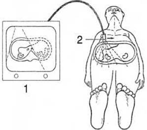
Fig. 16. THE PRINCIPLE OF sonography.
1 - ultrasound machine with a monitor; 2 - ultrasound transducer.
Piezoelectric ceramics, and more recently piezoelectric plastics and composites, are used to generate ultrasonic waves, converting electrical energy into mechanical energy. The shape of the ultrasonic beam is primarily determined by the size of the aperture from which it emerges. Electronic beam steering depends on the design of the ultrasound transducer (probe). The simplest and most common configuration is an array consisting of 100 adjacent transducer elements forming a group 50 mm long and 10 mm wide. To prevent ultrasonic wave interference, the generator operates in pulsed mode (approximately 1,000 pulses per second), alternating between pulse transmission and reception. Continuous-wave transmission and reception models are used in certain Doppler systems. Reflected waves are detected by the piezoelectric transducer, which converts mechanical vibrations into electrical impulses that are subsequently processed by a computer and transformed into an image (an echogram or ultrasound sonogram).
The main modalities of ultrasound Diagnostics are echography, sonography, and dopplerography.
Echography is a one-dimensional Ultrasound examination comprising the A-mode and M-mode techniques. In A-mode (amplitude mode), impulses reflected from individual Structure/83.html">Structural elements of an object form high-amplitude peaks along a straight baseline on the display. This allows measurement of the distances between various organ tissues, their depth, and the presence of foreign bodies or tumors. A-mode is utilized in ophthalmology and neurosonology. M-mode (motion mode) is used to examine moving Organs, particularly The Heart. In this technique, echoes reflected from the moving heart wall are recorded as a continuous curve. The shape and configuration of these curves provide insight into cardiac contraction patterns.

Fig. 17. Ultrasound scanner (A) and its various transducer shapes (B):
1 - standard (convex); 2 - linear; 3 - sector.
Sonography (ultrasonography), or B-mode (brightness mode), is based on two-dimensional scanning of organs and tissues using an ultrasound piezoelectric transducer moved across The surface of the region of interest (Fig. 16). The resulting image is displayed on a monitor or printed on paper as a pattern of bright dots. Bright white areas represent strong echoes reflected from echogenic structures (dense tissues, bones, calculi, etc.), whereas black areas indicate an absence of reflected signals, typical of parenchymal organs and fluids. Ultrasound transducers of various shapes are employed to examine different anatomical regions (Fig. 17).
Dopplerography is based on the Doppler effect—the shift in frequency of a reflected signal from a moving object. Dopplerography enables the detection of Blood flow and the calculation of blood velocity. Color-flow Doppler mapping facilitates the identification of Blood Vessels and abnormal flow patterns. Blood flowing toward the transducer is conventionally displayed in red, while blood flowing away is shown in blue. Color intensity increases with blood flow velocity. Microbubble-based contrast agents are used to enhance signal contrast.
Ultrasonography is one of the most widely used imaging modalities in clinical practice. While ultrasound scanners generally yield to computed tomography scanners in terms of diagnostic capability, the ultrasound Procedure itself is rapid, painless, and can be repeated as often as necessary. Ultrasound units occupy little space and require no radiation shielding. Ultrasound equipment is relatively inexpensive and cost-effective to operate. Ultrasound systems are widely utilized for examining both inpatients and outpatients presenting with various Pathologies of the abdominal and retroperitoneal organs, pelvic organs, Thyroid Gland, breasts, and other structures.
The primary advantage of ultrasound examination is that it does not involve ionizing radiation and produces no ionizing biological effects, making it entirely safe and exceptionally suitable for widespread use in pediatrics and obstetrics. Ultrasound has achieved its greatest impact in obstetrics. This modality allows for the prenatal detection of internal organ pathologies and developmental malformations, as well as the assessment of fetal status, biometry, and other critical diagnostic objectives throughout fetal development.

Fig. 18. The principle of radionuclide imaging.
1 - object of study (thyroid gland); 2 - gamma radiation; 3 - detector.
Last update: 08/08/2026
Editorial and Educational Adaptation: This material has been compiled based on the primary/original source text. The project team performed an editorial review, corrected technical inaccuracies, structured sections, and adapted the content for an educational format.
What was processed:
- elimination of formatting defects (OCR errors, structural breaks, corrupted characters);
- editorial organization of content;
- standardization of terminology in accordance with academic sources;
- verification of factual statements against the original source text.
All mentions of the author, publication year, and origin of the primary text have been preserved in accordance with the source.