Review of Medical Physiology - William F. Ganong 2002
Endocrine System, Metabolism, and Reproduction
Thyroid Gland
Clinical Correlations
The signs, symptoms, and complications of hypothyroidism and hyperthyroidism in humans, as predictable consequences of the Physiological effects of THYROID Hormones, are described above.
Hypothyroidism
The syndrome of hypothyroidism in adults is generally referred to as Myxedema, although this term is also used specifically to denote cutaneous changes associated with the syndrome. Hypothyroidism may be The ultimate outcome of various thyroid diseases or arise secondarily due to pituitary deficiency (pituitary hypothyroidism) or hypothalamic deficiency (hypothalamic hypothyroidism). Unlike the first condition, the latter two involve The Thyroid gland's response to a test dose of TSH. At least theoretically, hypothalamic hypothyroidism can be distinguished from pituitary hypothyroidism by the presence of an elevated plasma TSH level in response to a test dose of TRH. The TSH response to TRH is typically normal in hypothalamic hypothyroidism, whereas it is enhanced in hypothyroidism caused by primary thyroid disease and suppressed in hyperthyroidism due to negative feedback regulation by thyroid hormones on the pituitary.
In athyreotic individuals, the basal metabolic rate drops to around -40. The Hair becomes coarse and sparse, the Skin dry and yellowish (carotenemia), and cold intolerance is common. The voice becomes raspy and speech slows down—giving rise to the adage that myxedema is the only disease whose Diagnosis can be made over the telephone. Mental processes are sluggish, memory is impaired, and some patients develop severe psychiatric symptoms (myxedema madness). Plasma Cholesterol levels are elevated.
Cretinism
Children born with or developing hypothyroidism early in life are termed cretins. They exhibit dwarfism, mental retardation, a protuberant abdomen, and macroglossia (an enlarged Tongue that protrudes and cannot be contained within the Oral Cavity) (Fig. 18-14). Globally, congenital hypothyroidism is one of the most common preventable causes of mental retardation. The primary causes are summarized in Table 18-4. These include not only maternal iodine deficiency and various congenital Anomalies of the fetal Hypothalamus-pituitary-thyroid axis, but also maternal antithyroid Antibodies that cross the Placenta and damage the fetal thyroid gland. Because hormone T4 crosses the placenta, GROWTH AND DEVELOPMENT remain normal until birth provided the mother is euthyroid. If Treatment is initiated at birth, the prognosis for normal growth is favorable, and mental retardation can generally be avoided. For this reason, screening tests for congenital hypothyroidism are standard practice in most developed countries. However, if the mother is hypothyroid, particularly in settings of iodine deficiency, the neurological deficits are more complex and less responsive to postnatal treatment. In addition, these symptoms are compounded by deaf-mutism and rigidity (see above). Increased consumption of iodized salt has reduced the incidence of maternal iodine deficiency; nevertheless, this condition remains prevalent in many PARTS OF THE world, with an estimated 20 million people suffering from varying degrees of Brain Damage caused by intrauterine iodine deficiency.
Hyperthyroidism
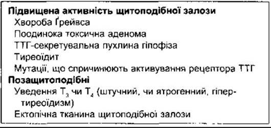
Hyperthyroidism (thyrotoxicosis) is characterized by nervousness, weight loss, hyperphagia, heat intolerance, increased pulse pressure, a fine tremor of outstretched fingers, warm and smooth skin, excessive sweating, and a basal metabolic rate ranging from +10 to as high as +100. The most common form of hyperthyroidism is Graves' disease (exophthalmic goiter), though it can be triggered by numerous other disorders, including rare TSH-secreting tumors of the anterior pituitary or unregulated activation of TSH receptors (Table 18-5). In Graves' disease, the thyroid gland is diffusely enlarged, and protrusion of the eyeballs, known as exophthalmos, is frequently observed (Fig. 18-15). This condition is an autoimmune disorder (see Chapter 27) in which circulating antibodies directed against the TSH receptor stimulate the receptor, leading to glandular hyperactivity. Four types of anti-thyroid antibodies have been identified. TSH receptor-stimulating antibodies (TSH-R [stim] Ab) are responsible for the Pathogenesis of Graves' disease. Other TSH receptor antibodies (TSH-R [block] Ab) exert an inhibitory rather than stimulatory effect. Additionally, there are antibodies against thyroglobulin (Tg Ab) and thyroid peroxidase (TPO Ab). Thyroglobulin antibody levels rise early in the disease, whereas in chronic autoimmune thyroiditis (Hashimoto's thyroiditis), anti-thyroid peroxidase levels become elevated at later stages. This condition involves cellular damage to the thyroid gland and may progress to hypothyroidism. In some cases of Hashimoto's thyroiditis, thyroid function is also suppressed by TSH-R [block] Ab. The exact mechanisms triggering The production of autoimmune anti-thyroid antibodies remain incompletely understood.
Class="center">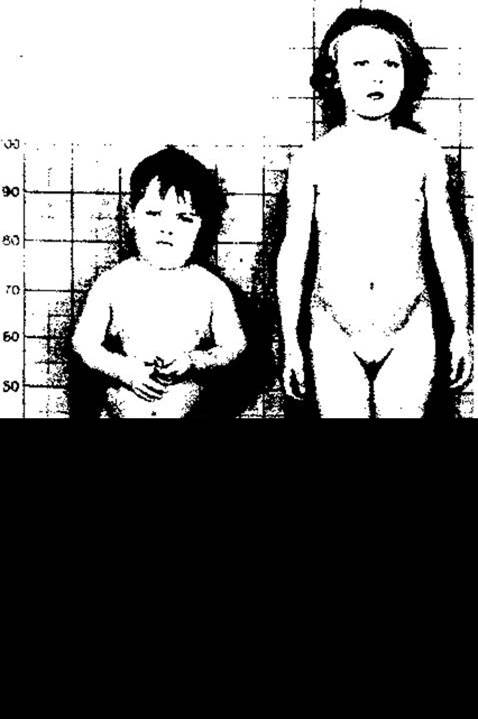
Fig. 18-14. Fraternal twins, age 8. The boy has congenital hypothyroidism (reproduced with permission from Wilkins L in Clinical Endocrinology I. Astwood EB, Cassidy CE [editors]. Grüne & Stratton, 1960).
Table 18-4. Causes of congenital hypothyroidism
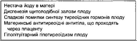
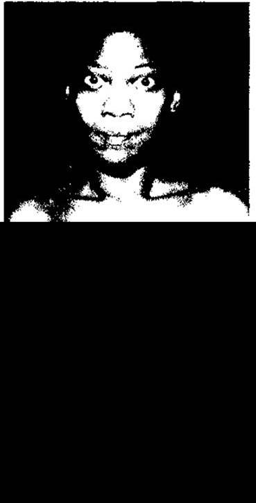
Fig. 18-15. Graves' disease. Note the goiter and exophthalmos (courtesy of PH Forsham).
In Graves' disease, thyroid hormone secretion is markedly stimulated, and elevated circulating levels of T4 and T3 suppress TSH secretion, resulting in decreased circulating TSH levels. High thyroid hormone levels tend to stimulate antibody production, whereas treatment-induced reductions in these levels aim to decrease, though not completely halt, antibody synthesis.
Table 18-5. Causes of hyperthyroidism
Iodine-deficiency goiter can reach massive proportions. Such "endemic goiter" has been recognized since antiquity. Before the widespread iodization of table salt, this condition was highly prevalent in Central and Eastern Europe and the Great Lakes region of the United States—areas known as "goiter belts," where heavy rainfall leached iodine from the soil, leaving local food supplies deficient in this element.
Radioactive Iodine Uptake
Iodine uptake serves as an index of thyroid activity and can be readily quantified using tracer doses of radioactive iodine isotopes that do not harm the thyroid gland. The tracer dose is administered orally, and its uptake by the thyroid is measured with a scintillation detector placed around the neck. Counts are also taken over the thigh region and subtracted from the neck counts to correct for extrathyroidal radioactivity in the neck. The most commonly used iodine isotope is 123I because its half-life is only 0.55 days, compared with 8.1 days for 131I and as long as 60 days for 125I. However, diagnostic Applications of radioactive iodine have become uncommon due to the routine availability of plasma T4, T3, and TSH assays. Furthermore, widespread use of iodized salt expands the iodide pool, leading to low fractional uptake. Nevertheless, radioactive iodine uptake testing remains valuable for understanding thyroid physiology. Normal uptake patterns are illustrated in Fig. 18-16. In hyperthyroidism, iodide is rapidly incorporated into T4 and T3, and these hormones are released at an accelerated rate. Consequently, thyroid radioactivity rises sharply, then plateaus and begins to decline by 24 hours, whereas normal uptake is still increasing at that time. In hypothyroidism, uptake is sluggish.
Large quantities of radioactive iodine destroy thyroid tissue through radiation-induced Cell death. Radioiodine therapy is useful for certain types of thyroid Cancer and can be utilized to treat benign tumors; however, particularly in young patients, the potential risks of radiation-induced carcinogenesis and germline Mutations must be carefully considered.
Radioactive iodine isotopes are major fission products. When these fission products are released into the atmosphere following a nuclear power plant accident or atomic bomb detonation, the isotopes disperse widely across distances because they are more volatile than other fallout components. Potassium iodide prophylaxis is routinely administered in radioactive fallout zones to expand the iodide pool and minimize thyroidal radioiodine uptake.
Antithyroid Drugs
Most drugs that suppress thyroid function act either by interfering with the iodine-trapping mechanism or by blocking the organic binding of iodine within the gland. In either case, the resulting decrease in circulating thyroid hormone concentrations stimulates TSH secretion, leading to goiter formation. Numerous monovalent anions compete with iodide for The Active Transport mechanism.
Exophthalmos in this condition results from edema of the extraocular Muscles and, to a lesser extent, the Connective Tissue within the rigid bony walls of the Orbit, which forces the Eyeball forward. Although typically associated with hyperthyroidism, thyroid hormones are not directly responsible for The Development of exophthalmos. This symptom may improve or resolve following Treatment of the underlying disease and can manifest before, during, or after the thyrotoxic state. In some thyrotoxic patients, exophthalmos continues to progress even after thyroidectomy. Furthermore, this sign is occasionally observed in euthyroid patients with Hashimoto's thyroiditis, patients with myxedema, and those with atypical thyroid disorders. It is now well established that exophthalmos stems from autoimmune injury to the extraocular muscles and orbital connective tissue mediated by cytotoxic antibodies directed against shared Antigens present in both eye muscles and the thyroid gland. This condition responds progressively to corticosteroids and other immunosuppressive agents.
Thyrotoxicosis places a significant burden on The Cardiovascular system, particularly in elderly individuals, in whom most or even all symptoms are cardiovascular. The incidence of atrial fibrillation increases and peripheral resistance decreases as a consequence of cutaneous vasodilation, resulting in marked Heart Failure in patients (see Chapter 33).
Resistance to Thyroid Hormones
Certain mutations in the Gene encoding human TRs are associated with resistance to the action of T3 and T4. More common is resistance to thyroid hormones in peripheral Tissues and the anterior pituitary. Patients with such anomalies are generally not clinically hypothyroid because they maintain plasma levels of T4 and T3 high enough to overcome the resistance, while TRs remain unchanged. Plasma TSH levels are inappropriately high for the elevated levels of circulating T3 and T4, and thus should be lowered using exogenous thyroid hormones. Some patients exhibit resistance to thyroid hormones restricted to the Pituitary Gland. They are characterized by an elevated metabolic rate and increased plasma levels of T3 and T4 with a normal, unsuppressible TSH level. Other patients may present with peripheral resistance coupled with normal pituitary sensitivity. They exhibit a lowered metabolic rate despite normal plasma levels of T3, T4, and TSH, and require large doses of thyroid hormones to increase their metabolic rate.
An interesting finding is that attention deficit hyperactivity disorder, a condition frequently observed in hyperactive and impulsive children, is much more prevalent among individuals with resistance to thyroid hormones than in the general population. This suggests that TRβ may play a specific role in brain development (see above).
Iodine Deficiency
When dietary iodine intake falls below 50 µg/day, the synthesis of thyroid hormones becomes insufficient, and their secretion declines. As a consequence of increased TSH secretion, thyroid hypertrophy develops, driven by enhanced transport into the thyroid gland, thereby suppressing uptake to the level where the T/S ratio reaches unity. Such a reduction in iodide transport can be overcome by ingesting supplementary iodide. These anions include chlorate, pertechnetate, periodate, biiodate, nitrate, and perchlorate. Thiocyanate—another monovalent anion—inhibits iodine transport, yet does not accumulate within the gland itself. The activity of perchlorate is ten times greater than that of thiocyanate.
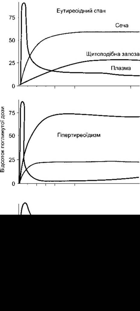
Fig. 18-16. Distribution of radioactive iodine in individuals with a relatively low dietary iodine intake. Percentages are shown as a function of time following the ingestion of a radioactive iodine dose. In hyperthyroidism, plasma radioactivity decreases rapidly and then increases again due to the release of labeled T4 and T3 by the thyroid gland.
Thiouylenes—a group of compounds structurally related to thiourea—inhibit the iodination of monoiodotyrosine (organification of iodine) and block coupling reactions. Two members of this group used clinically are propylthiouracil and methimazole (Fig. 18-17). Tyrosine iodination is inhibited because propylthiouracil and methimazole compete with tyrosine residues for iodine, undergoing iodination themselves. In addition, propylthiouracil, but not methimazole, inhibits 5-DI, thereby reducing The conversion of T4 to T3 in numerous extra-thyroidal tissues. Both drugs can also ameliorate thyrotoxicosis by suppressing The Immune System and, consequently, the production of TSH-R [stim] Ab. They may also inhibit the Biosynthesis or affect The Structure of thyroglobulin.
Propylthiouracil and methimazole affect coupling at doses lower than those affecting iodination. They do not block the iodine-concentrating mechanism. Due to increased TSH secretion, initial radioactive iodine uptake is actually enhanced during therapy, and the T/S ratio may reach 250. However, because the coupling process is inhibited, iodine does not accumulate; nevertheless, 24 hours after isotope administration, uptake normalizes. Another substance that inhibits thyroid function under certain conditions is iodine itself. The Role of iodine in thyroid physiology is unique in that while a certain amount of iodide is required for normal glandular function, both excessively low and excessively high amounts lead to abnormal thyroid function. Normally in humans, large doses of iodides act directly on the thyroid gland, resulting in a moderate and temporary inhibition of iodide organification and, consequently, hormone synthesis. This inhibition is known as the Wolff-Chaikoff effect. It is prolonged when iodide transport is enhanced, which explains why thyrotoxic patients are more sensitive to iodide than normal individuals. Susceptibility to this effect is also increased in the presence of a defect in the organification mechanism, and therefore inhibition is accentuated in individuals with a partially resected or ablated thyroid gland or with thyroiditis. There are at least two additional mechanisms by which excess I inhibits thyroid function. It diminishes the action of TSH on the gland by reducing the cAMP response to this hormone and inhibits thyroglobulin proteolysis. There is no direct effect on the concentration mechanism of I, but its overall uptake is low due to the inhibition of organification and, to a lesser extent, because the circulating iodide pool is so large that the administered tracer is immediately diluted. In thyrotoxicosis, iodides accumulate in the colloid, and the vascularity of the hyperplastic gland decreases, which plays an important role in preparing thyrotoxic patients for surgery.
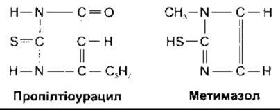
Fig. 18-17. Common thiouylenes.
Naturally Occurring Goitrogens
Thiocyanates are occasionally ingested with food, and certain foods contain relatively large amounts of naturally occurring goitrogenic substances. Cruciferous vegetables, particularly rutabagas, cabbages, and turnips, contain progoitrin—a substance converted into goitrin, an active antithyroid agent (Fig. 18-18). The plant activator of progoitrin is heat-labile, but since activators are also present in the gut (likely of bacterial origin), goitrin is produced even from cooked vegetables. The consumption of goitrin in a normal, balanced diet is insufficient to cause harm, yet vegetarians and individuals with unusual diets may develop "cabbage goiter." Other, yet unidentified plant goitrogens may be responsible for occasional small "goiter epidemics" reported from various parts of the world.
Use of Thyroid Hormones in Nonthyroidal Diseases
When the pituitary-thyroid axis is normal, doses of exogenous thyroid hormones do not markedly affect METABOLISM due to a compensatory decline in endogenous secretion resulting from suppressed TSH secretion. In euthyroid individuals, oral doses of T4 that suppress endogenous thyroid function can be estimated from the doses that normalize plasma TSH levels in athyreotic adults, which amount to 100–125 µg/day. Suppression of TSH secretion by exogenous T4 or pituitary disease eventually leads to thyroid atrophy. The atrophic gland initially responds sluggishly to TSH, and following prolonged suppression, some time is required for its normal responsiveness to return. The adrenal cortex and certain other Endocrine glands respond in a similar manner. When deprived of the support of their tropic hormones for some time, they become atrophic and are capable of only a weak response to their tropic hormones until the hormone acts upon the gland again.
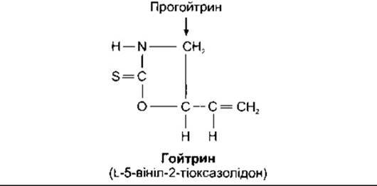
Fig. 18-18. Naturally occurring goitrogen in cruciferous vegetables.
The Use of thyroid hormones for weight reduction is valuable only if the patient is willing to pay the price of increased nervousness and heat intolerance. Furthermore, appetite must be restrained to prevent a compensatory increase in food intake.
Last update: 10/08/2026
Editorial and Educational Adaptation: This material has been compiled based on the primary/original source text. The project team performed an editorial review, corrected technical inaccuracies, structured sections, and adapted the content for an educational format.
What was processed:
- elimination of formatting defects (OCR errors, structural breaks, corrupted characters);
- editorial organization of content;
- standardization of terminology in accordance with academic sources;
- verification of factual statements against the original source text.
All mentions of the author, publication year, and origin of the primary text have been preserved in accordance with the source.