Human Anatomy - Kotsan I. Ya. 2009
Sense Organs
Organ of Vision (Eye)
Eyeball
The eyeball (bulbus oculi) is located within the orbital cavity and has a spherical shape, slightly flattened in the anteroposterior direction. Its anterior surface is more convex than the posterior. The eyeball features anterior and posterior poles.
The anterior pole (polus anterior) is the most anteriorly protruding point on the anterior surface of the eyeball, located at the center of the anterior surface of the cornea at its apex.
The posterior pole (polus posterior) is the most posteriorly protruding point on the back of the eyeball, situated slightly lateral to the exit point of the Optic nerve. The line connecting the anterior and posterior poles is called the optical or external axis of the eyeball (axis bulbi externus). It averages 24 mm in length (Fig. 266). The segment of this axis located between the posterior surface of the cornea and the retina is called the internal axis of the eyeball (axis bulbi internus). The length of the internal axis averages 21.5 mm. If the internal axis is longer, light rays, after refraction in the eyeball, are focused in front of the retina. In this case, clear Vision is possible only at close range (myopia or nearsightedness). The focal length in nearsighted individuals is shorter than the internal axis of the eyeball.
Class="center">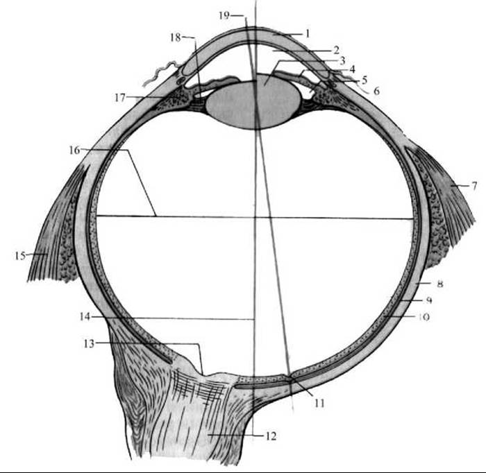
Fig. 266. Horizontal section of the eyeball (diagram).
Showing The change in the curvature of the lens during contraction and relaxation of the ciliary Muscle (after M. R. Sapin)
1 — cornea; 2 — anterior chamber of the eyeball; 3 — lens; 4 — iris; 5 — posterior chamber of the eyeball; 6 — conjunctiva; 7 — lateral rectus muscle; 8 — sclera; 9 — choroid; 10 — retina; 11 — fovea centralis; 12 — optic nerve; 13 — optic cup; 14 — external axis of the eyeball; 15 — medial rectus muscle; 16 — transverse axis of the eyeball; 17 — ciliary body; 18 — zonule of Zinn (suspensory ligament); 19 — visual axis
If the internal axis of the eyeball is relatively short, light rays after refraction are focused behind the retina. In this case, distance vision is better than near vision (hyperopia or farsightedness). The focal length in farsighted individuals is longer than the internal axis of the eyeball.
In addition to the external and internal axes of the eyeball, there is also the so-called visual axis (axis opticus), which extends from the observed object to the central fovea of the retina (the area of sharpest vision). Passing through the central points of the cornea and the lens, the visual axis intersects the internal axis of the eyeball at an acute angle.
Lines connecting the anterior and posterior poles along the circumference of the eyeball form meridians (meridiani), while a plane perpendicular to the optical (external) axis forms the equator (equator), which divides the eyeball into anterior and posterior halves. The horizontal diameter of the equator is slightly shorter than the external axis of the eyeball (23.6 mm), and its vertical diameter is even smaller (23.3 mm).
The eyeball consists of a capsule and a core. The capsule of the eyeball is formed by three tunics: an outer fibrous tunic, a middle vascular tunic, and an inner tunic—the retina. The core makes up the bulk of the eyeball. It is represented by the lens, the vitreous body, and the aqueous humor, which fills the anterior and posterior Chambers of the eye.
Last update: 08/08/2026
Editorial and Educational Adaptation: This material has been compiled based on the primary/original source text. The project team performed an editorial review, corrected technical inaccuracies, structured sections, and adapted the content for an educational format.
What was processed:
- elimination of formatting defects (OCR errors, structural breaks, corrupted characters);
- editorial organization of content;
- standardization of terminology in accordance with academic sources;
- verification of factual statements against the original source text.
All mentions of the author, publication year, and origin of the primary text have been preserved in accordance with the source.