Human Anatomy - Kotsan I. Y. 2009
Sense Organs
The Organ of Vision (Eye)
In the course of evolution, the visual system has traversed a complex pathway from light-sensitive Cells on the body surface of primitive animals to a sophisticated organ capable of capturing a light beam and focusing it onto specialized photoreceptor cells that perceive both monochrome and color images. Having achieved remarkable perfection, the human visual organ reproduces images of the external world by transforming light stimuli into nerve impulses, the higher-level analysis of which is performed in the Cerebral Cortex. The Organ of Vision (organum visus), or the eye (oculus, Greek ophthalmos), consists of the Eyeball and accessory structures (Fig. 265).
Class="center">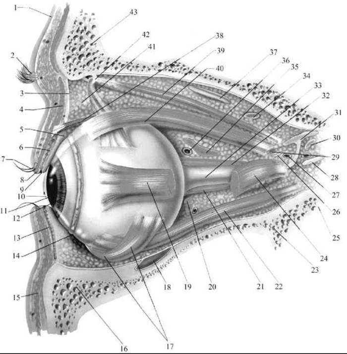
Fig. 265. The Organ of vision (lateral view)
1 — Skin; 2 — eyebrow; 3 — orbital septum; 4 — orbicularis oculi Muscle; 5 — superior conjunctival fornix; 6 — meibomian gland; 7 — eyelash; 8 — gland of Zeiss; 9 — duct of meibomian gland; 10 — cornea; 11 — pupil; 12 — iris; 13 — inferior tarsal Cartilage; 14 — inferior conjunctival fornix; 15 — orbicularis oculi muscle; 16 — Maxilla; 17 — inferior oblique muscle; 18 — infraorbital nerve; 19 — lateral rectus muscle; 20 — posterior ciliary artery; 21 — medial rectus muscle; 22 — inferior rectus muscle; 23 — Sphenoid bone; 24 — lateral rectus muscle; 25 — Palatine bone; 26 — nasociliary nerve; 27 — ophthalmic venous plexus; 28 — lateral branch of Oculomotor nerve; 29 — ophthalmic artery; 30 — lacrimal nerve; 31 — ROOT of oculomotor nerve; 32 — long ciliary muscle; 33 — Optic nerve; 34 — Trochlear nerve; 35 — orbital artery; 36 — orbital vein; 37 — Facial Nerve; 38 — levator palpebrae superioris muscle; 39 — superior oblique muscle; 40 — superior rectus muscle; 41 — adipose tissue; 42 — trochlea; 43 — Frontal bone
Last update: 08/08/2026
Editorial and Educational Adaptation: This material has been compiled based on the primary/original source text. The project team performed an editorial review, corrected technical inaccuracies, structured sections, and adapted the content for an educational format.
What was processed:
- elimination of formatting defects (OCR errors, structural breaks, corrupted characters);
- editorial organization of content;
- standardization of terminology in accordance with academic sources;
- verification of factual statements against the original source text.
All mentions of the author, publication year, and origin of the primary text have been preserved in accordance with the source.