Special Histology and Embryology: Practical Course - V. K. Naphpanyuk 2001
Sense Organs
Organ of Vision
The Eyeball (oculus) is the peripheral part of the visual analyzer, which consists of the eyeball (bulbus oculi) containing photoreceptor Cells and connected to the Brain via the Optic nerve, along with accessory structures including the eyelids, Lacrimal Apparatus, and extraocular striated Muscles.
The eyeball is formed by three tunics: 1) the fibrous tunic, consisting of the sclera and cornea; 2) the vascular tunic with its components: the choroid proper, the ciliary body, and the iris; 3) the internal (sensory) tunic of the eyeball — the retina.
Three main functional systems are distinguished within the eyeball:
1) the dioptric, or light-refracting system (cornea, aqueous humor of the anterior and posterior chambers, lens, vitreous body);
2) the accommodation and adaptation system (iris, ciliary body with the suspensory ligament);
3) the receptor, light-perceiving system (retina).
Slides for Study
Slide 1. Angle of the eye. Cytology/practical/75.html">Meridional section of the anterior segment of the eyeball (Fig. 5).
Low power. Examine and sketch the slide. Identify the anterior and posterior hemispheres of the eye.
The anterior hemisphere of the eye includes the lens (lens), the posterior and anterior Chambers of the eye filled with aqueous humor.
The posterior hemisphere of the eye, or fundus, contains the vitreous body (corpus vitreum).
In the region of the fundus, the optic nerve (nervus opticus) emerges from the eyeball.
The sclera (sclera) is formed by dense Fibrous Connective Tissue, consisting of Collagen and elastic fibers interspersed with fibrocytes and melanocytes. In the anterior part of the fibrous tunic, the sclera transitions into the cornea. The thickness of the sclera is 0.3-0.4 mm posteriorly and 0.6 mm near the cornea.
The cornea (cornea). The anterior surface of the cornea is covered by stratified squamous non-keratinized epithelium, which continuous with the conjunctival epithelium. The posterior surface of the cornea is covered by simple squamous epithelium, which continuous with the epithelium of the anterior surface of the iris.
The stratified squamous non-keratinized epithelium of the anterior surface of the cornea rests on the anterior limiting lamina (lamina limitans anterior), while the simple squamous epithelium of the posterior part rests on the posterior limiting lamina (lamina limitans posterior), which appears as a thick homogeneous layer (10 µm). Between the limiting laminae lies the proper substance of the cornea (substantia propria), which consists of collagen fibers (micrograph 2).
Class="center">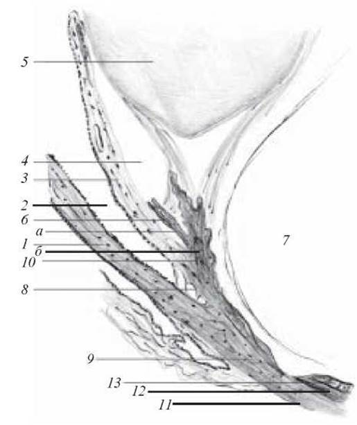
Fig. 5. Angle of the eye. Hematoxylin-eosin staining. X 56:
1 — cornea; 2 — anterior chamber of the eyeball; 3 — iris; 4 — posterior chamber of the eyeball; 5 — lens; 6 — ciliary zonule (zonule of Zinn); 7 — vitreous body; 8 — pectinate ligament; 9 — venous sinus of the sclera; 10 — ciliary body (a — ciliary processes; b — ciliary Muscle); 11 — sclera; 12 — choroid; 13 — retina
At the corneoscleral junction (limbus, limbus), the posterior limiting lamina breaks up into the trabecular meshwork (reticulum trabeculare). The spaces between the trabeculae are lined with simple squamous epithelium. The venous sinus (canal of Schlemm) is located in the limbus region. On the inner side, the choroid adheres to the sclera, recognized by its black coloration due to the presence of abundant pigment.
The vascular tunic (tunica vasculosa bulbi). It is formed by the choroid proper (chorioidea), ciliary body (corpus ciliare), and iris (iris).
The choroid proper provides Nutrition to the retina and, from superficial to deep, comprises the following layers:
1. Suprachoroidal layer (lamina suprachorioidea) is formed by loose fibrous connective tissue. It contains numerous elastic fibers, fibroblasts, and pigment cells (melanocytes).
2. Vascular layer (lamina vasculosa) consists of intertwined Arteries and Veins. Between them lies loose fibrous connective tissue containing A large number of pigment cells. Individual bundles of smooth myocytes are also located here.
3. Choriocapillary layer (lamina choriocapillaris) is formed by Blood capillaries of various calibers, some of which belong to sinusoids. Flattened fibroblasts are situated between the capillaries.
4. Basal complex (complexus basalis) is a very thin strip (1-4 µm) located between the choroid and the pigmented layer of the retina, consisting of three layers: a) outer elastic layer; b) inner fibrous layer; c) basal lamina.
At its anterior part, the vascular tunic proper continues into the ciliary body.
The ciliary body (corpus ciliare) is divided into two parts: a) the internal part — the ciliary crown (corona ciliaris); b) the external part — the ciliary ring (orbiculus ciliaris). Ciliary processes (procesus ciliares), to which the fibers of the ciliary zonule attach, extend from The surface of the ciliary crown toward the lens. The main part of the ciliary body, with the exception of the processes, is formed by the ciliary muscle (m. ciliaris). When the ciliary muscle contracts, the ciliary zonule relaxes, the lens takes on a more convex shape, and the eye accommodates for closer Vision.
The iris (iris) is a pigmented, disc-shaped Structure with a variable-sized aperture in the center — the pupil. The pupil can dilate or constrict due to the presence of the pupillary dilator muscle (m. dilatator pupillae) and the pupillary sphincter muscle (m. sphincter pupillae). Five layers are distinguished in the iris: a) the anterior epithelium (epithelium anterius iridis), which covers the anterior surface of the iris; b) the external limiting (avascular) layer (stratum externum limitans); c) the vascular layer (stratum vasculosum); d) the internal limiting layer (stratum internum limitans); e) the posterior pigmented epithelium (epithelium posterior pigmentosum).
The lens (lens) consists of lens fibers (fibra lentis), anterior to which lies a single-layered lens epithelium (epithelium lentis). Externally, the lens is surrounded by a capsule (capsula lentis), which is a thickened basement membrane.
Label the following on the diagram: 1) iridocorneal angle (angulus iridocornealis); 2) cornea (cornea); 3) sclera (sclera); 4) limbus (limbus); 5) ciliary body (corpus ciliare); 6) ciliary muscle (m. ciliaris); 7) ciliary body processes (procesus ciliaris); 8) venous sinus of the sclera (Schlemm's canal) (sinus venosus sclerae); 9) lens (lens); 10) anterior chamber (camera anterior); 11) posterior chamber (camera posterior).
Slide 2. Cornea of the eye (Fig. 6).
Low magnification. Position the slide so that the anterior epithelium of the cornea is at the top.
High magnification. Examine the layers of the cornea: beneath the anterior epithelium lie the anterior limiting lamina, the proper substance (stroma), the posterior limiting lamina, and the posterior epithelium (simple squamous). Note the absence of Blood Vessels in the cornea.
Label the following on the diagram: 1) anterior (stratified squamous) epithelium of the cornea; 2) anterior limiting lamina; 3) proper substance of the cornea (stroma); 4) posterior limiting lamina; 5) endothelium of the anterior chamber (simple squamous epithelium).
The retina (retina) is the inner tunic of the eye, the larger part of which (pars optica retinae) is light-sensitive and contains photoreceptor cells.
A smaller part covers the inner side of the ciliary body (pars ciliaris retinae) and the posterior surface of the iris (pars iridica retinae), which is devoid of photoreceptors.
The retina consists of three radially arranged Neurons (external — photoreceptor, middle — associative, and internal — ganglionic) and Two Types of neurons integrated into radial chains: at the level of contact between the First and Second neurons (horizontal neurons), as well as at the junction of the second and third neurons (amacrine neurons).

Fig. 6. Cornea of the eye. Hematoxylin-eosin staining. x 200:
1 — anterior epithelium of the cornea; 2 — anterior limiting lamina; 3 — proper substance of the cornea; 4 — posterior limiting lamina; 5 — endothelium
The neuronal Cell bodies and their processes form the layers of the retina. The photosensory layer (stratum photosensorium) is formed by rods and cones — specialized dendrites of rod and cone neurosensory cells (micrograph 3). The outer nuclear layer (stratum nucleare externum) consists of The Cell bodies of the neurosensory cells. The outer plexiform layer (stratum reticulare externum) is formed by the axons of the neurosensory cells and their synapses with the dendrites of neurons in the subsequent inner nuclear layer. The inner nuclear layer (stratum nucleare internum) comprises the cell bodies of bipolar, horizontal, and amacrine neurons. The inner plexiform layer (stratum reticulare internum) consists of the axons of bipolar neurons and their synapses with the dendrites of multipolar neurons. The cell bodies of multipolar neurons form the ganglionic layer (stratum ganglionare), and their axons form the nerve fiber layer (stratum neurofibrarum). Coming together, these axons form the optic nerve. The exit point of the optic nerve is called the blind spot because it contains no light-sensitive elements. The cell bodies of radial glial cells (gliocyti radiales) are also located in the inner nuclear layer, and their processes form the outer limiting membrane (stratum limitans externum). Situated between the photosensory and outer nuclear layers, the inner limiting membrane covers the retina from the inside.
Rods and cones possess an outer segment (segmentum externum), consisting of membrane discs (discus membranaceus), and an inner segment (segmentum internum), which contains the Endoplasmic reticulum and Mitochondria. The inner and outer segments are connected by a cilium (cilium). A cone differs from a rod in the shape of its outer segment and the attachment of some of its membrane discs to the Plasmalemma.
The central fovea of the retina is the site of highest visual acuity because, in the region of the fovea, all retinal layers except the neurosensory layer are displaced to the side, allowing light to strike the rods and cones without scattering.
Slide 3. Human retina (Fig. 7).
The structure of the retina should be studied under high magnification.
The lowermost pigmented layer is the retinal pigment epithelium. It is followed by the pink-colored photosensory layer. Next lies a layer containing numerous nuclei — the outer nuclear layer. Between it and the inner nuclear layer is the pink outer plexiform layer, and deeper than the inner nuclear layer lies the inner plexiform layer. Beyond the inner plexiform layer is the ganglionic layer, which consists of large multipolar neurons. Further on is the nerve fiber layer, on the inner surface of which lies the inner limiting membrane. The outer limiting membrane is located between the layer of rods and cones and the outer nuclear layer.
Label the following on the diagram: 1) inner glial limiting lamina; 2) nerve fiber layer; 3) ganglion cell layer; 4) inner plexiform layer; 5) inner nuclear layer; 6) outer plexiform layer; 7) outer nuclear layer; 8) outer glial limiting lamina; 9) layer of rods and cones; 10) layer of pigmented cells; 11) processes of pigmented cells.

Fig. 7. Human retina. Hematoxylin-eosin staining. x 400:
1 — inner glial limiting lamina; 2 — nerve cell layer; 3 — ganglion cell layer; 4 — inner plexiform layer; 5 — inner nuclear layer; 6 — outer plexiform layer; 7 — outer nuclear layer; 8 — outer glial limiting lamina; 9 — layer of rods and cones; 10 — layer of pigmented cells; 11 — processes of pigmented cells

Electron micrograph 2. Proper substance of the cornea. х 18,500:
1 — fibroblast; 2 — collagen protofibrils sectioned longitudinally and transversely (from Rodin's atlas)

Electron micrograph 3. Outer segment of a retinal cone cell. х 100,000:
1 — cell membrane; 2 — intermediate junctions of individual membrane "packets" of the outer segment; 3 — closed ends of membrane structures; 4 — open ends of membrane structures (sites of cell membrane invagination) (after V. L. Borovjagin)
Control Questions
1. Sense Organs. General characteristics. Classification.
2. Developmental sources of the main Structural components of the eyeball.
3. Main Tunics of the eye, structural features.
4. Principal functional apparatuses of the eye.
5. Cornea: structure, histophysiology.
6. STRUCTURE OF THE lens. Function of the vitreous body.
7. Structure and function of the iris.
8. Ciliary body, its structure and function.
9. Features of the ultrastructure of retinal neurosensory cells, its neuronal composition.
10. Microscopic and submicroscopic structure of the retina.
11. Morphology and function of the retinal pigment epithelium.
12. Accessory structures of the eye.
13. Structure of the choroid.
14. Structural Features of the sclera.
Situational Problems
1. Is corneal epithelium regeneration possible in case of injury? If so, at the expense of which cells?
2. In The Development of "night blindness" (impaired twilight vision), the function of which cells is disrupted, and What is the underlying mechanism?
3. Which analyzer is damaged in a person with an injury to the occipital region of the HEAD?
Sample Examination Questions
1. Sources of Development of the main structural Components of the eyeball.
2. Structural features of the main membranes of the eye.
3. Neuronal COMPOSITION OF THE retina, and features of the ultramicroscopic structure of retinal neurosensory cells.
4. Functional apparatus of the eye.
Last update: 10/08/2026
Editorial and Educational Adaptation: This material has been compiled based on the primary/original source text. The project team performed an editorial review, corrected technical inaccuracies, structured sections, and adapted the content for an educational format.
What was processed:
- elimination of formatting defects (OCR errors, structural breaks, corrupted characters);
- editorial organization of content;
- standardization of terminology in accordance with academic sources;
- verification of factual statements against the original source text.
All mentions of the author, publication year, and origin of the primary text have been preserved in accordance with the source.