Biological Membranes - A. N. Ogurtsov 2012
Biomembrane Electrogenesis
Mechanisms of Action Potential Generation
Neurons
Neurons generate and propagate action potentials, which are processes driven by the coordinated opening and closing of specific voltage-Gated Ion Channels along the neuron.
Although the Morphology of Different types of neurons varies considerably, all of them feature four functionally distinct parts (Figure 123):
1) Cell body (soma or perikaryon);
2) axon;
3) axon terminals;
4) dendrites.
Class="center">
Figure 123 - Schematic diagram of the main morphological components of mammalian neurons: a - multipolar interneuron; b - motor neuron
The Cell body contains The Nucleus and serves as the site of synthesis for virtually all Proteins and membrane Components of the neuron. While some proteins are synthesized in dendrites, Protein Synthesis does not occur in axons and axon terminals.
A specific transport process—in which motor proteins such as dyneins and kinesins move essential cargo (proteins and membrane components) along the cytoskeletal microtubules located inside the axon—ensures the delivery of these "cargoes" from their synthesis site in the cell body to their destination in the axon and axon terminals.
Axons, ranging in diameter from 1 µm in human Brain neurons to 1 mm in the giant squid axon, are specialized for transmitting nerve impulses (propagating action potentials).
As shown above (Section 12.1), an Action Potential consists of a series of sharp Membrane Potential changes (spikes) shifting from approximately -60 mV (Resting Potential) to around +50 mV (a total swing of 110 mV). This membrane depolarization is followed by rapid repolarization, which restores the membrane potential back to its resting value (Figure 124).
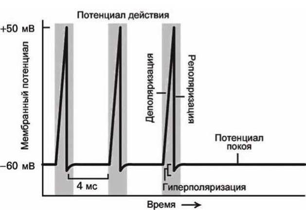
Figure 124 - Action potential waveforms
The action potential is generated at the axon hillock, the region where the axon connects to the cell body, and propagates along the axon toward the axon terminals—the branching structures at the end of the axon that form synapses connecting neurons to other Cells.
Action potentials propagate at an average speed of 100 m/s. Since human axons can reach a length of one meter, the propagation of a Nerve Impulse takes only a few milliseconds.
When an action potential reaches a synapse at the axon terminal of a presynaptic cell, voltage-gated calcium channels open, Ca2+ ions enter the terminal, and the local calcium concentration in the Cytosol of the presynaptic axon spikes.
This rise in calcium ion concentration triggers regulated exocytosis: synaptic vesicles containing Neurotransmitters fuse with the axonal membrane at the synaptic cleft, releasing neurotransmitters into the cleft (Figure 125). The regulation of neurotransmitter exocytosis will be discussed in more detail below in Section 16.3.
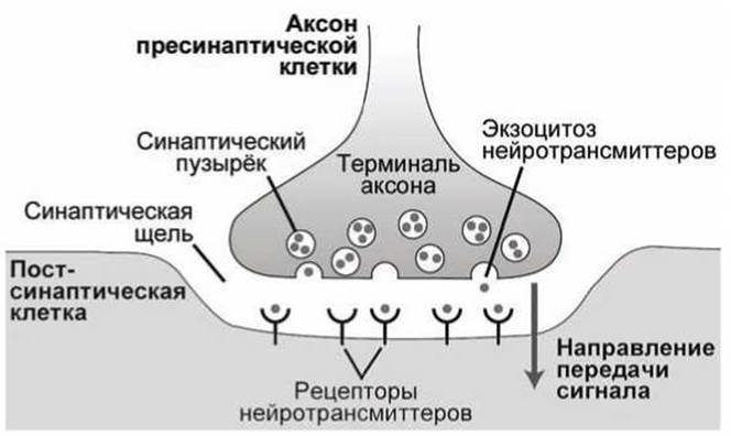
Figure 125 - Structure of a chemical synapse
Within approximately 0.5 ms, neurotransmitters diffuse across the synaptic cleft and bind to receptors on the postsynaptic membrane, altering the membrane potential at that specific synaptic site. A single Central Nervous system axon can form synapses with numerous other neurons, transmitting nerve impulses to all of them simultaneously.
Most neurons possess multiple dendrites that branch out from the cell body and specialize in receiving chemical signals from the axons of other neurons. Dendrites convert these signals into electrical impulses and deliver them to the cell body.
The cell body itself can also form synapses and, consequently, receive signals directly. Central nervous system neurons, in particular, feature long dendrites with complex branching structures. This allows them to form synapses with thousands of other neurons and receive signals from them accordingly (Figure 123a).
Depolarization or hyperpolarization generated in the dendrites spreads toward the axon hillock. If the amplitude of membrane depolarization at the axon hillock is sufficiently large, an action potential (nerve impulse) will be triggered and subsequently propagated along the axon.
Last update: 13/08/2026
Editorial and Educational Adaptation: This material has been compiled based on the primary/original source text. The project team performed an editorial review, corrected technical inaccuracies, structured sections, and adapted the content for an educational format.
What was processed:
- elimination of formatting defects (OCR errors, structural breaks, corrupted characters);
- editorial organization of content;
- standardization of terminology in accordance with academic sources;
- verification of factual statements against the original source text.
All mentions of the author, publication year, and origin of the primary text have been preserved in accordance with the source.