LEHNINGER PRINCIPLES OF BIOCHEMISTRY - VOL. 1. THE FOUNDATIONS OF BIOCHEMISTRY: STRUCTURE AND CATALYSIS - 2011
PART I. STRUCTURE AND CATALYSIS
12. BIOSIGNALING
12.6. Ion Channels
Ion Channels play a central role in electrical signaling in excitable Cells
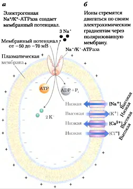
Certain cells of Multicellular Organisms are "excitable": they can detect an external signal, convert it into an electrical signal (specifically, altering the Membrane Potential), and propagate it further. Excitable cells play key roles in nerve conduction, Muscle contraction, hormone secretion, sensory perception, learning, and memory. The excitability of sensory cells, Neurons, and myocytes relies on signal transducers—ion channels that, in response to diverse stimuli, provide a regulated pathway across Cell/30.html">The Plasma Membrane for the movement of inorganic ions such as Nа+, К+, Са2+, and Сl-. Ion channels are gated (Chapter 11); they can open or close depending on whether the corresponding receptor is activated by binding to a specific Ligand (e.g., a neurotransmitter) or by A change in the transmembrane electrical potential Vm. The Na+/К+-ATPase creates a charge imbalance across the plasma membrane by pumping 3Nа+ out of The Cell for every 2К+ brought in (Fig. 12-24, a), making the inside negative relative to the outside. The membrane is then said to be polarized.
Class="center">Figure 12-24. Transmembrane electrical potential. (a) The electrogenic Na+/К+-ATPase establishes a transmembrane electrical potential of -60 mV (negative inside). (b) Blue arrows indicate the direction in which ions tend to move across an animal cell plasma membrane under the combined influence of chemical and electrical gradients. The chemical gradient drives Nа+ and Са2+ inward (leading to membrane depolarization) and К+ outward (leading to hyperpolarization). The electrical gradient drives Сl- outward, against its concentration gradient (causing depolarization).
Key Conventions.
Vm is negative when the charge inside the cell is negative relative to the charge outside. For a typical animal cell, Vm ranges from -60 to -70 mV. ■
Because ion channels are generally selective for either anions or cations, but not both, ion flow through a channel causes a redistribution of charge across the Two Sides of the membrane, thereby altering Vm. The influx of positively charged ions, such as Nа+, or the efflux of negatively charged ions (Сl-) depolarizes the membrane, causing Vm to approach zero. Conversely, the efflux of К+ hyperpolarizes the membrane, making Vm more negative. These ion fluxes through channels are passive, in contrast to The Active Transport mediated by the Na+/К+-ATPase.
The direction of spontaneous ion flow across a polarized membrane is determined by the Electrochemical Potential of that ion across the membrane, which consists of two components: the concentration difference (C) of the given ion across the membrane and the electrical potential difference (typically expressed in millivolts). The energy (∆G) that drives the spontaneous movement of a cation (say, Nа+) through an ion channel into the cell is a function of The ratio of the ion concentrations on the two sides of the membrane (Cinside/Coutside) and the electrical potential difference (∆Ψ or Vm):
∆G = RTIn (Сinside/Сoutside) + ZFVm (12-1)
where R is the gas constant, T is the absolute Temperature, Z is the charge of the ion, and F is the Faraday constant. Note that the sign of the ion's charge determines the sign of the second term in Equation 12-1. In a typical neuron or myocyte, the cytosolic concentrations of Nа+, К+, Са2+, and Сl- differ markedly from their concentrations in the extracellular fluid (Table 12-6). For these concentrations and a Resting Potential of -60 mV, Equation 12-1 predicts that the opening of a Na+ or Са2+ channel will result in the spontaneous influx of Nа+ (and membrane depolarization), whereas the opening of a K+ channel will lead to the spontaneous efflux of К+ (and membrane hyperpolarization) (Fig. 12-24, b).
Table 12-6. Ion Concentrations in Cells and Extracellular Fluids (mM)
К+ |
Na+ |
Са2+ |
Сl- |
|
Cell Type |
Inside Outside |
Inside Outside |
Inside Outside |
Inside Outside |
Squid axon |
100 20 |
50 440 |
≤0.4 10 |
40-150 560 |
Frog muscle |
124 2.3 |
10.4 100 |
<0.1 2.1 |
1.5 78 |
These ions continue to move through the channel only as long as the combination of the concentration gradient and the electrical potential provides a driving force, according to Equation 12-1. For example, as Nа+ moves down its concentration gradient, it depolarizes the membrane. When the membrane potential reaches +70 mV, The Effect of this membrane potential (resisting further Nа+ influx) exactly balances the effect of the concentration gradient (driving Nа+ inward). At this equilibrium potential (E), the net driving force (∆G) is zero. The equilibrium potential is unique to each type of ion because their concentration gradients differ.
The number of ions that must move to noticeably alter the membrane potential is negligibly small compared with the concentrations of Na+, К+, and Сl- inside and outside the cell; consequently, the ion fluxes that occur during signaling in excitable cells have essentially no effect on the bulk concentrations of these ions. However, because the intracellular concentration of Са2+ is normally very low (~10-7 M), a Ca2+ influx can significantly alter the cytosolic calcium ion concentration.
At any given moment, a cell's membrane potential is the net result of how many channels of which type are open. In most cells at rest, more K+ channels are open than Na+, Cl-, or Ca2+ channels, and thus the resting potential is closer to E for K+ (-98 mV) than for any other ion. When Na+, Cl-, or Ca2+ channels open, the membrane potential shifts toward E for that ion. The time-regulated opening and closing of ion channels, and the resulting transient Changes in membrane potential, form The basis of electrical signaling by which The Nervous system stimulates Skeletal Muscle contraction, heartbeat, or secretory cell release of signal molecules. Furthermore, many Hormones exert their effects by altering membrane potentials in target cells. These mechanisms are not restricted to animal animals; ion channels play vital roles in environmental response in Bacteria, protists, and plants.
To illustrate the action of ion channels in cell-to-cell signaling, we will describe the mechanisms by which a neuron propagates an impulse along its length and across a synapse to the next neuron (or myocyte), using acetylcholine as the neurotransmitter.
Voltage-Gated Ion Channels Create Action Potentials in Neurons
In the nervous system, signaling is carried out by a network of neurons—specialized cells that conduct an electrical impulse (Action Potential) from one end of the cell (the cell body) along an elongated cytoplasmic extension (the axon). The electrical signal triggers the release of neurotransmitter molecules at the synapse, passing the signal to the next cell in the circuit. Three types of voltage-gated ion channels are essential for this signaling mechanism. Running the length of the axon are voltage-gated Na+ channels (Fig. 12-25), which are closed when the membrane is at rest (Vm = -60 mV) but open briefly when the membrane is locally depolarized in response to acetylcholine (or another neurotransmitter). Also situated along the axon are voltage-gated K+ channels, which open slightly later than the Na+ channels in response to the depolarization induced by the opening of the Na+ channels. Thus, the depolarizing influx of sodium ions sweeping down the axon is rapidly followed by a repolarizing efflux of potassium ions. At the remote (distal) end of the axon are voltage-gated Ca2+ channels. When the waves of depolarization and repolarization generated by the Na+ and K+ channels reach these terminals, the Ca2+ channels open, allowing Ca2+ to flow in from the extracellular space and trigger the release of acetylcholine, which transmits the signal to the next nerve cell (firing an action potential!) or muscle (contracting!).
Voltage-gated Na+ channels are highly selective for sodium ions over other cations (100-fold or greater) and support a very high throughput rate (>107 ions/s). Once opened by activation, Na+ channels rapidly undergo inactivation driven by the membrane depolarization—the channels close within a millisecond and remain refractory for many milliseconds. Because K+ channels open in response to the depolarization caused by the opening of Na+ channels, the resulting potassium efflux locally repolarizes the membrane. A wave of depolarization sweeps along the axon as local depolarization triggers the sequential opening of nearby Na+ channels and then K+ channels (Fig. 12-25). The opening of each Na+ channel is followed by a brief refractory period during which the channel cannot reopen; this ensures the unidirectional Propagation of the action potential from the neuron cell body toward the axon terminal (step (1) in Fig. 12-25).
Figure 12-25. The roles of voltage-gated and ligand-gated ion channels in Nerve Impulse transmission. Initially, the plasma membrane of the presynaptic neuron is polarized (interior negative) through the action of the electrogenic Na+/K+-ATPase, which pumps 3 Na+ ions out for every 2 K+ ions pumped in (see Fig. 12-24). (1) A stimulus acting on the neuron initiates an action potential propagating along the axon (white arrow) away from the cell body. The opening of a single voltage-gated Na+ channel permits Na+ influx; the resulting local depolarization triggers the opening of an adjacent Na+ channel, and so on. The unidirectional travel of the action potential is ensured by the brief refractory period following the opening of each voltage-gated Na+ channel. (2) When the wave of depolarization reaches the axon terminal, voltage-gated Ca2+ channels open, allowing Ca2+ to enter the presynaptic neuron. (3) This raises the intracellular Ca2+ concentration, triggering the exocytotic release of the neurotransmitter acetylcholine into the synaptic cleft. (4) Acetylcholine binds to a receptor on the postsynaptic neuron, causing an acetylcholine-gated ion channel to open. (5) Extracellular Na+ and Ca2+ flow through this channel, depolarizing the postsynaptic cell. The electrical signal thus reaches the postsynaptic cell body and travels along its axon to a third neuron through the same sequence of events.
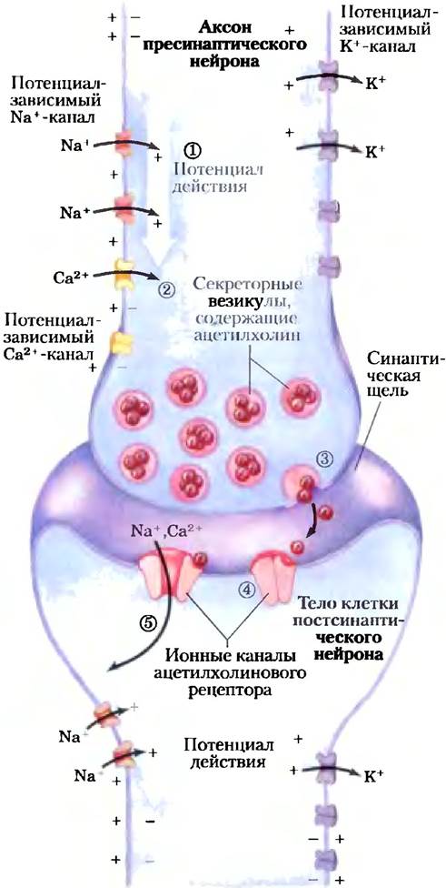
When a wave of depolarization reaches voltage-gated Ca2+ channels, they open (stage (2)), allowing Calcium Ions to enter the cell from the extracellular space. The resulting surge in cytoplasmic Ca2+ concentration triggers the exocytosis of acetylcholine into the synaptic cleft (stage (3)). Acetylcholine then diffuses across to the postsynaptic cell (another neuron or a myocyte), where it binds to its receptor and initiates depolarization. In this manner, the signal is propagated to the next cell in the "chain." Thus, ligand- and voltage-gated ion channels transmit signals in one of two ways: either by altering the cytosolic concentration of an ion (such as Ca2+), which then acts as a secondary messenger (with the primary messenger being a hormone or neurotransmitter), or by altering Vm and thereby affecting other Membrane Proteins sensitive to Vm. Both mechanisms operate when an electrical signal passes from one neuron to the next.
In Section 11.3, we discussed the Structure and MECHANISM OF ACTION of the voltage-gated potassium channel in considerable detail (see Figs. 11-48, 11-49, and 11-50). Here, we focus more closely on the function of sodium channels. The core component of a sodium channel is a large polypeptide (comprising 1,840 amino acid residues) made up of four domains arranged around a central pore (Fig. 12-26a, b), through which sodium ions cross the membrane. Channel Specificity for sodium ions is conferred by a "pore" formed by the loops connecting transmembrane helices 5 and 6 of each domain. Helix 4 in each domain contains a high concentration of positively charged Arg residues; this segment is thought to shift within the membrane in response to Changes in the transmembrane potential from -60 mV (the resting potential) to -30 mV. The movement of helix 4 triggers the opening of the channel, which forms the basis of its voltage-gated mechanism (Fig. 12-26c).
Figure 12-26. Voltage-gated neuronal Na+ channels. While sodium channels from different Tissues and organisms are constructed from various subunits, The primary function is carried out by the major α subunit. (a) The α subunit is a large protein composed of four homologous domains (I through IV, shown separately here for clarity), each containing six transmembrane helices (1 through 6). Helix 4 in each domain (blue) is voltage-sensitive, whereas helix 6 (orange) appears to form the activation gate. The pore region (red), formed by the loops between helices 5 and 6, acts as the selectivity filter, and the segment between domains III and IV (green) serves as the inactivation gate. (b) The four domains wrap around a central transmembrane pore lined by polar amino acid residues. The four pores (red) converge near the outer surface of the membrane to form a selectivity filter, which has an identical structure in all sodium channels. This filter allows the channel to distinguish sodium ions from other ions of similar size. The inactivation gate (green) closes (dashed line) shortly after the activation gate opens. (c) The voltage-sensing mechanism relies on the realignment of helix 4 (blue) perpendicular to the plane of the membrane upon changes in the transmembrane potential. As shown at the top of the figure, the large positive charge on helix 4 causes this helix to be pulled inward when the inner membrane potential (Vm) becomes negative. Upon depolarization, this electrostatic attraction weakens, allowing helix 4 to return to its relaxed state (bottom). This conformational shift is communicated to the activation gate (orange), driving the conformational changes that open the channel upon depolarization.
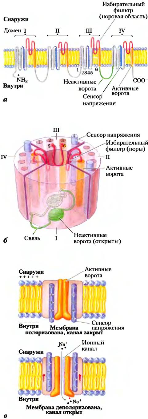
Channel inactivation appears to proceed via a "ball-and-chain" mechanism. A protein domain on the cytoplasmic face of the sodium channel (the inactivation gate, or "ball") is tethered to the channel by a short polypeptide loop (the "chain"; Fig. 12-26b). This domain floats freely on its chain when the channel is closed, but upon channel opening, it binds to a newly exposed complementary site within the channel pore, thereby blocking it. The duration of the open state is presumably determined by the length of the chain: the longer the chain, the longer the channel remains open. Other regulated ion channels likely undergo inactivation via similar mechanisms.
The Acetylcholine Receptor Is a Ligand-Gated Ion Channel
The nicotinic acetylcholine receptor mediates electrical signal transmission at certain types of synapses as well as at neuromuscular junctions (between motor neurons and muscle fibers), signaling Muscles to contract. (Nicotinic and muscarinic acetylcholine receptors were originally distinguished by the sensitivity of the former to nicotine and the latter to the fungal alkaloid muscarine; these receptors possess distinct structures and perform different Functions.) Acetylcholine released by a presynaptic or motor neuron diffuses a few micrometers to the plasma membrane of the postsynaptic neuron or myocyte, where it binds to the acetylcholine receptor. This binding induces a conformational change in the receptor molecule, forcing the ion channel to open. The resulting influx of cations depolarizes the plasma membrane, which in Muscle tissue leads to fiber contraction. The acetylcholine receptor permits the passage of Ca2+ and K+ ions, while excluding other cations and all anions. The flux of sodium ions through the acetylcholine receptor channel is non-saturating (meaning that the ion flow rate is linearly dependent on the extracellular Na+ concentration), and the conduction rate is exceptionally high—approximately 2 • 107 ions per second under physiological conditions.

Like other gated ion channels, the acetylcholine receptor opens in response to a signal molecule and features an internal timing mechanism that closes the channel after a few milliseconds. Consequently, the acetylcholine signal is transient, which, as we have seen, is a crucial feature of efficient electrical signaling. Although we understand the structural rearrangements that lead to the opening of the acetylcholine receptor, the precise mechanism of "desensitization"—whereby the channel pore remains closed even in the prolonged presence of acetylcholine—is not yet fully understood.
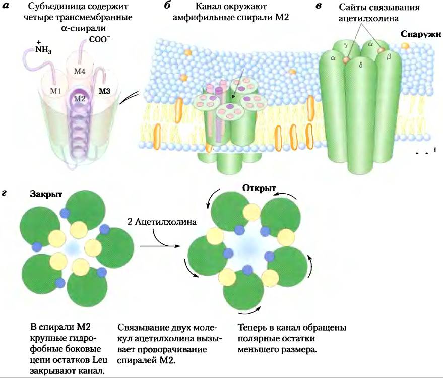
The nicotinic acetylcholine receptor is composed of five subunits: one β, one γ, one δ, and two identical α subunits, each containing an acetylcholine-binding site. All five subunits share a similar Amino Acid Sequence and tertiary structure, and each features four transmembrane helical segments (M1 through M4) (Fig. 12-27a). The five subunits surround a central pore lined by their M2 helices (Fig. 12-27b, c). In the regions projecting beyond the cytoplasmic and extracellular surfaces, the pore is about 20 Â wide, but it narrows within The Lipid Bilayer. Near the center of the lipid bilayer, a ring of bulky hydrophobic leucine side chains from the M2 helices are packed so tightly together that they physically block ion permeation (Fig. 12-27d). The allosteric Conformational changes induced by acetylcholine binding to the two α subunits trigger a slight Rotation of the M2 helices, swinging these hydrophobic residues away from the center of the channel and opening a pathway for ion conduction.
Figure 12-27. The acetylcholine ion channel. (a) Each of the five homologous subunits (α2βγδ) contains four transmembrane helices (M1 through M4). The M2 helix is amphiphilic, whereas the others consist predominantly of hydrophobic amino acid residues. (b) The five subunits surround a central transmembrane pore lined by polar residues of the M2 helices. Rings of negatively charged amino acid residues are located at the extracellular and cytoplasmic entrances of the channel. (c) A model of the acetylcholine receptor based on Electron Microscopy and X-ray crystallographic data of a related protein (molluscan acetylcholine-binding protein). (d) Top view of a cross-section through the center of the M2 helix, showing the side chains of five leucine residues (yellow; one from each M2 helix) projecting into the channel lumen, rendering it too narrow for the passage of Ca2+, Na+, or K+ ions. When both acetylcholine-binding sites (one on each α subunit) are occupied, a conformational change ensues: the M2 helices rotate slightly, moving the five leucine residues out of the channel center and replacing them with smaller polar residues (blue). This opens the channel, allowing the passage of Ca2+, Na+, or K+ ions.
Neurons Contain Receptor Channels That Respond to Various Neurotransmitters
Animal cells, particularly those of the nervous system, harbor a vast array of ion channels that are voltage-gated, ligand-gated, or regulated by both potential and chemical stimuli. The neurotransmitters 5-hydroxytryptamine (serotonin), glutamate, and Glycine can act via receptor channels structurally related to the acetylcholine receptor. Serotonin and glutamate induce the opening of cation channels (K+, Na+, Ca2+), whereas glycine opens Cl- channels. Cation and anion channels differ very slightly in their Amino acid sequences along the hydrophilic pore lining. Cation channels feature negatively charged Glu and Asp side chains at key positions. When several of these acidic residues were experimentally replaced
with basic residues, the cation channel was converted into an anion channel.
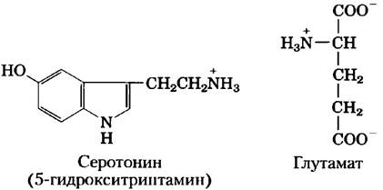
Depending on which ion permeates the channel, the neurotransmitter ligand will either depolarize or hyperpolarize the target cell. A single neuron typically receives input from several (or many) other neurons, with each releasing its characteristic neurotransmitter that exerts either a depolarizing or hyperpolarizing effect. Thus, the Vm of the target cell reflects the integrated input from numerous neurons (Fig. 12-1d). The cell responds by firing an action potential only if the integrated input produces a net depolarization of sufficient magnitude.
The receptor channels for acetylcholine, glycine, glutamate, and γ-aminobutyric acid (GABA) are gated by extracellular ligands. Intracellular secondary messengers—such as cAMP, cGMP (3',5'-cyclic GMP, a close analog of cAMP), IP3 (Inositol-1,4,5-trisphosphate), Ca2+, and ATP—regulate a different class of ion channels which, as we will see in Section 12.10, participate in Sensory Signal Transduction in Vision, Olfaction, and gustation.
Toxins Target Ion Channels
Many potent naturally occurring toxins exert their effects by targeting ion channels. For instance, as mentioned in Section 11.3, dendrotoxin (from black mamba venom) blocks Voltage-Gated Potassium Channels, tetrodotoxin (produced by pufferfish) targets Voltage-Gated Sodium Channels, and cobrotoxin disrupts acetylcholine receptor function. Why did evolution favor ion channels—rather than key metabolic Enzymes—as the primary targets for such toxins?
Ion channels are exceptionally powerful biological amplifiers. A single channel allows roughly 10 million ions to pass per second. Consequently, transmitting a signal requires a relatively small number of ion channel proteins per neuron. As a result, a minute quantity of a high-affinity toxin molecule can profoundly disrupt neural signal transmission in an Organism. Achieving a comparable physiological effect by targeting metabolic enzymes, whose cellular concentrations are typically much higher than those of ion channel proteins, would require a vastly greater number of toxin molecules.
Summary of Section 12.6 Gated Ion Channels
■ Ion channels gated by membrane potential or chemical ligands are key players in signal transmission in neurons and other cell types.
■ Voltage-gated Na+ and K+ channels in neuronal membranes propagate action potentials along the axon as a wave of depolarization (Na+ influx) followed by repolarization (K+ efflux).
■ Voltage-gated channels open when a transmembrane peptide bearing a high charge density (due to Arg or other charged amino acid residues) shifts in a plane perpendicular to the membrane.
■ When an action potential reaches the distal end of a presynaptic neuron, it triggers the release of a neurotransmitter. The neurotransmitter (e.g., acetylcholine) diffuses toward the postsynaptic neuron (or a myocyte at the Neuromuscular Junction), binds to a specific receptor on the plasma membrane, and induces changes in Vm.
■ The acetylcholine receptor of neurons and myocytes is a chemically gated ion channel; binding of acetylcholine leads to conformational changes that open the channel to the passage of Na+ and Ca2+ ions.
■ Neurotoxins produced by many organisms target the ion channels of Nerve Cells, which is why they act rapidly and are lethal.
Last update: 06/08/2026
Editorial and Educational Adaptation: This material has been compiled based on the primary/original source text. The project team performed an editorial review, corrected technical inaccuracies, structured sections, and adapted the content for an educational format.
What was processed:
- elimination of formatting defects (OCR errors, structural breaks, corrupted characters);
- editorial organization of content;
- standardization of terminology in accordance with academic sources;
- verification of factual statements against the original source text.
All mentions of the author, publication year, and origin of the primary text have been preserved in accordance with the source.