LEHNINGER PRINCIPLES OF BIOCHEMISTRY - VOLUME 1. THE FOUNDATIONS OF BIOCHEMISTRY: STRUCTURE AND CATALYSIS - 2011
PART I. STRUCTURE AND CATALYSIS
12. BIOSIGNALING
12.5 Multivalent Adaptor Proteins and Membrane Rafts
The Study of signaling systems discussed thus far leads to two generalizations: (1) protein Kinases that phosphorylate Tyr, Ser, and Thr residues play a central role in signal Transduction; and (2) reversible Protein-Structure/156.html">Protein Interactions, triggered by the reversible phosphorylation of Tyr, Ser, and Thr residues in signaling Proteins, enable these proteins to interact with others. In fact, many signaling proteins are multivalent in the sense that they can interact with several different proteins simultaneously, forming multiprotein signaling complexes. In this section, we present several Examples illustrating the General Principles of protein phosphorylation-dependent interactions in signaling pathways.
Protein Modules Bind Phosphorylated Tyr, Ser, or Thr Residues in Partner Proteins
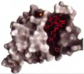
The Grb2 protein in the Insulin signaling pathway (Figs. 12-15 and 12-19) binds via its SH2 domain to other proteins that contain accessible p-Tyr residues. The Human Genome encodes at least 87 SH2-containing proteins, many of which are known to participate in signal transduction. The binding site for the p-Tyr residue is located in a deep pocket of the SH2 domain, where each oxygen atom of the phosphate group participates in hydrogen bonding or Electrostatic Interactions; the positive charges of two Arg residues play a prominent role in binding. Subtle differences in The structure of SH2 domains in various proteins account for the Specificity of their interactions with different p-Tyr-containing proteins. The specificity of these interactions is determined by three to five residues on the C-terminal side of the p-Tyr residue (Fig. 12-21).
Class="center">Figure 12-21 Structure of an SH2 domain and its interaction with a p-Tyr residue in a partner protein (PDB ID 1SHC). The surface of the SH2 domain is outlined in gray. The phosphorus atom of the phosphate group in the interacting p-Tyr is shown as an orange sphere; most of the residue is shaded. The next few residues toward the C-terminus of the partner protein are highlighted in red. The SH2 domain interacts with the p-Tyr (designated as position 0 as the phosphorylated residue) and with the next three residues toward the C-terminus (designated as +1, +2, +3). Some SH2 domains (Src, Fyn, Hck, Nck) tend to interact with negatively charged residues at positions +1 and +2; others (PLC-y1, SHP-2) have an extended hydrophobic cavity that "selects" aliphatic residues at positions +1 through +5. These differences define subclasses of SH2 domains specific for different partners.
Phosphotyrosine-binding (PTB) domains also bind to p-Tyr in partner proteins, but they differ from SH2 domains in their primary sequences and three-dimensional structures. The human genome encodes 24 proteins containing PTB domains, including IRS-1, which we previously encountered as a scaffold protein in insulin signaling (Fig. 12-15). The p-Tyr residues required for the binding of SH2 and PTB domains in partner proteins are generated by Tyrosine kinases and removed by phosphoprotein Phosphatases (PTPases).
Other signaling protein kinases, including PKA, PKC, PKG, and members of the MAPK cascade, phosphorylate Ser or Thr residues in their target proteins, which in some cases acquire The ability to interact with partners via the phosphorylated residue, triggering downstream steps. An alphabetical catalog of domains that bind p-Ser or p-Thr residues has already been compiled, and undoubtedly many more such domains remain to be discovered. Each domain interacts best with a specific sequence surrounding the phosphorylated residue, representing families of highly specific recognition sites capable of binding a specific subset of phosphorylated proteins. The domain-binding partner may even reside within the same protein. Phosphorylation of certain kinases inhibits their activity by favoring the interaction of an SH2 domain with a p-Tyr in another domain of the same enzyme. For example, the soluble Tyr protein kinase Src, phosphorylated at a key Tyr residue, is inactive because for activity its SH2 domain must bind to a substrate rather than to its own internal p-Tyr (Fig. 12-22a). Glycogen synthase kinase 3 (GSK3) is inactive when phosphorylated at a Ser residue within its autoinhibitory domain (Fig. 12-22b). Dephosphorylation of this domain allows the enzyme to bind and phosphorylate target proteins.
In addition to the three most frequently phosphorylated amino acid residues, There is a fourth structure around which supramolecular complexes of signaling proteins assemble: the phosphorylated HEAD group of membrane phosphatidylinositols. Many signaling proteins contain domains similar to SH3 and PH (pleckstrin Homology) domains, which tightly bind PIP3 protruding from the inner monolayer of Cell/30.html">The Plasma Membrane. Proteins that bind to this head group begin to accumulate in the region of the membrane where the enzyme PI3K generates it (such as in response to an insulin signal).
Figure 12-22 Mechanism of autoinhibition in Src and GSK3. (a) In the active form of the Tyr kinase Src, the SH2 domain binds to the p-Tyr of the substrate, and the SH3 domain binds to a Proline-rich region of the substrate, positioning the active center of the kinase parallel to several Tyr residues on the substrate (top). When Src is phosphorylated at a specific Tyr residue (bottom), the SH2 domain binds to the internal p-Tyr instead of the substrate p-Tyr, preventing productive binding of the kinase to its protein substrate; thus, the enzyme is autoinhibited. (b) In the active form of glycogen synthase kinase 3 (GSK3), the internal p-Ser-binding site can bind the p-Ser residue in the substrate (glycogen synthase) and therefore positions the kinase to phosphorylate adjacent Ser residues (top). Phosphorylation of the internal Serine residue allows this internal segment of the kinase to occupy the p-Ser-binding site, blocking substrate binding (bottom).

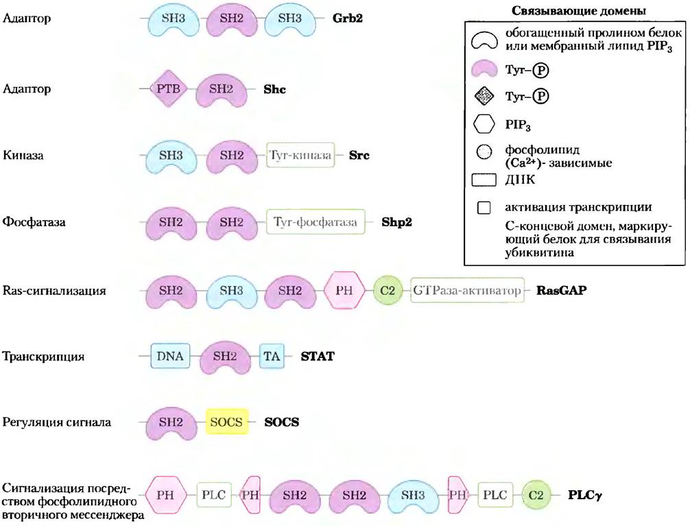
Most plasma Membrane Proteins involved in signal transduction contain one or more protein- or phospholipid-binding domains; many have three or more such domains and can thus interact with multiple signaling proteins simultaneously. Figure 12-23 illustrates just a few of the many known multivalent signaling proteins. Many complexes include components whose specialized domains are responsible for membrane binding. Because a significant portion of signal transduction occurs at the inner surface of the plasma membrane, it is natural that the localization of molecules that must collide with each other for further signal propagation is restricted to a two-dimensional space—the membrane surface. The probability of collision in a two-dimensional space is far higher than in the three-dimensional space of the Cytosol.
Figure 12-23 Selected binding modules of signaling proteins. Each protein is represented by a line (N-terminus on the left); symbols indicate the localization of conserved binding domains (for their specificity, see text; PH denotes pleckstrin homology; other Abbreviations are defined in the text); catalytic activity is shown by green rectangles. Each protein is named by its C-terminus. These signaling proteins interact with phosphorylated proteins or Phospholipids in numerous combinations to form integrated signaling complexes.
In summary, the study of numerous signaling proteins and their multiple binding domains has revealed a striking picture of cellular signal transduction pathways. An initial signal triggers the phosphorylation of a receptor or target protein, Setting into motion large multiprotein complexes held together on scaffolds formed by multivalent adaptor proteins. Some of these complexes contain multiple protein kinases that sequentially activate one another, forming a phosphorylation cascade and providing substantial Amplification of the initial signal. Interactions between protein kinases in the cascade do not occur via random collisions in three-dimensional space. For example, in the MAPK cascade, an entire family of adaptor proteins serves to bind all three protein kinases (MAPK, MAPKK, and MAPKKK), bringing them into close proximity, ensuring proper orientation, and imparting an allosteric character to their interaction so that sequential phosphorylation occurs even in response to very weak stimuli.
Phosphotyrosine phosphatases (PTPases) remove phosphate groups from p-Tyr residues, reversing the effects of phosphorylation. Some of these phosphatases are receptor-like membrane proteins that are likely regulated by as-yet-unidentified extracellular factors; other PTPases are soluble and contain SH2 domains. Additionally, animal Cells contain protein phosphoserine and protein phosphothreonine phosphatases, which reverse the effects of Ser- and Thr-specific protein kinases. Signal transduction occurs via protein "circuits" efficiently established by signaling receptors in response to effectors and capable of being switched off instantaneously through the Hydrolysis of a single phosphoester bond.
The multivalency of signaling proteins enables the assembly of vast arrays of diverse signaling module combinations, each presumably corresponding to specific signals, cell types, and metabolic states. The multitude of protein kinases and phosphoprotein-binding domains, each with its own specificity (required consensus sequence in the substrate), generates numerous combinations and exceptionally complex signaling circuits.
Membrane Rafts and Caveolae Can Segregate Signaling Proteins
Membrane rafts (p. 543) are Regions of the membrane bilayer enriched in Sphingolipids, sterols, and certain proteins, many of which are attached to the bilayer via GPI anchors.
The adrenergic receptor migrates into rafts containing G proteins, adenylyl cyclase, PKA, and a specific protein phosphatase, PP2A, which together form a highly integrated signaling unit. Isolating all the elements required to initiate and terminate a response to a signal within a small patch of the plasma membrane allows The Cell to rapidly increase the concentration of a secondary messenger in a strictly confined space.
Certain receptor tyrosine kinases (such as EGF-R and PDGF-R) appear to localize in rafts, and such segregation presumably has functional significance. When Cholesterol is depleted from rafts by Treatment with cyclodextrin (which binds cholesterol and extracts it from membranes), the rafts disintegrate, and certain signaling pathways are impaired.
If a receptor tyrosine kinase within a raft is phosphorylated, but the phosphotyrosine phosphatase that removes this phosphorylation resides in a different raft, the dephosphorylation of the Tyr kinase will be slowed down or entirely prevented. Interactions between adaptor proteins may be strong enough to pull a signaling protein into a raft or push receptors out of a raft. For example, EGF receptors in isolated fibroblasts are typically concentrated in specialized rafts called caveolae (Fig. 11-21), but upon treatment with EGF, the receptors leave the raft. This migration depends on The activity of the receptor protein kinase; mutant receptors lacking this activity remain in the raft upon EGF treatment. Caveolin, an integral membrane protein localized in caveolae, is phosphorylated at a Tyr residue in response to insulin and, following phosphorylation, may allow the now-activated EGF receptor to pull its binding partners into the raft. The segregation of signaling proteins into rafts adds another "dimension" to the already complex processes initiated by extracellular signals.
Summary of Section 12.5 Multivalent Adapter Proteins and Membrane Rafts
■ Many signaling proteins contain domains that bind to phosphorylated Tyr, Ser, or Thr residues in other proteins; the binding specificity for each domain is determined by the Amino acid sequences flanking the phosphorylated residue in the substrate.
■ SH2 and PTB domains bind to proteins containing p-Tyr residues; other domains bind p-Ser and p-Thr residues in various contexts.
■ SH3 and PH domains bind the membrane phospholipid PIP3.
■ Many signaling proteins are multivalent, containing multiple distinct binding modules. By combining the substrate specificity of different protein kinases with the specificity of domains that bind phosphorylated Tyr, Ser, or Thr residues, alongside phosphatases that can rapidly shut down the signaling pathway, cells assemble a vast array of multi-protein signaling complexes.
■ Membrane rafts and caveolaes compartmentalize groups of signaling proteins within specialized microdomains of the plasma membrane, facilitating their interactions and enhancing the efficiency of signal transduction.
Last update: 06/08/2026
Editorial and Educational Adaptation: This material has been compiled based on the primary/original source text. The project team performed an editorial review, corrected technical inaccuracies, structured sections, and adapted the content for an educational format.
What was processed:
- elimination of formatting defects (OCR errors, structural breaks, corrupted characters);
- editorial organization of content;
- standardization of terminology in accordance with academic sources;
- verification of factual statements against the original source text.
All mentions of the author, publication year, and origin of the primary text have been preserved in accordance with the source.