Review of Medical Physiology - William F. Ganong 2002
Functions of the Nervous System
Regulation of Posture and Movement
General Principles
Two Types of muscular activity occur in the body: reflex (or involuntary) and voluntary. Rhythmic movements such as swallowing, chewing, scratching, and walking could also be classified as reflex activities since they are largely involuntary, yet they remain subject to voluntary adjustment and control.
Much remains unknown regarding The regulation of voluntary movements. To execute them—for instance, within a limb—the Brain must generate a movement plan, program the simultaneous coordination of corresponding movements across multiple joints, and regulate these actions by comparing the initial plan with the actual outcome. The motor system "learns" through action execution, and movement performance is refined via repetition, promoting synaptic plasticity. A GENERALIZED SCHEME OF motor activity regulation is likely depicted in Fig. 12-1. Commands for voluntary movements originate in the cortical association areas. Movement programming occurs within the cortex, Basal Ganglia, and lateral Regions of the cerebellar hemispheres as a consequence of increasing electrical activity preceding movement. Information from the basal ganglia and cerebellar hemispheres is relayed via the thalamus to the premotor and motor cortices. Motor commands from the motor cortex travel primarily through the corticospinal and corresponding corticobulbar pathways to the motor Neurons of the Brainstem. However, collaterals of these pathways and several direct connections extending from the motor cortex to brainstem nuclei also transmit impulses to the motor neurons of the brainstem and Spinal Cord, thereby mediating voluntary movements. Movement alters the streams of sensory nerve impulses originating from Muscles, tendons, joints, and Skin. This feedback information, essential for ensuring the precision and smoothness of movements, is channeled directly to the motor cortex and the spinocerebellum, which subsequently transmits information to the brainstem nuclei. The primary brainstem pathways involved in posture and coordination are the rubrospinal, reticulospinal, tectospinal, vestibulospinal, and corresponding pathways directed toward brainstem motor neurons.
Class="center">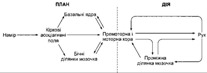
Fig. 12-1. Regulation of voluntary movements.
Innervation of the Axial and Distal Musculature
When examining the regulation of motor activity, it is also important to note that within the brainstem and spinal cord, medial or ventral pathways are responsible for controlling the Muscles of the Trunk and proximal limb segments, whereas lateral pathways control the muscles of the distal limb segments. Axial musculature maintains body posture and executes simple, primitive movements, while the muscles of the distal limb segments perform fine and precise movements. Accordingly, neurons in the medial part of the anterior horn innervate proximal limb muscles (flexors, in particular), whereas neurons in its lateral part innervate distal limb muscles. Similarly, the anterior corticospinal tract and medially located descending pathways originating from the brainstem (tectospinal, reticulospinal, and vestibulospinal tracts) regulate proximal muscles and body posture, whereas the lateral corticospinal and rubrospinal tracts regulate distal limb muscles—specifically, in the case of the lateral corticospinal tract, the execution of precise and fine voluntary movements. Phylogenetically, medial pathways are older, while lateral pathways are newer.
Other Terms
Because the fibers of the lateral corticospinal tract form the pyramids within the Medulla Oblongata, the corticospinal pathways were designated as the pyramidal system. The remaining descending Pathways of the brainstem and spinal cord that do not pass through the pyramids and are associated with postural regulation are termed the extrapyramidal system. However, the anterior corticospinal tract does not pass through the pyramids, and many pyramidal fibers perform other Functions; furthermore, the extrapyramidal system likewise encompasses various pathways associated with a multitude of different functions. Consequently, the terms pyramidal and extrapyramidal are not entirely accurate and should presumably be avoided.
In addition, the motor system is frequently subdivided into upper and lower motor neurons. Damage to lower motor neurons—the spinal and cranial motor neurons that directly innervate muscles—results in flaccid paralysis, Muscle atrophy, and loss of Reflexes. Spastic paralysis syndrome and hyperactive stretch reflexes occurring without muscle atrophy are the consequences of upper motor neuron destruction (neurons within the BRAIN AND SPINAL cord that activate motor neurons). However, it must be borne in mind that there are Three types of upper motor neurons. Damage to pathways regulating posture causes spastic paralysis, whereas damage restricted to the Corticospinal and Corticobulbar pathways leads to paresis rather than paralysis, leaving the affected musculature predominantly hypotonic. Cerebellar damage results in impaired coordination. Therefore, the term upper motor neuron is also misleading.
Last update: 10/08/2026
Editorial and Educational Adaptation: This material has been compiled based on the primary/original source text. The project team performed an editorial review, corrected technical inaccuracies, structured sections, and adapted the content for an educational format.
What was processed:
- elimination of formatting defects (OCR errors, structural breaks, corrupted characters);
- editorial organization of content;
- standardization of terminology in accordance with academic sources;
- verification of factual statements against the original source text.
All mentions of the author, publication year, and origin of the primary text have been preserved in accordance with the source.